UNIVERSIDADE DE LISBOA
FACULDADE DE MEDICINA VETERINÁRIA
DIROFILARIA IMMITIS AND ANGIOSTRONGYLUS VASORUM: EPIDEMIOLOGY AND IMPACT OF MAJOR HEARTWORMS IN CARNIVORES IN PORTUGAL
Ana Margarida Pignateli Vasconcelos de Assunção Alho
Orientadores: Professor Doutor Luís Manuel Madeira de Carvalho Professora Doutora Silvana Maria Duarte Belo Professor Doutor Peter Deplazes
Tese especialmente elaborada para obtenção do grau de Doutor em Ciências Veterinárias na Especialidade de Sanidade Animal
Júri:
Presidente: Professor Doutor Rui Manuel de Vasconcelos e Horta Caldeira Vogais: Professor Doutor Adolfo Paz-Silva
Professor Doutor Luís Miguel Martins Lucas Cardoso Professor Doutor Luís Manuel Madeira de Carvalho
Professora Doutora Isabel Maria Soares Pereira da Fonseca de Sampaio Professor Doutor José Augusto Farraia e Silva Meireles
“I have been impressed with the urgency of doing. Knowing is not enough; we must apply. Being willing is not enough; we must do.”
Leonardo Da Vinci
This work is dedicated to my family, for all their support, optimism and understanding.
ACKNOWLEDGEMENTS
This work would have not been accomplished without the collaboration and help of many people and institutions, to which I address my sincere thanks. In particular, I would like to thank to:
My supervisor, Professor Luís Madeira de Carvalho, for his strong friendship, invaluable understanding and encouragement during this Parasitological journey. For all his confidence, knowledge and strength to go forward. A deep thank for guiding and advising me in my scientific career and for being a permanent reference.
My co-supervisor Professor Silvana Belo, who gave me the opportunity to embrace a national Project on Dirofilaria, that triggered my access into the scientific research community. For all her patience, enthusiastic help, critical discussions and prompt support during this intensive partnership between IHMT and FMV.
My co-supervisor Professor Peter Deplazes, for his brilliant guidance and support, constructive criticism and for welcoming me in his Laboratory. My deep thanks to the Institute of Parasitology, University of Zurich, and to all the friendly staff involved throughout this path. Professor José Meireles for his strong encouragement, advises and priceless stories during our exhaustive field work. For his inestimable and true friendship!
Professor Luís Cardoso, for his infinite support and commitment, guiding me in this hard residency programme at the European Veterinary Parasitology College (EVPC). For his inestimable friendship and for his knowledge, the constructive criticism, brainstorming sessions and enlightened advices. For believing in me!
Dr. Manuela Schnyder for her profound knowledge and wise guidance.
Professor Domenico Otranto, for all the opportunities and powerful collaborations provided. For being such a great personal and professional reference.
Professor Laura Rinaldi for her friendship and for inspiring me every day.
To Doctor Ana Amaro, Doctor Fátima Amaro, Doctor Hans-Peter Fuehrer, Professor Isabel Fonseca, Professor Jorge Correia, Lídia Gomes, Professor Manuela Calado, Dr. Roland Schaper, for their friendship, support and scientific collaboration.
To CIISA, my second home, and to Professor Luís Costa, for all his support and prompt help. To Dr. Hugo Pissarra for all the long conversations and wise advices.
To my EVPC residency colleagues Adnan Hodzic, Alessio Giannelli, Simone Manzocchi, Vito Colella and COST action network friends for sharing this passion for Parasitology during this tough journey.
Sinclair Owen for officially being the first reader of every publication. For his patience, critic and clever revision and for helping me so much during these hardworking times.
Clara Lima for her friendship and for sharing with me so many impossible projects.
Carla Motolla and Joana Dias for their unique and invaluable friendship. For all their support, great advices, motivation, endless conversations and for truly being always by my side!
Rui Seixas for his deep complicity and permanent friendship since the first day at the Faculdade de Medicina Veterinária, in 2005. For all his help, intelligence, critical analysis and advices. For the dozens of challenges that we have faced together and for being such a great best friend! All my Family, in particular my parents, my sister, Tita and Daniel for their unconditional support, love and attention. For being my example of incessant willingness to do better! All those that I did not mention, but have somehow been with me during this professional and personal journey. Thanks to all!
FUNDING
The present work was funded by the PhD Research Grant SFRH/BD/85427/2012 and by the research projects PTDC/SAU-SAP/113523/2009 and UID/CVT/00276/2013 (Centre for Interdisciplinary Research in Animal Health, CIISA, Faculdade de Medicina Veterinária, Universidade de Lisboa), supported by Fundação para a Ciência e a Tecnologia (FCT), Portugal.
Additional funding was given from the Institut für Parasitologie, Universität Zürich, as well as from Instituto de Higiene e Medicina Tropical, Universidade Nova de Lisboa, for laboratory materials and reagents. The TD1303 COST Action “European network for neglected vectors and vector-borne infections” provided funding for scientific missions, training schools and annual network conferences.
Title: Dirofilaria immitis and Angiostrongylus vasorum: epidemiology and impact of major heartworms in carnivores in Portugal
ABSTRACT
Cardiopulmonary nematodes, Dirofilaria immitis and Angiostrongylus vasorum, are severe and life-threatening parasites that are increasingly reported throughout Europe. However, in Portugal, accurate data on both illnesses is scarce, hampering the awareness and the implementation of effective prevention and control strategies. A multidisciplinary study was therefore designed to characterize and assess the current situation regarding dirofilariosis and angiostrongylosis in carnivores from Portugal.
A national survey was conducted to assess the prevalence and distribution of D. immitis and A. vasorum in canine and red fox populations in Portugal, confirming the occurrence of both diseases in canids either from northern, central and southern regions. An overall prevalence of 11.9% and 0.7% dogs, and 8.5% and 12.7% foxes were positive to D. immitis and A. vasorum, respectively. Additionally, a high prevalence of D. immitis was found in pinnipeds kept at an oceanographic park in the Algarve region where the South African fur seal was also reported as a new host for D. immitis infection. DNA of the endosymbiont bacterium Wolbachia pipientis was detected in dogs naturally infected with D. immitis in Portugal. The transmission risk period for Dirofilaria spp. in Portugal was estimated, showing that although transmission is markedly seasonal, the necessary climatic factors are starting earlier and lasting longer than the summer. A new minimally invasive surgical technique was developed to extract D. immitis adult worms from the hearts of dogs through transjugular catheterization with a non-traumatic snare. A questionnaire conducted on Portuguese pet owners showed that the majority deworm their dogs at irregular and consequently ineffective intervals and their level of knowledge about parasites and parasitic diseases is low.
Although exposure may differ depending on the region of Portugal, the likelihood of cardiopulmonary dirofilariosis and angiostrongylosis is considerable nationwide. Active surveillance, increasing awareness and regular preventive measures are crucial to control cardiopulmonary parasitic diseases in carnivores in the country.
Título da Tese: Dirofilaria immitis e Angiostrongylus vasorum: epidemiologia e impacto dos principais nemátodes cardiopulmonares em carnívoros em Portugal
RESUMO
A dirofilariose e a angiostrongilose são doenças parasitárias crescentemente notificadas em toda a Europa, representando uma ameaça grave para a saúde animal. São causadas respetivamente pelos nemátodes cardiopulmonares Dirofilaria immitis (agente transmitido por mosquitos culicídeos) e Angiostrongylus vasorum (agente transmitido por moluscos gastrópodes). Os fatores subjacentes a esta expansão são diversos, destacando-se a globalização, as alterações climáticas, a conjuntura socioeconómica, espécies invasoras com capacidade vetorial, urbanização de hospedeiros silvestres, bem como novos métodos de diagnóstico. Dados concretos sobre a prevalência e distribuição são cruciais para o controle de doenças animais e, no caso de D. immitis e Dirofilaria repens, também para o controlo de doenças potencialmente zoonóticas. Contudo, em Portugal, a informação existente à data de início deste trabalho era escassa e pontual. Com o intuito de clarificar esta situação e colmatar esta lacuna, foi delineado o presente trabalho para caracterizar a situação da dirofilariose e da angiostrongilose em Portugal.
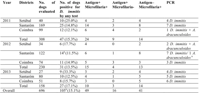
Como primeiro objetivo pretendeu-se avaliar a prevalência atual e áreas de distribuição de Dirofilaria spp. e A. vasorum em canídeos domésticos e selvagens em Portugal. Relativamente à dirofilariose, foram testados 696 canídeos domésticos de três distritos do centro de Portugal (Coimbra, Santarém e Setúbal), durante 2011, 2012 e 2013. Para avaliação das espécies de Dirofilaria circulantes, foram utilizadas técnicas parasitológicas diretas, serológicas e moleculares. Setúbal registou a maior prevalência (24,8%), seguido de Coimbra (13,8%) e Santarém (13,2%), observando-se uma prevalência média global de 15,1%. A espécie D. immitis foi detetada nos três distritos, durante os três anos de estudo, com uma prevalência crescente. Em 2014, dada a escassez de dados epidemiológicos nas regiões transfronteiriças de Portugal e Espanha, este estudo foi alargado a mais sete distritos (i.e., Beja, Bragança, Castelo Branco, Évora, Faro, Guarda e Portalegre), envolvendo mais 248 canídeos. Beja foi o distrito que registou maior número de casos de infeção por D. immitis (8.9%), seguido pela Guarda (6,7%), Faro (2,7%) e Castelo Branco (2,5%). Não foram registados casos positivos de infeção por D. immitis em Bragança, Évora e Portalegre, e não foram observadas outras espécies de Dirofilaria em todo o País. No total observou-se uma prevalência global de 11,9% de cães positivos a D. immitis nos dez distritos, um valor superior comparativamente ao reportado anteriormente.
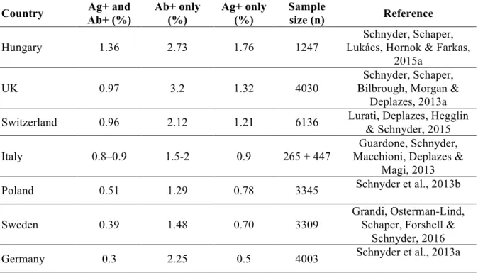
No que respeita à angiostrongilose, foi efetuado o primeiro rastreio serológico nacional de infeção por A. vasorum. Foram testados 906 canídeos domésticos provenientes de 16 distritos (i.e., Beja, Braga, Bragança, Castelo Branco, Coimbra, Évora, Faro, Guarda, Lisboa, Portalegre, Porto, Santarém, Setúbal, Viana do Castelo, Vila Real e Viseu). Foi utilizada a nova técnica de imunoabsorção enzimática (ELISA) de elevada sensibilidade para deteção de antigénios circulantes de A. vasorum e de anticorpos específicos contra este parasita. Observou-se um total de 0,7% de animais positivos simultaneamente para antigénio e anticorpo, dispersos pelas zonas norte, centro e sul de Portugal, indicando a presença de infeções ativas de A. vasorum distribuídas pelo País. Adicionalmente observou-se 1,3% de animais positivos apenas para anticorpo de A. vasorum, sugerindo uma exposição prévia ao parasita. No geral, esta prevalência foi semelhante à registada noutros países europeus. Este rastreio permitiu não só confirmar a endemicidade de A. vasorum em cães de diferentes zonas geográficas de Portugal, como colmatar a lacuna de conhecimento deste parasita em Portugal.
Atendendo ao facto de os cães militares constituírem um grupo de risco de exposição a doenças transmitidas por vetores pela natureza da sua atividade e longos períodos de permanência no exterior, foi efetuado um rastreio de infeção por D. immitis e A. vasorum à população de canídeos da Força Aérea Portuguesa. Foram testados 100 animais, mantidos em bases militares localizadas em Aveiro, Beja, Leiria, Lisboa, Setúbal, Madeira e Açores. Dos animais testados, 5% apresentaram anticorpos específicos contra A. vasorum (Aveiro, Lisboa e Setúbal) e 1% antigénios circulantes de A. vasorum. Não foram observados casos de infeção para D. immitis. Este rastreio permitiu alargar a informação sobre a presença e distribuição geográfica de D. immitis e A. vasorum em Portugal.
Com o objetivo de avaliar a prevalência de D. immitis e A. vasorum em canídeos silvestres, foi efetuado um rastreio serológico a 118 raposas (Vulpes vulpes) abatidas durante a época oficial de caça, provenientes de oito distritos de Portugal, distribuídos por duas regiões: Norte (Aveiro, Braga, Bragança, Porto, Viana do Castelo e Vila Real) e Sul (Évora e Setúbal). Observou-se uma prevalência de 8,5% de animais positivos para D. immitis. Adicionalmente, foi encontrada uma prevalência de 12,7% de animais positivos para A. vasorum, dos quais 5,9% simultaneamente positivos para antigénios e anticorpos de A. vasorum, 5,1% positivos exclusivamente para antigénios de A. vasorum e 1,7% positivos exclusivamente para anticorpos de A. vasorum. Os animais positivos para dirofilariose cardiopulmonar e angiostrongilose foram detetados nas regiões Norte e Sul de Portugal, ampliando a distribuição destes agentes a nível silvestre e destacando o seu potencial papel como reservatório para carnívoros domésticos.
Embora os hospedeiros definitivos da dirofilariose sejam principalmente canídeos domésticos e selvagens, D. immitis apresenta baixa especificidade de hospedeiro vertebrado, sendo capaz de infetar outras espécies de mamíferos silvestres, incluindo aquáticos. Durante a execução deste trabalho, surgiu a possibilidade de rastrear uma população de pinípedes mantidos num parque oceanográfico no Algarve. Foi encontrado DNA de D. immitis em três espécies distintas (focas comuns, leões-marinhos Californianos e Otárias Sul-Africanas), com uma prevalência global de 43.8%, e foi pela primeira vez documentada a Otária Sul Africana (Arctocephalus pusillus pusillus) como hospedeiro de D. immitis.
Tendo em conta as múltiplas implicações da bactéria Wolbachia spp. na patogénese da dirofilariose, outro dos objetivos deste projeto consistiu na pesquisa deste agente em canídeos domésticos, naturalmente infetados com D. immitis em Portugal. Utilizando métodos de deteção molecular, foi encontrado DNA de Wolbachia pipientis em 52,6% de amostras testadas, tendo-se deste modo efetuado a descrição deste endosimbionte em populações caninas em Portugal.
Adicionalmente, foram utilizadas ferramentas geoespaciais para avaliação do risco de transmissão de dirofilariose em Portugal, utilizando o modelo graus-dia. Recorreu-se aos valores médios diários de temperaturas registados na última década (de 2003 a 2013), em cinco estações meteorológicas: Faro, Funchal, Lisboa, Porto e São Miguel. Consideraram-se como pré-requisitos um limiar de temperatura de 14°C, 130 unidades de desenvolvimento de Dirofilaria spp., e uma expetativa máxima de vida dos vetores de 30 dias. A região da Madeira foi a que registou um maior número de dias com condições compatíveis à transmissão de D. immitis (média de 209,9 dias/ano), seguida de Faro (175,2 dias/ano), Lisboa (163,5 dias/ano), Açores (140 dias/ano) e Porto (117,2 dias/ano). Observou-se também na última década um período médio de risco de transmissão desta parasitose de 8 meses/ano na Madeira, 6,9 meses/ano em Faro, 6,4 meses/ano em Lisboa, 5,6 meses/ano nos Açores e de 5 meses/ano no Porto.
Dada a elevada virulência de D. immitis e complicações tromboembólicas associadas, outro dos objetivos deste estudo consistiu na elaboração de uma nova abordagem terapêutica minimamente invasiva. Para isso, desenvolveu-se um método não traumático de remoção percutânea de vermes adultos de D. immitis, baseado na utilização de fio coronário para extração de espécimes de vermes da artéria pulmonar e ventrículo direito, que se revelou eficaz. Outros dos objetivos deste trabalho consistiu em caracterizar as práticas de desparasitação e o conhecimento dos proprietários de animais de companhia sobre doenças parasitárias, tendo sido efetuado um questionário aos utentes do Hospital da Faculdade de Medicina Veterinária da Universidade de Lisboa. Observou-se que apesar da generalidade dos proprietários desparasitar
o seu animal, esta prática é efetuada a intervalos irregulares e consequentemente ineficazes: apenas 11,8% dos cães são desparasitados internamente com a periodicidade aconselhada (i.e., mínimo trimestral) e apenas 28,4% estão protegidos continuamente ao longo do ano contra artrópodes. Adicionalmente 37% refere não efetuar a recolha de dejetos dos seus cães em todos os locais públicos, contribuindo para a contaminação ambiental. O nível de conhecimento público sobre parasitas e doenças parasitárias ainda é reduzido no País, com 88% dos donos afirmando nunca ter ouviu falar de "verme do coração" e 85% de "zoonose".
Como corolário deste trabalho, foi efetuada uma caracterização da situação epidemiológica nacional da dirofilariose e da angiostrongilose cardiopulmonar em canídeos nos últimos vinte anos.
Conclui-se que apesar do risco de exposição dos carnívoros ser variável de acordo com a região geográfica de Portugal, a probabilidade de infeção por D. immitis e A. vasorum é considerável em todo o País, ainda que se observe uma prevalência de D. immitis muito superior à de A. vasorum.
Tendo em conta as atuais alterações climáticas e sociodemográficas, prevê-se um aumento do risco de infeção por estes agentes em áreas endémicas e não endémicas. Atendendo à gravidade da patologia provocada por D. immitis e A. vasorum e considerando o potencial zoonótico e impacto em saúde pública da Dirofilaria spp., estes dados alertam para a necessidade de sensibilização da comunidade médico-veterinária e da população em geral, para a realização de uma profilaxia anti-parasitária dirigida e regular em animais de companhia em Portugal.
Palavras-chave: Angiostrongylus vasorum, Dirofilaria immitis, epidemiologia, Portugal,
INDEX
Chapter 1 – Bibliographic review and research objectives ...1
1.1. General introduction to major canine heartworms ...2
1.2. Introduction and historical perspective of the parasite Dirofilaria spp. ...2
1.3. Epidemiology of Dirofilaria spp. ...3
1.3.1. General distribution ...3
1.3.2. Climate-matching model and seasonality ...5
1.3.3. Risk factors and populations at risk ...6
1.4. Biology of Dirofilaria spp. ...7
1.4.1. Life-cycle ...7
1.4.2. Host range and specificity ...9
1.4.3. Role of wildlife hosts ...9
1.4.4. Morphological characteristics of Dirofilaria immitis and Dirofilaria repens...9
1.4.5. Role of the symbiotic bacteria Wolbachia spp. ...10
1.5. Pathophysiology and clinical signs of Dirofilaria spp. ...10
1.5.1. Respiratory and cardiovascular signs caused by Dirofilaria immitis ...11
1.5.2. Other clinical signs caused by Dirofilaria immitis ...12
1.5.3. Clinical signs caused by Dirofilaria repens ...12
1.6. Diagnosis of canine dirofilariosis ...12
1.6.1. Parasitological diagnosis ...12 1.6.2. Immunological diagnosis ...14 1.6.3. Molecular diagnosis ...14 1.6.4. Laboratory diagnosis ...14 1.6.5. Imaging findings ...15 1.6.6. Pathological findings ...15
1.6.7. Difficulties and misconceptions in diagnosing canine dirofilariosis ...16
1.7. Treatment of canine dirofilariosis ...17
1.7.1. Anthelmintic therapy ...17
1.7.2. Supportive therapy ...19
1.7.3. Surgical intervention for Dirofilaria immitis worm’s extraction ...20
1.7.4. Prognosis ...20
1.8. Control and prevention of Dirofilaria spp. infections ...20
1.9. Public health relevance of Dirofilaria immitis and Dirofilaria repens ...21
2.1 Introduction and historical perspective of the parasite Angiostrongylus vasorum ...23
2.2 Epidemiology of Angiostrongylus vasorum ...23
2.2.1 General distribution ...23
2.2.2 Climate-matching model and seasonality ...25
2.2.3 Risk factors and populations at risk ...26
2.3 Biology of Angiostrongylus vasorum ...27
2.3.1 Life-cycle ...27
2.3.2 Host range and specificity ...29
2.3.3 Role of wildlife hosts ...29
2.3.4 Morphological characteristics of Angiostrongylus vasorum ...30
2.4 Pathophysiology and clinical signs of Angiostrongylus vasorum ...30
2.4.1 Respiratory and cardiovascular signs ...31
2.4.2 Coagulopathy signs ...32
2.4.3 Neurological signs ...33
2.5 Diagnosis of canine cardiopulmonary angiostrongylosis ...33
2.5.2 Immunological diagnosis ...35
2.5.3 Molecular diagnosis ...35
2.5.4 Laboratory diagnosis ...36
2.5.5 Imaging findings ...36
2.5.6 Pathological findings ...37
2.5.7 Difficulties and misconceptions in diagnosing canine angiostrongylosis ...38
2.6 Treatment of canine cardiopulmonary angiostrongylosis ...38
2.6.1 Anthelmintic therapy ...38
2.6.2 Supportive therapy ...39
2.6.3 Prognosis ...40
2.7 Control and prevention of Angiostrongylus vasorum infections ...41
2.8 Public health relevance of Angiostrongylus cantonensis and Angiostrongylus costaricensis ...42
3.1 Background and research objectives ...45
Chapter 2 - Epidemiological survey of Dirofilaria immitis and Angiostrongylus vasorum in domestic dogs in Portugal ...50
2.1 Chapter 2, Scientific publication 1 - Prevalence and seasonal variations of canine dirofilariosis in Portugal ...51
2.2 Chapter 2, Scientific publication 2 - Cardiopulmonary and gastrointestinal parasites in dogs – epidemiological study in shelters from continental Portugal ...67
2.3 Chapter 2, Scientific publication 3 - Molecular characterization of Dirofilaria spp. circulating in Portugal ...70
2.4 Chapter 2, Scientific publication 4 - Seroprevalence of circulating Angiostrongylus vasorum antigen and parasite-specific antibodies in dogs from Portugal ...87
2.5 Chapter 2, Scientific publication 5 - Seroprevalence of vector-borne pathogens and molecular detection of Borrelia afzelii in military dogs from Portugal ...99
Chapter 3 - Epidemiological survey of Dirofilaria immitis and Angiostrongylus vasorum in wild carnivores in Portugal ...112
3.1 Chapter 3, Scientific publication 1 - Serological survey of the heartworms Dirofilaria immitis and Angiostrongylus vasorum in red foxes (Vulpes vulpes) from Portugal ...113
3.2 Chapter 3, Scientific publication 2 - Dirofilaria immitis in pinnipeds and new host record ...115
Chapter 4 – DNA detection of the endosymbiont Wolbachia pipientis in Dirofilaria infected dogs in Portugal ...127
4.1 Chapter 4, Scientific publication 1 - Detection of Wolbachia in Dirofilaria infected dogs in Portugal ...128
Chapter 5 – Assessment of the transmission risk of Dirofilaria spp. in Portugal ...136
5.1 Chapter 5, Scientific publication 1 - Transmission risk of Dirofilaria spp. in Portugal ...137
Chapter 6 - A new mechanical therapeutic approach for removal of Dirofilaria immitis
...139
6.1 Chapter 6, Scientific publication 1 - A homemade snare: an alternative method for mechanical removal of Dirofilaria immitis in dogs ...140
Chapter 7 - Parasite control practices and public perception of pet owners in Portugal ...153
7.1 Chapter 5, Scientific publication 1 - Parasite control practices in companion animals: a survey of dog and cat owners ...154
Chapter 8 - The current situation of two major canid heartworms in Portugal ...173
8.1 Chapter 8, Scientific publication 1 - Dirofilaria immitis and Angiostrongylus vasorum: the current situation of two major canid heartworms in Portugal ...174
Chapter 9 - General discussion and conclusions ...193
9.1 General discussion ...194
9.2 Limitations of the study and future perspectives ...203
9.3 Conclusions ...204
LIST OF FIGURES
Chapter 1 ...1
Figure 1 - Geographical distribution of canine dirofilariosis ...5 Figure 2 - Life cycle of Dirofilaria immitis ...8 Figure 3 - Adult nematodes of Dirofilaria immitis collected at the necropsy of a pinniped and a domestic dog ...11 Figure 4 - Microfilariae of Dirofilaria immitis in a fresh blood smear, stained by the Modified Knott's technique and stained by the acid phosphatase, highlighting the anal and excretory pores ...13 Figure 5 - Predicted geographical distribution of Angiostrongylus vasorum ...26
Figure 6 - Life cycle of Angiostrongylus vasorum ...28 Figure 7 - Adult nematodes of Angiostrongylus vasorum collected at the necropsy of a domestic dog ...31 Figure 8 – Tail of a first stage larvae of Angiostrongylus vasorum collected using Baermann migration-sedimentation test, highlighting the characteristic sinus wave s-shaped curve with a dorsal spine in the tail ...34 Figure 9 - Overall prevalence of canine dirofilariosis in dogs from Portugal ...195
Chapter 2, Scientific publication 1 ...51
Figure 1 - Microfilaria of Dirofilaria immitis after acid phosphatase histochemical staining ...58 Figure 2 - Gel electrophoresis of filarial PCR products in 1.5% agarose gel ...59 Figure 3 - Dirofilaria immitis evolution trend during 2011, 2012 and 2013 in Coimbra, Santarém and Setúbal districts of Portugal ...59
Chapter 2, Scientific publication 3 ...70
Figure 1 - Alignment of heterozygous ITS1 sequences of Dirofilaria immitis from Portuguese canine samples ...77 Figure 2 - Alignment of heterozygous ITS2 sequences of Dirofilaria immitis from Portuguese canine samples ...78
Chapter 2, Scientific publication 4 ...87
Figure 1 - Occurrence of Angiostrongylus vasorum detected by ELISA in 906 dogs from Portugal ...91
Chapter 2, Scientific publication 5 ...99
Figure 1 - Regional occurrence (presence or absence) of vector-borne pathogens and Angiostrongylus vasorum in military working dogs from the seven air bases in mainland Portugal (Aveiro, Beja, Leiria, Lisboa and Setúbal) and on the Atlantic archipelagos of Azores and Madeira ...105
Chapter 3, Scientific publication 2 ...115
Figure 1 - Microfilaria of Dirofilaria immitis detected using the modified Knott’s technique ...119 Figure 2 - Adult nematodes of Dirofilaria immitis collected at the necropsies of South African fur seals ...121
Figure 3 - Macroscopic appearance of the lungs in the necropsy of a South African fur seal, highlighting an extensive pulmonary congestion and pulmonary haemorrhages ...122 Figure 4 - Histopathology of the lungs of a South African fur seal, stained with haematoxylin and eosin ...122
Chapter 4, Scientific publication 1 ...127
Figure 1 - Examples of PCR detection of Wolbachia DNA in blood samples of dogs from Portugal ...132
Chapter 6, Scientific publication 1 ...140
Figure 1 - An echocardiographic image of the right ventricle and pulmonary artery, in a short axis view, right parasternal section, in a right lateral decubitus ...144 Figure 2 - Mechanical heartworm removal device used during the procedure ...146 Figure 3 - Heartworm surgical extraction under fluoroscopy guidance ...147 Figure 4 - Retracted worms: the 15 specimens of Dirofilaria immitis extracted with the homemade snare from the right side of the heart and pulmonary artery ...148
LIST OF TABLES
Chapter 1 ...1
Table 1 - Recommended treatment and management protocol for Dirofilaria immitis infections in dogs by the American Heartworm Society ...19
Table 2 - Seroprevalence of Angiostrongylus vasorum in dogs from European countries ...24
Chapter 2, Scientific publication 1 ...51
Table 1 - Detection of Dirofilaria immitis in shelter dogs in regions of Portugal by serological and direct techniques ...55
Chapter 2, Scientific publication 3 ...70
Table 1 - Performance of ITS1 versus ITS2-PCR in 720 dog samples ...76
Table 2 - Prevalence of filarial infection according to the diagnostic assays performed ...76
Table 3 - Agreement between ITS2-PCR in relation to direct and serological methods ...77
Chapter 2, Scientific publication 4 ...87
Table 1 - Serological results of 906 dog samples from Portugal tested for the presence of Angiostrongylus vasorum circulating antigens and for specific antibodies against Angiostrongylus vasorum ...92
Chapter 2, Scientific publication 5 ...99
Table 1 - Serological specific antibody detection against vector-borne pathogens (VBP) in military working dogs from Portugal (including molecular detection of Borrelia spp.) ...104
Chapter 3, Scientific publication 2 ...115
Table 1 - Test results of the 16 pinnipeds surveyed to Dirofilaria immitis ...119
Table 2 - Data of the four necropsies of seals in which Dirofilaria immitis nematodes were detected ...120
Chapter 4, Scientific publication 1 ...127
Table 1 - Correlation of D. immitis detection by Knott’s and Witness® Dirofilaria tests ...132
Chapter 7, Scientific publication 1 ...154
Table 1 - Endoparasite control practices performed in adult dogs and cats ...160
Table 2 - Endoparasiticides used in adult dogs and cats ...161
Table 3 - Ectoparasite control practices in adult dogs and cats ...162
Table 4 - Ectoparasiticides used in adult dogs and cats ...163
Chapter 8, Scientific publication 1 ...174
Table 1 - Review on the occurrence of Dirofilaria immitis infection in dogs in Portugal ...178
Table 2 - Review on the occurrence of Angiostrongylus vasorum infection in dogs in Portugal ...180
LIST OF ABBREVIATIONS AND SYMBOLS
% Percentage
AHS American Heartworm Society
APHS Acid phosphatase histochemical staining APP Acute phase protein
BID bis in die, twice a day CK-MB Creatine kinase-MB
CPA Cardiopulmonary angiostrongylosis CPD Cardiopulmonary dirofilariosis CRP C-reactive protein
CT Computed tomography
cTnI Cardiac troponin I
CVBDs Canine vector-borne diseases
DIC Disseminated intravascular coagulation DNA Deoxyribonucleic acid
DPI Days post infection
e.g. For example
EDTA Ethylenediaminetetraacetic acid ELISA Enzyme-linked immunosorbent assay FDA Food and Drug Administration
g Gravity
GDD Growing degree-days
HRCT High resolution computerized tomography scanning
Kg Kilogram L1 First-stage larvae L2 Second-stage larvae L3 Third-stage larvae L4 Fourth-stage larvae L5 Fifth-stage larvae mg Milligram
MKT Modified Knott's technique
ml Millilitre
MLs Macrocyclic lactones
mm Millimetre
MPI Months post infection MRI Magnetic resonance imaging
ºC Celsius
PCR Polymerase chain reaction per os Oral administration
SCD Subcutaneous dirofilariosis SCI Science Citation Index
UK United Kingdom
USA United States of America VCS Vena cava syndrome
vs Versus
WSP Wolbachia surface protein
CHAPTER 1
1.1 General introduction to major canine heartworms
Dirofilariosis and angiostrongylosis are highly virulent and life-threatening dog diseases, which have increasingly been reported throughout Europe, with overlapping areas of endemicity (reviewed by Traversa, Di Cesare & Conboy, 2010). Numerous factors underlie this apparent expansion, including faster and incremented global transports and trade network, with the concurrent movement or relocation of infected animals from endemic to non-endemic areas. Additionally, political and demographic changes, new urban areas, irrigated crops (ideal for mosquito breeding) may have also affected its distribution. Moreover, climate changes and urbanization of vulpine reservoir populations represent other factors contributing to the spread of these diseases (reviewed by Colwell, Dantas-Torres & Otranto, 2011; Otranto et al., 2013). Despite improved diagnostic methods, increasing awareness and effective preventives, canine heartworms are increasingly diagnosed and becoming more prevalent in areas previously of low risk. Considering all the anthropogenic factors, coupled with climate changes currently happening, it is expected that animals’ exposure to infection will increase in the future, not only in endemic areas, but also in those with suitable conditions but not yet colonized. However, there is limited data and insufficient understanding of the spread of infection to draw conclusions or to predict further range expansions. No national or international surveillance mechanisms are in place to determine the prevalence and distribution of dirofilariosis and angiostrongylosis, currently considered important emerging diseases (reviewed by Helm, Morgan, Jackson, Wotton & Bell, 2010; Simón et al., 2012; Elsheikha, Holmes, Wright, Morgan & Lacher, 2014).
In an increasingly globalized world, travel and migratory flows coupled with progressive environmental alterations and human driven factors pose a great challenge to animal and public health care, which is forced to adapt to different needs regarding early diagnosis, treatment, disease prevention and control strategies for (re)emerging diseases.
1.2 Introduction and historical perspective of the parasite Dirofilaria spp.
Dirofilariosis encompasses a group of parasitosis caused by species of the genus Dirofilaria, transmitted by Culicidae mosquitoes. The name of the Genus Dirofilaria is derived from the Latin words “Dirus” meaning cruel/horrible and “filum” meaning thread, in respect to its filiform appearance (reviewed by Deplazes, Eckert, Mathis, von Samson-Himmelstjerna & Zahner, 2016).
Among all the species of Dirofilaria, Dirofilaria immitis represents the most important one in veterinary medicine, given its virulence and increasing incidence (reviewed by Simón et al., 2012). Dirofilaria immitis (Leidy, 1856) (Filarioidea, Onchocercidae) is the causative agent of canine and feline cardiopulmonary dirofilariosis (CPD), also known as cardiovascular dirofilariosis or heartworm disease, and the responsible for pulmonary dirofilariosis in humans. Dirofilaria (Nochtiella) repens is another relevant species, responsible for subcutaneous dirofilariosis (SD) in dogs and cats, and for subcutaneous and ocular dirofilariasis in humans (reviewed by Simón et al., 2012). Domestic and wild canids and felids constitute the natural hosts of these nematodes, although infection may also occur in other mammalian species (reviewed by McCall, Genchi, Kramer, Guerrero & Venco, 2008b; Simón et al., 2012).
Despite all the advances regarding disease pathology, host-parasite relationship, pathogenesis and parasite survival mechanisms, dirofilariosis remains a priority subject of study for researchers and clinicians. Furthermore, the involvement of mosquito vectors in their life cycle makes the transmission highly susceptible to climate changes, with prevalence undergoing fast and significant fluctuations in diverse geographic regions over the last decades (reviewed by Morchón, Carretón, González-Miguel & Mellado-Hernández, 2012; Simón et al., 2012).
1.3 Epidemiology of Dirofilaria spp. 1.3.1 General distribution
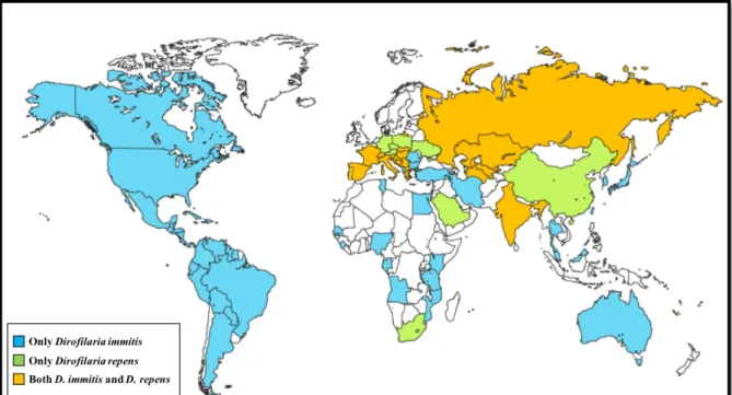
Dirofilaria immitis is present in tropical and temperate regions throughout the world, including canine populations from Africa, America, Asia, Australia and Europe (Genchi, Rinaldi, Cascone, Mortarino & Cringoli, 2005; reviewed by Simón et al., 2012). Dirofilaria repens is exclusive to the Old World, reported in Africa, Asia and Europe (Fig. 1). In the European continent, dirofilariosis profile is described by the presence of both D. immitis and D. repens, with some countries reporting the coexistence of both species (reviewed by European Scientific Counsel Companion Animal Parasites [ESCCAP], 2012; Simón et al., 2012). In endemic regions of southern Europe and United States of America (USA), local prevalence in dogs may reach 50%. Nevertheless, comparisons of the current and past epidemiological data show significant changes in the distribution pattern and prevalence of dirofilariosis, highlighting the establishment of new foci and increasing prevalence throughout the world (reviewed by Simón et al., 2012). Epidemiological surveys and recent clinical reports describe a significant expansion of canine autochthonous infections by D. immitis and/or by D. repens, particularly in central and northern European countries, areas where dirofilariosis was not reported or only
sporadic cases were documented (Genchi et al., 2005; Svobodova & Misonva, 2005; Duscher et al., 2009; Overgaauw & Van Dijk, 2009; Pantchev et al., 2009).
Portugal is regarded as a country where canine dirofilariosis by D. immitis is endemic, given its geographical situation in southern Europe, and its favourable climate for vector development, breeding and survival. According to Pereira da Fonseca, Madeira de Carvalho, Carvalho and Carvalho-Varela (1991), reviewed by Araújo (1996) dirofilariosis was prevalent in dogs from regions of southern Portugal, such as Algarve (12%), Alentejo (16.5%), Ribatejo (16.7%); regions of northern Portugal, such as Beira Litoral (4.2%) and Entre Douro e Minho (6.9%); and in Madeira Island (30%), with an overall prevalence of 14.1% microfilaremic dogs. However, this study was exclusively based on microfilariae detection, without differentiating D. immitis from other apathogenic or weakly pathogenic canine filarial species such as Acanthocheilonema (syn. Dipetalonema) dracunculoides and Acanthocheilonema (syn. Dipetalonema) reconditum, that are known to occur in Portugal. Additionally, in Azores, no cases of dirofilariosis were detected and no microfilaraemia was found in a survey on guard dogs (Medeiros, 1995). In a retrospective study performed in Madeira, a total of 22% dogs were microfilaremic, of which 89% of the microfilariae were histochemical identified as D. immitis and the remaining as A. reconditum and A. dracunculoides (Clemente, 1996). In northern Portugal, in the municipality of Alijó, 4.4% of the tested dogs were microfilaremic, of which 80% of the microfilariae were identified by acid phosphatase histochemical staining (APHS) as A. dracunculoides, 14% as D. immitis and 6% as A. reconditum; in the municipality of Sabrosa, 11.8% of the surveyed dogs were microfilaremic, all identified as A. dracunculoides (Santos, Cardoso & Rodrigues, 2000). All these studies were based exclusively on microfilariae detection, thereby underestimating occult infections.
Regarding wild canids, 3.2% red foxes (Vulpes vulpes) were found infected by D. immitis in northern-centre locations (Eira, Vingada, Torres & Miquel, 2006) and 11.8% in southern and central districts of Portugal (Carvalho-Varela & Marcos, 1993).
Figure 1 - Geographical distribution of canine dirofilariosis. Adapted from Simón et al.,
(2012).
1.3.2 Climate-matching model and seasonality
The transmission of D. immitis and D. repens depends on several factors, such as: sufficient numbers of infected and microfilaremic dogs, competent mosquito species and suitable climatic conditions that allow the extrinsic incubation of the parasites in the vector mosquitoes (Medlock, Barrass, Kerrod, Taylor & Leach, 2007).
Forecast models based on growing degree days (GDD) have been used to predict the occurrence and seasonality of D. immitis and D. repens in different parts of the world (Slocombe, 1989; Genchi et al., 2005; Genchi, Rinaldi, Mortarino, Genchi & Cringoli, 2009). The model is based on evidence that there is a threshold temperature of 14ºC below which Dirofilaria development will not occur in mosquitoes. Furthermore, there is a requirement of 130 GDD for larvae to reach infectivity and a maximum life expectancy of 30 days for mosquitoes (Lok & Knight, 1998; Slocombe, 1989). Models have allowed to estimate the number of annual Dirofilaria generations, as well as the length of infection risk periods in several regions of Europe (Genchi et al., 2005; 2009; Genchi, Kramer & Rivasi, 2011). Overall, the length of the transmission season is critically dependent on the accumulation of sufficient heat to incubate larvae to the infective stage in the mosquito (Lok & Knight, 1998). In the northern hemisphere, the peak months for transmission are typically July and August. Although the transmission decreases in the winter months, the risk never reaches zero, due to the presence of microenvironments in urban areas. Early predictions estimated that dirofilariosis would have the conditions to be
introduced into central and northern Europe, where it was previously not reported, which is now demonstrated (Svobodová, Svobodová, Genchi & Forejtek, 2002; Jacsó et al., 2009; Kartashev et al., 2011; Tasić-Otašević, Trenkić Božinović, Gabrielli & Genchi, 2015; Fuehrer et al., 2016). Indeed, the ongoing climate changes are lengthening the annual periods of mosquito activity and shortening larval developmental stages, with a consequent increase in the transmission in several areas.
Moreover, it’s important to consider the introduction of new species of competent mosquitoes, like Aedes albopictus (the Asian tiger mosquito), a highly adaptable species. This vector is native from south-eastern Asia and western Pacific, but has already spread to Africa, America and Europe, becoming adapted to colder climates (Roiz, Rosà, Arnoldi & Rizzoli, 2007). Other examples of invasive species introduced in Europe are Aedes koreicus and Aedes japonicus, which are enhancing the risk of spreading D. immitis in endemic and non-endemic areas (Montarsi et al., 2015; Silaghi, Beck, Capelli, Montarsi & Mathis, 2017).
1.3.3. Risk factors and populations at risk
As other mosquito-borne diseases, the distribution of dirofilariosis is prevalent in regions that show high average temperatures throughout (or part of) the year and high humidity. The last condition can be provided by the existence of abundant rainfall or by the proximity to water sources, such as irrigated cultivation areas, wetlands and rice fields. Generally, the prevalence in rural and peri-urban areas is higher than in urban ecosystems. This is usually explained by a set of factors: the largest number of irrigated fields in rural areas that attract vectors and allow their proliferation; the abundant presence of wildlife animals that may carry the infection and spread the disease; the highest number of stray dogs in rural areas along with sporadic or incorrect pet chemoprophylaxis, contributing to the perpetuation of the disease.
The Atlantic islands (Madeira and the Canaries) constitute an optimal environment for the transmission of dirofilariosis as the average temperatures tend to be moderate or high, and marine environment allows high humidity rates (reviewed by Simón et al., 2012; Diosdado et al., 2016).
In what concerns risk populations, both outdoor and indoor pets are at risk of dirofilariosis. Nevertheless, outdoor animals or those who spend more time outside (e.g., shepherd, military, hunting or guard dogs) particularly during peak mosquito hours, in areas of high endemicity, are naturally at an increased risk of infection (reviewed by Simón et al., 2012; American Heartworm Society [AHS], 2014).
1.4 Biology of Dirofilaria spp. 1.4.1 Life-cycle
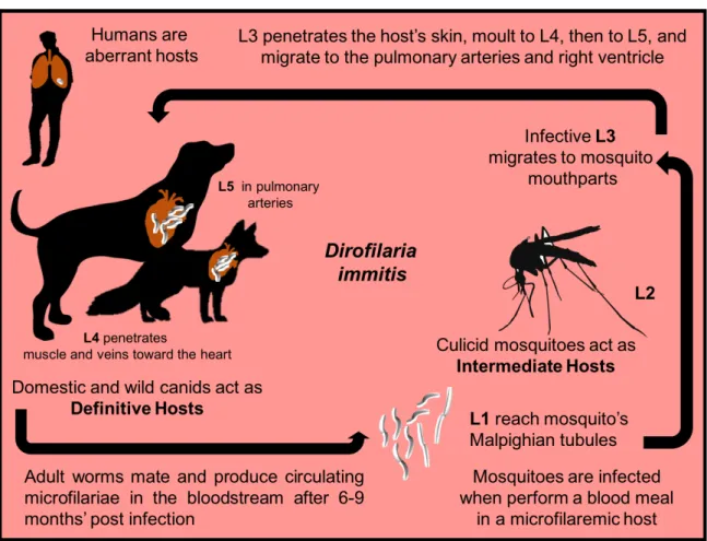
The life cycle of D. immitis and D. repens includes a definitive vertebrate host (domestic and wild canid as the main host of infection) and a vector (mosquitoes Diptera: Culicidae) (Cancrini & Kramer, 2001). In dogs, the life cycle of D. immitis is long (usually ranging from 7 to 9 months) (Kotani & Powers, 1982). It starts when a susceptible mosquito performs a blood meal on a microfilaremic host and becomes infected (Fig. 2). Approximately 24 hours after, the first-stage larvae (L1) reach the mosquito’s Malpighian tubules, where they moult into second-first-stage larvae (L2) within 8 to 10 days’ post infection (dpi), and from L2 into third-stage larvae (L3) in approximately 3 days (Taylor, 1960). Then, infective L3 migrates to the mouthparts of the vector, where they reside until the following blood meal. The time required for the development of microfilariae into the infective stage in the mosquito is temperature dependent (e.g., 10 to 14 days at 27°C and 80% of relative humidity; with cooler temperatures, the time required for the development is longer and will only progresses above 14°C) (Kartman, 1953; Slocombe, 1989). During a blood meal, infective L3 larvae (with approximately 1 mm long) migrates to the proboscis and invades the definitive host through the wound generated by the blood-feeding mosquito (Venco, Genchi & Simón, 2011). Then L3 moult to fourth-stage larvae (L4) between 3 and 12 dpi and penetrates muscle and veins toward the heart and lungs (Kotani & Powers, 1982; Lichtenfels, Pilitt, Kotani, & Powers, 1985). The subsequent moult takes place between 50-70 dpi, producing the pre-adult worms (fifth-stage larvae, L5), that arrive in the pulmonary arteries and right ventricle at between 70-85 dpi, grow and reach sexual maturity at 120 dpi. Adult D. immitis worms mate and produce circulating unsheathed microfilariae in the bloodstream after 6-9 months’ post infection (mpi) (Kotani & Powers, 1982; McCall et al., 2008b). The concentration of microfilariae in the peripheral blood has periodicity along the day, reaching highest density values in late afternoon and early evening, in accordance with the maximum activity of the intermediate host. Microfilariae and adult worms may last in dogs for 2 and 7 years, respectively (Venco et al., 2011). Some infected dogs do not develop microfilaraemia, an event, possibly explained by single-gender worm infections and host immune response factors (Simón & Genchi, 2000).
Figure 2 - Life cycle of Dirofilaria immitis (original).
Regarding D. repens, although adult worms can be found in the abdominal cavity and muscular fasciae (Genchi et al., 2011), they usually inhabit the subcutaneous tissues of definitive hosts where they achieve sexual maturity at 6-9 mpi (Manfredi, Di Cerbo & Genchi, 2007).
Numerous species of culicid mosquitoes are involved in the transmission of D. immitis and D. repens, as revised by Cancrini and Kramer (2001) and Cancrini and Gabrielli (2007). Nearly 70 species of culicid mosquitoes, mainly belonging to the genera Culex, Aedes, Anopheles, Culiseta and Coquillettidia have been identified as potential vectors of animal and human dirofilariosis. However, only in few cases its real vectorial capacity has been proven. Epidemiological studies conducted on vectors in distinct continents have shown a lower prevalence of Dirofilaria spp. in mosquitoes in comparison to vertebrate hosts (ranging from 1% to 10%), with the higher values detected in Culex theileri. In Europe, Culex pipiens is considered the main vector of both Dirofilaria spp. Nevertheless, Aedes vexans, Aedes punctor, Aedes albopictus, Aedes caspius and Anopheles maculipennis are also species in which vector capacity has been documented in European countries (Manrique-Saide et al., 2010; Yildirim et al., 2011; reviewed by Otranto et al., 2013).
In mainland Portugal, Culex theileri, Culex pipiens, Anopheles maculipennis s.l., Anopheles atroparvus, Aedes caspius and Aedes detritus s.l. were found naturally infected with D. immitis
(Ferreira et al., 2015). This is worrying if we consider that out of the 41 species of mosquitoes identified in continental Portugal (Ribeiro, Ramos, Pires & Capela, 1988), An. maculipennis s.l., Cx. pipiens s.l., Cx. theileri and Ae. caspius were the most abundant and broadly distributed (Almeida et al., 2008) and are all proven vectors of D. immitis.
1.4.2 Host range and specificity
CPD and SCD primarily affect members of the family Canidae, representing its main reservoir of infection. Nevertheless, D. immitis and D. repens demonstrate poor vertebrate host specificity, being able to infect several mammalian species, such as black bears, California sea lions, domestic cats, polecats, cougars, ferrets, jaguars, leopards, lions, seals, ocelots and tigers. Humans may also be infected, representing aberrant hosts. From an epidemiological point of view, cats are considered to play a minor role in the transmission cycle of dirofilariosis as its microfilaraemia is low and of short duration (Barriga, 1982; McCall et al., 2008b).
1.4.3 Role of wildlife hosts
Several species of wild carnivores were found infected by D. immitis and D. repens, including coyotes (Canis latrans), dingoes (Canis lupus dingo), red (Vulpes vulpes) and grey (Urocyon cinereoargenteus) foxes, jackals (Canis aureus) and wolves (Canis lupus lupus), with variable prevalences (Franson, Jorgenson & Boggess, 1976; Marconcini, Magi, Macchioni & Sassetti, 1996; Mañas, Ferrer, Castellà & López-Martín, 2005; Magi et al., 2008; Cirović et al., 2014; Gavrilović, Blitva-Robertson, Özvegy, Kiskároly & Becskei, 2014; Ionică et al., 2016). Indeed, carnivores with peridomestic habits are known to constitute an excellent sentinel for the spread of D. immitis (Sacks & Caswell-Chen, 2003; Wixcom, Green, Corwin & Fritzell, 1991).
1.4.4 Morphological characteristics of Dirofilaria immitis and Dirofilaria repens
Dirofilaria immitis adult worms are threadlike nematodes, presenting six small papillae surrounding the mouth opening, with females measuring 250-300 mm in length and 1-1.3 mm in diameter and males measuring 120-200 mm in length and 0.7-0.9 mm in diameter (Manfredi et al., 2007). D. immitis microfilariae (after fixation with 2% formalin) measure 290-330 µm in length and 5-7 µm in diameter, and have a straight posterior end and a conical-shaped cephalic extremity.
Dirofilaria repens adult worms are smaller than D. immitis, with females measuring 100-170 mm in length and 4.6-6.3 mm in diameter, and males measuring 50-70 mm in length and 3.7-4.5 mm in diameter (Manfredi et al., 2007). D. repens microfilariae also reside in the bloodstream. Nonetheless, they are longer, measuring between 350-385 µm in length and 7-8 µm in diameter (after fixation with 2% formalin), with a hook-shaped tail and a blunt cephalic extremity (Venco et al., 2011).
1.4.5 Role of the symbiotic bacteria Wolbachia spp.
Both D. immitis and D. repens host a symbiotic intracellular bacterium that belongs to the order Rickettsiales, genus Wolbachia (Sironi et al., 1995). This bacterium is found in all filarial developmental stages, i.e., located in the genital organs of females and on the lateral cords of both males and females, as well as in microfilariae and in the larvae in the vector. As Wolbachia spp. are involved in the moulting and embryogenesis of filariae (Bandi, Dunn, Hurst & Rigaud, 2001), these bacteria are vital for the development of larvae and for the survival of adult worms in vertebrate hosts (McGarry, Egerton & Taylor, 2004). Indeed, in the 1990s, it was shown that tetracycline treatment of filarial-infected animals led to the interruption of worm development in the host, disruption of microfilarial production and long-term survival of adults (reviewed by McCall et al., 2008a). Wolbachia spp. is also an important player in the interaction with the immune system of the infected host, associated with the upregulation of pro-inflammatory cytokines, neutrophil recruitment and increasing specific immunoglobulins (Taylor, Bandi & Hoerauf, 2005). This ground-breaking discovery resulted in a profound shift regarding a better understanding of the filarial biology, including the pathological mechanisms caused on the hosts, but most particularly, on the treatment approach, with the valuable association of an antibiotic in the treatment of dirofilariosis (reviewed by Simón et al., 2012).
1.5 Pathophysiology and clinical signs of Dirofilaria spp.
Canine CPD is a life-threatening disease caused by the presence of D. immitis adult worms (Furlanello, Caldin, Vezzoni, Venco & Kitagawa, 1998; McCall et al., 2008b) and by their antigenic products (Kramer, Simón, Tamarozzi, Genchi & Bazzocchi, 2005). It has a chronic progression, with numerous dogs showing no clinical signs for months or years, unless there is a large worm burden (until 250 worms) and/or animals undergo strenuous exercise.
1.5.1 Respiratory and cardiovascular signs caused by Dirofilaria immitis
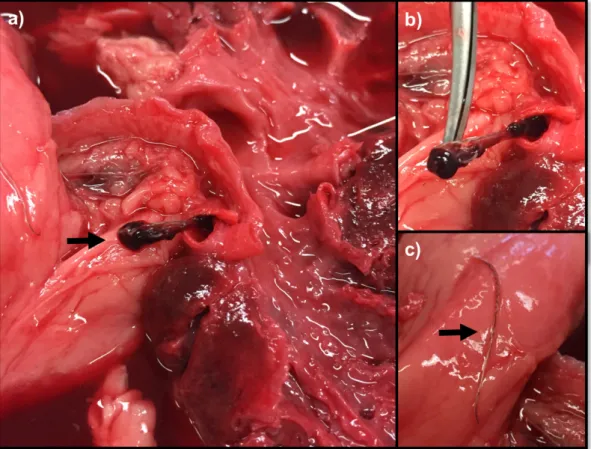
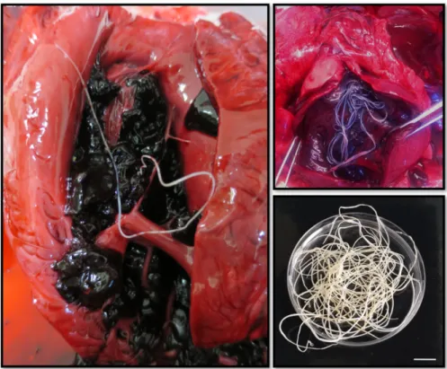
The main location of D. immitis adult worms is in the pulmonary arteries followed by the right ventricle (Fig. 3), so the first effects occur in the pulmonary arteries and lungs and, secondarily, in the heart. Clinical signs develop gradually, often beginning with a chronic unproductive cough (that increases with exercise), later associated with dyspnoea, regurgitation and lethargy. Infected dogs may also exhibit intolerance to exercise or syncope related with increased physical activity or excitement. Physical examination may reveal evidence of weight loss, right-sided heart murmur of tricuspid insufficiency, split-second heart sound and cardiac gallop (reviewed by Atkins, 2010). Whenever right heart failure is present, jugular venous ingurgitation and pulsation, along with hepatosplenomegaly and ascites may occur. Pulmonary manifestations include cough, dyspnoea, pulmonary crackles, muffled lung sounds and eventually cyanosis. If pulmonary thromboembolism occurs, dyspnoea may worsen and fever and haemoptysis may be noted. Although sudden death is rare, it can occur due to cardiorespiratory insufficiency or severe thromboembolism (Venco et al., 2011).
Figure 3 - Adult nematodes of Dirofilaria immitis collected at the necropsy of a pinniped
1.5.2 Other clinical signs caused by Dirofilaria immitis
Dirofilaria immitis may cause renal dysfunction and glomerulonephritis induced by the deposition of immune complexes triggered by the antigens of adult worms and larval stages (Abramowsky, Powers, Aikawa & Swinehart, 1981; Paes-de-Almeida, Ferreira, Labarthe, Caldas & Mc-Call, 2003). Dogs may also develop eczematous dermatitis (secondary to kidney disorders) and eosinophilic pneumonia, due to an eosinophilic reaction against microfilarial antigens. Other organs may also be affected by ectopic localisations, such as the eyes, brain, liver and peritoneal cavity, with related pathologies.
1.5.3 Clinical signs caused by Dirofilaria repens
SD is usually subclinical, although manifestations may occur associated with the presence of adult worms of D. repens between subcutaneous and deep connective tissue layers. The primary clinical sign is the presence of one or more skin non-inflammatory nodule(s), ranging from 0.5– 3 cm, located in different anatomical sites. Other manifestations include pruritus, erythema, papules, focal or multifocal alopecia and hyperkeratosis (Tarello, 2011). Extra-dermic signs may occur, such as anorexia, vomiting, lethargy, fever, lymphadenomegaly and conjunctivitis. Intra-vitreous infection is rare in dogs, but may be caused by D. repens (Guterbock, Vestre & Tood, 1981).
1.6 Diagnosis of canine dirofilariosis
The diagnosis of Dirofilaria infections may be done through the microscopic detection and morphological identification of circulating blood microfilariae; by the detection of circulating adult worm antigens and/or antibodies (only available for D. immitis) in serum; or by the detection of DNA of Dirofilaria using molecular methods.
1.6.1 Parasitological diagnosis
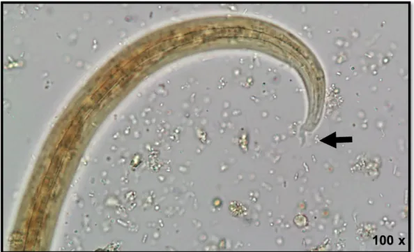
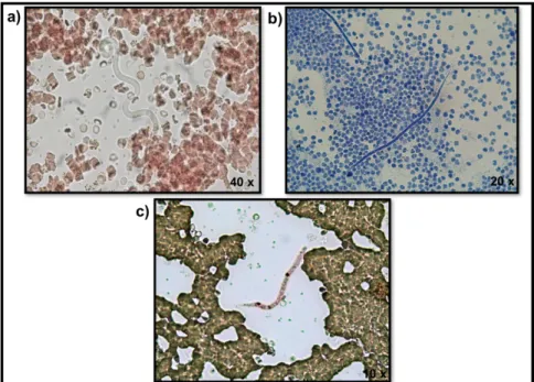
The most commonly used method for microfilariae identification is the Modified Knott's technique (MKT) (Fig. 4). It is an easy, quick and inexpensive diagnostic concentration method. Briefly, 1 ml of EDTA blood is mixed with 9 ml of 2% formalin and centrifuged for 5 minutes at 500×g. After that, the supernatant is poured off, one drop of blue methylene is added and the sediment is observed under the light microscope (Venco et al., 2011; Magnis et al., 2013). Given the variety of canine filarial species presenting blood microfilariae [(with D. immitis, D.
repens, A. dracunculoides and A. reconditum as the most important in Europe (Genchi et al., 2011)] it is essential to perform a morphometric analysis to obtain a correct diagnosis and select the appropriate treatment. Magnis et al., (2013) validated morphometric criteria for the identification of microfilariae in the dog’s blood using the MKT, allowing a clear distinction between D. immitis (302 µm average length, 6 µm average width, with a conical front end and a straight rear end), D. repens (369 µm average length, 9 µm average width, with a conical front end and curved caudal end), A. dracunculoides (259 µm average length, 5 µm average width, with a round front and straight caudal end) and A. reconditum (265 µm average length, 5 µm average width, with a blunt front end and a small hook in the rear end). Due to the overlapping size ranges of A. dracunculoides and A. reconditum, biochemical or molecular methods are required to distinguish these two species (Magnis et al., 2013).
Acid phosphatase histochemical staining is an additional method that allows the distinction of microfilariae according to their anatomical regions according to the acid phosphatase activity (Chalifoux & Hunt, 1971). Nowadays this method is inclusively available as a commercial kit (Peribáñez et al., 2001). Dirofilaria immitis microfilariae has two acid phosphatase activity zones (near the anal and excretory pores) (Fig. 4c), whereas D. repens has only one (near the anal pore and eventually some inner body complex). Other canine filariae, such as A. dracunculoides exhibits three areas of enzymatic activity (anal pore, excretory pore and internal body in-between these two pores) and A. reconditum exhibits a diffuse staining (Chalifoux & Hunt, 1971; Balbo & Abate, 1972).
Figure 4 - Microfilariae of Dirofilaria immitis in: a) fresh blood smear; b) stained by the
Modified Knott's technique; c) stained with the acid phosphatase, highlighting the anal and excretory pores (original).
1.6.2 Immunological diagnosis
Serological methods are other useful diagnostic techniques that allow the detection of amicrofilaremic infections, not detectable by MKT or APHS. Highly specific and sensitive enzyme linked immunosorbent assays (ELISA) or immunochromatography-based assays that detect circulating adult worm antigens of D. immitis females are commercially available for the diagnosis of cardiopulmonary dirofilariosis (Simón, Genchi, Prieto & Allende, 2001; Venco et al., 2011). However, commercial antigen-detection kits for D. immitis were tested with sera from dogs infected with A. vasorum, and cross-reactions were detected (Schnyder & Deplazes, 2012), which is of some concern.
1.6.3 Molecular diagnosis
Other diagnostic options are the amplification of microfilaria DNA by PCR (Favia, Lanfrancotti, della Torre, Cancrini & Coluzzi, 1997a; Favia Tringali, & Cancrini, 1997b). A duplex real-time PCR was developed to detect and differentiate infection by D. immitis and D. repens in dogs and mosquitoes and a multiplex PCR was described for the simultaneous detection of canine filarioids (Latrofa et al., 2012a,b).
1.6.4 Laboratory diagnosis
Marked alterations in haematology and biochemistry are usually seen when acute changes take place or in the late stage of the disease. Leukocytosis, eosinophilia, neutrophilia and non-regenerative normocytic normochromic anaemia is frequently seen. Thrombocytopenia can also be found, particularly when disseminated intravascular coagulation (DIC) is present, as well as azotaemia and hyperbilirubinemia. An increase in the acute phase protein (APPs), such as C-reactive protein (CRP) occurs and is useful for staging the disease and monitoring recovery after treatment. The concentration of cardiac troponin I (cTnI), myoglobin and creatine kinase MB (CK-MB) increase in dogs with high parasite burden, due to myocardial injury, and may also be used as early markers of cardiac damage. Similarly, D-dimer concentrations increase after thromboembolism, being useful on the diagnosis of pulmonary thromboembolism in dogs (Carretón et al., 2011).
1.6.5 Imaging findings
Thoracic radiograph, echocardiography and electrocardiography provide insights regarding the clinical status, severity and prognosis of cardiopulmonary disease secondary to heartworm infection. Characteristic (nearly pathognomonic) radiographic features are enlarged, tortuous, truncated peripheral intralobar and interlobar branches of the pulmonary arteries (particularly on the diaphragmatic lobes), accompanied by pulmonary parenchymal disease, right heart cardiomegaly in advanced stages, and pleural effusion following right heart congestive failure (Rawlings, 1986; Bowman & Atkins, 2009). Echocardiography allows the visualization of the worms, which are seen as two parallel hyperechoic lines in the main pulmonary artery, interlobe branches, right heart atrium and ventricle (Badertscher, Losonsky, Paul & Kneller, 1988). Besides, allows the assessment of cardiac anatomy and functional capacity, providing conclusive confirmation of the vena cava syndrome (VCS) when heartworms are located in the tricuspid valve. However, echocardiography is not an efficient method of making this diagnosis, particularly in lightly infected dogs, as the worms are frequently limited to the peripheral branches of the pulmonary arteries, thus beyond the echocardiographic field of view.
Electrocardiography can reveal alterations in the electrical axis and rhythm (deviations to the right side of the axis and atrial fibrillation) in dogs in terminal stages that exhibit severe enlargement of the right atrium (Venco et al., 2011).
1.6.6 Pathological findings
The first lesions occur on the walls of the pulmonary arteries and will lead to the development of subsequent pulmonary and cardiac pathology, observed in the last stage of infection (Venco, 2007). Worms induce mechanical trauma in the pulmonary arteries, causing a proliferative pulmonary endarteritis, with intravascular villus formation. Additionally, smooth muscle proliferation and consequent lumen narrowing and reduction of their compliance occur. All these changes in the arterial walls coupled with the release of inflammatory mediators lead to pulmonary hypertension. These events generate an overload on the right side of the heart, culminating in cor pulmonale, congestive right heart insufficiency and consequent hypertrophy and dilation. If congestive heart failure develops, venous ingurgitation, peripheral oedema, hydrothorax, hydropericardium and ascites may occur. Hepatomegaly may also happen, leading to liver insufficiency, jaundice and coagulation disorders. Additionally, worms’ death may cause a severe inflammation that could induce thromboembolism (Calvert & Rawlings, 1985; Rawlings, 1986). An acute and fulminant multisystemic condition that may also develop is
VCS, predominantly occurring in small dogs. This happens when a mass of worms displaces from the pulmonary arteries to the right ventricle, blocking the tricuspid valve and circulating blood, causing an overload of the right atrium and the caudal vena cava. This leads to the death of the animal due to hemolysis and DIC, other severe multisystemic condition (Furlanello et al., 1998).
1.6.7 Difficulties and misconceptions in diagnosing canine dirofilariosis
Although filarial species can be identified by microfilarial size and morphology, these features are challenging, particularly when low parasitaemia or mixed infections are present. Besides, both MKT and APHS only work when heartworm-infected dogs have a detectable microfilaraemia. This happens only in 2/3 of the cases, as 30-40% of infected dogs remain or become amicrofilaremic despite a persisting infection with adults (reviewed by Deplazes et al., 2016). Moreover, APHS reagents have a limited shelf life and require fresh samples to yield interpretable results (Peribáñez et al., 2001).
Diagnoses exclusively based on circulating antigen, may give a false negative result in infections with low parasite burden. Furthermore, in some dogs, antigen–antibody complexes may entrap antigens, hampering the immunological detection. As circulating antigens are only detectable when D. immitis reached the adult stage, antigen testing should not be carried out earlier than 7 months after exposure to infection. For D. repens, there are still no available immunologic methods.
The combination of the serological techniques with MKT or APHS allows an accurate detection of dirofilariosis. A positive microfilaria test with a positive antigen test confirms an infection with D. immitis. A positive antigen test without circulating microfilariae indicates an amicrofilaremic or occult infection by D. immitis that may be due to pre-patency, unisex infection by female worms, drug-induced sterility of adult filariae, or even immune-mediated clearance of microfilariae (Genchi, Venco & Genchi, 2007). A positive microfilaria test with a negative antigen test may indicate an infection caused by a species apart from D. immitis that may be confirmed by MKT or molecular techniques. If it is D. immitis microfilariae, it might be due to antigen-antibody complex formation that can interfere with the antigen detection (Little et al., 2014), low female worm burden, or the persistence of microfilariae following the natural or the pharmacological death of adults (Atkins, 2003).
Overall, diagnostic techniques must be taken together (testing for both microfilaria and antigens) and interpreted along with the results from clinical examination (thoracic radiography and echocardiography) to achieve a reliable diagnosis. Annual testing of dogs is important not
only to ensure that prophylaxis is correctly being performed but also to provide early treatment with lower pathological effects when an infection is present (AHS, 2014).
1.7 Treatment of canine dirofilariosis
The main goal of D. immitis treatment is to eliminate all forms of the parasite (e.g., adults and larval stages) and improve the animal’s clinical conditions and welfare, with minimal complications (AHS, 2014). This can be achieved pharmacologically using a multimodal approach, combining melarsomine dihydrochloride (an adulticide drug) with macrocyclic lactones (MLs) (microfilaricide) and doxycycline (antibiotic against Wolbachia spp. organisms). Mechanical heartworm removal is also indicated as a method of eliminating as many adult worms as possible before pharmacological treatment is initiated (reviewed by Nelson, 2012; AHS, 2014). Despites the range of therapeutic options, it’s important to underline the complexity and inherent risk associated with the treatment of cardiopulmonary dirofilariosis, given the massive worm destruction in the bloodstream and its multiple side effects (reviewed by Simón et al., 2012). Before starting therapy, the staging and risk of thromboembolic complications of each animal should be assessed, considering age, dog size, parasite load, severity of pulmonary disease and possibility to restrict dog’s physical activity (Venco, McCall, Guerrero & Genchi, 2004).
Treatment of SD is indicated in dogs suffering from clinical signs (e.g., dermal swelling, nodules, pruritus) and to decrease the risk of infection to other dogs and humans near the infected animal. Worms of D. repens can also be removed from the subcutaneous nodules. Additionally, imidacloprid 10% combined with moxidectin 2.5% (spot-on) has proven to be effective against adult D. repens worms and microfilariae, with monthly treatments (Petry et al., 2015).
1.7.1 Anthelmintic therapy
The only adulticidal drug available and approved by the Food and Drug Administration (FDA) for D. immitis is melarsomine dihydrochloride, an arsenic compound. According to the American Heartworm Society (AHS), the three-dose protocol of melarsomine (one injection of 2.5 mg/kg followed one month later by two injections of 2.5 mg/kg, 24 hours apart) is the recommended regimen regardless the stage or severity of the disease (with the exception of VCS) (Table 1). The three-dose protocol has proven to have an overall increased safety and efficacy (98% vs 90%) in comparison to the two-injection protocol (two injections of 2.5 mg/kg,
![Table 1- Recommended treatment and management protocol for Dirofilaria immitis infections in dogs (American Heartworm Society [AHS], 2014)](https://thumb-eu.123doks.com/thumbv2/123dok_br/19187785.948493/38.892.164.769.150.831/recommended-treatment-management-protocol-dirofilaria-infections-american-heartworm.webp)