CLINICAL SCIENCE
Respiratory exercise program for elderly individuals
with asthma
Ludmila Tais Yazbek Gomieiro,IAndre´ia Nascimento,IILuciana Kase Tanno,IRosana Agondi,IJorge Kalil,I Pedro Giavina-BianchiI
IClinical Immunology and Allergy Division, Faculdade de Medicina da Universidade de Sa˜o Paulo, Sa˜o Paulo, SP/Brazil.IIHealth Technologies Assessment
Unit - Institute of Education and Sciences, Hospital Alema˜o Oswaldo Cruz, Sa˜o Paulo, SP/Brazil.
INTRODUCTION:Asthma in older adults is frequently underdiagnosed, as reflected by approximately 60% of asthma deaths occurring in people older than age 65.
OBJECTIVE:The present study evaluates the effects of a respiratory exercise program tailored for elderly individuals with asthma. We are not aware of any other reports examining breathing exercises in this population.
METHODS:Fourteen patients concluded the 16-week respiratory exercise program. All the patients were evaluated with regard to lung function, respiratory muscle strength, aerobic capacity, quality of life and clinical presentation.
RESULTS:After 16 weeks of this open-trial intervention, significant increases in maximum inspiratory pressure and maximum expiratory pressure (27.6% and 20.54%, respectively) were demonstrated. Considerable improvement in quality of life was also observed. The clinical evaluations and daily recorded-symptoms diary also indicated significant improvements and fewer respiratory symptoms. A month after the exercises were discontinued, however, detraining was observed.
DISCUSSION: In conclusion, a respiratory exercise program increased muscle strength and was associated with a positive effect on patient health and quality of life. Therefore, a respiratory training program could be included in the therapeutic approach in older adults with asthma.
KEYWORDS: Asthma; Respiratory exercise program; Maximum inspiratory pressure; Maximum expiratory pressure; Respiratory training program.
Gomieiro LTY, Nascimento A, Tanno LK, Agondi R, Kalil J, Giavina-Bianchi P. Respiratory exercise program for elderly individuals with asthma. Clinics. 2011;66(7):1165-1169.
Received for publication onJanuary 12, 2011;First review completed onFebruary 21, 2011;Accepted for publication onMarch 21, 2011
E-mail: [email protected]
Tel.:+55 11 30713189
INTRODUCTION
Asthma is an ancient, multifactorial, and complex chronic inflammatory disease with high prevalence worldwide and significant associated morbidity.1,2
Unfortunately, the literature regarding asthma in senior citizens is sparse, even though the topic is important in light of the expected worldwide increase in the elderly popula-tion during the 21st century.3 Aging is more than just a scientific issue; it is also a social and economic issue.
Older patients with asthma, who are more likely to die from the disease, account for 15% of the hospital admissions and 60% of all asthma-related deaths.4
The diagnosis of asthma in older adults has frequently been overlooked. Even when it is discovered, it is often undertreated because the disease is frequently masked by
other health problems, such as heart disease and emphy-sema.5-7Often, older patients have poor recognition of their
asthma symptoms.8
Respiratory impairment tends to be more intense in older people with asthma due to both pre-existing co-morbidities and the anatomical and functional changes associated with the natural aging process.9,10
Hyperinsuflation shortens the respiratory muscles and has a considerable impact on their function. There is a consequent reduction in muscle strength and endurance, parameters that are already impaired in these patients due to aging.11,12 To compensate for impaired ventilation, asthmatic patients also contract their inspiratory accessory muscles and engage in forced expiration, which results in the characteristic upper-thoracic breathing. Respiratory muscle fatigue and energy consumption are associated with a higher incidence of respiratory failure in elderly indivi-duals.
A number of studies have demonstrated that exercise programs and motor and respiratory physiotherapy improve aerobic capacity, breathing pattern, muscle strength, and quality of life in senior citizens.13 Exercise
Copyrightß2011CLINICS– This is an Open Access article distributed under
programs tailored for asthmatic patients have a positive effect both on physical, physiological, and psychological parameters and on social and personal relationships.14
However, these interventions have not been evaluated in asthmatic senior citizens.
Respiratory muscle strength can be directly measured through the following static pressures: the maximum expiratory pressure (Pemax), and the maximum inspiratory pressure (Pimax). Dynamic maneuvers, such as maximum voluntary ventilation, are also used to estimate respiratory muscle strength. These measures play an important role in the diagnosis and prognosis of various pulmonary and neuromuscular disorders and have also been associated with fitness, morbidity, and mortality in general.
The aim of the study was to measure the effects of an exercise program for asthmatic senior citizens on clinical presentation, pulmonary function, maximum inspiratory pressure, maximum expiratory pressure, cardiopulmonary capacity and quality of life.
METHODS
This was an open trial carried out between January 2008 and December 2009. A sample size of 20 patients, with a loss of 15%, was determined be sufficient to demonstrate a 20% improvement in the primary outcomes (Alfa = 0.05; Beta = 0.1). The Project was approved by the Ethics Committee, and all the patients signed a free and informed consent form.
A total of 21 physically independent patients (men and women) were invited to participate in the respiratory exercise program, but only 14 were able to follow the study to completion. The main reason for inability to participate in the study was a lack of available time. All the patients were aged 60 or older and had been diagnosed with moderate or severe asthma according to the Global Initiative for Asthma (GINA) criteria.1
The exclusion criteria included patients with uncontrolled asthma, uncontrolled systemic chronic diseases, vocal cord dysfunction, exercise-induced asthma, physical impairment, smoking, and respiratory infections in the prior month.
Respiratory Exercise Program
During the 16-week program, the patients participated in 1-hour sessions that took place twice a week in the morning. The sessions focused on breathing exercises that strength-ened and lengthstrength-ened the skeletal muscles of the thorax and abdomen. All of the exercises were associated with diaphragmatic breathing and pursed-lip breathing. In pursed-lip breathing, air is inspired through the nose, causing the abdomen to inflate; therefore, air is exhaled through the mouth with semi-open lips. The patients were encouraged to breathe out ‘‘the entire air contents of the lungs’’. At the beginning of each session, the participants performed exercises to stretch the muscles of the thoracic wall. These exercises were followed by breathing exercises and exercises to strengthen the abdominal and diaphrag-matic muscles. Most of the sessions concluded with ‘‘playful’’ breathing exercises, followed by approximately five minutes of relaxation.
Peak Expiratory Flow (PEF) was determined before and after the respiratory exercises using a Mini-Wright pulmon-ary function-monitoring device.
Clinical Evaluation
Based on the asthma severity, which was determined according to the GINA criteria,1 the patients were treated with inhaled corticosteroids combined with a long-acting beta-2 agonist, as needed, over a run-in period of at least one month. The physician in charge of patient evaluation prescribed the maintenance therapy and drug dosage, and the treatment was kept unchanged throughout the study period.
The patients were instructed and allowed to use short-acting beta-2-agonists if they experienced exacerbation of their asthma symptoms. During routine or additional examinations, the physician decided whether or not to use oral corticosteroid pulse therapy to treat more severe asthmatic crises. Throughout the study period, the treat-ment was kept unchanged.
The physician responsible for the clinical monitoring evaluated each patient on four different occasions. The first evaluation was prior to the start of the exercise program, and examinations followed at 8, 16 (the end of exercise program) and 20 weeks (four weeks after the end of the exercise program). During these evaluations, the patients completed standard clinical forms used by our service to monitor asthmatic patients.
On these occasions, the patients underwent a 6-minute walking test to evaluate their aerobic capacity. Briefly, the walk test was performed on a straight path of approxi-mately 30.5 meters with two cones marking the beginning and the end of the distance. The patients were instructed to walk as fast as possible without running for as many laps around the cones as possible. The patients were asked to stop after 6 minutes, and the monitor measured the total distance walked in meters.
The quality of life was evaluated using a questionnaire that had been previously translated and validated in Brazil.15The patients completed the questionnaire prior to the start of the training program, at the end of the program (16 weeks) and 4 weeks after the end of the exercise program (20 weeks). The questionnaire focused on five areas: physical limitations, which included questions about daily and leisure activities; frequency and severity of symptoms; treatment compliance; socio-economic status; and psycho-social issues.
The patients were also instructed to record their symp-toms in a diary on a daily basis.
Evaluation based on complimentary exams
All the patients underwent lung function tests and maximum inspiratory and expiratory pressure measure-ments prior to the start of the exercise program, at 8 and 16 weeks (during the program and at the end of the program, respectively) and 4 weeks after the end of the exercise program.
The spirometry was performed between 7:00 and 9:00 am using a Koko spirometer. The spirometry was performed and interpreted according to the ATS/ERS 2005 consen-sus.16In addition to the routine flow and volume measures (CVF and VEF1), we also measured the maximum voluntary ventilation (MVV).
repeated three consecutive times until the maneuvers were performed correctly with a minimum percent difference. The highest value was recorded; the Pimax and Pemax were the main outcomes.
During the determination of the Pimax, the patient was asked to perform an initial maximum expiration until reaching the expiratory reserve volume. They were then requested to breathe in through the mouth with maximum effort and to sustain their peak effort for at least one second. This maneuver was repeated three consecutive times; the patient was allowed small intervals between the maneuvers. In the case of the Pemax, the patient performed a maximum inspiration until reaching the total lung capacity, after which the patient breathed out through the mouth with maximum effort and sustained their peak level for at least one second. This maneuver was repeated three consecutive times; the patient was allowed small intervals between the maneuvers.
Thus, the previously described parameters (except for the quality of life questionnaire) were evaluated at 4 different times: prior to the start of the breathing exercise program (the 1stevaluation), after 8 weeks (the 2ndevaluation), after 16 weeks (the 3rdevaluation) and 4 weeks after the end of the exercise program. The patients were requested to complete the quality of life questionnaire during the 1st, 3rd, and 4thevaluations.
Statistical Analysis
After the data had been checked for consistency, descriptive and inferential statistical analyses were per-formed. The description of the qualitative variables was based on absolute and relative frequencies, and the description of the quantitative variables was based on central trend (average and/or median) and dispersion measurements (minimum, maximum, and standard devia-tion).
An analysis of variance (ANOVA) with repeated mea-surements model followed by multiple comparisons with the Bonferroni method was used to compare changes in the quantitative results over the course of the study (e.g., spirometry parameters and domains of the quality of life scale). An analysis of variance with repeated measures model and the variable gender was used to determine whether patient gender was associated with different developments in the quality of life domains, the Pimax and the Pemax.
The Friedman Test was used to compare changes in the percentage of patients who reported respiratory symptoms throughout the evaluations.
A statistical significance level of p#0.05 was used. The statistical analysis was performed using the SPSS computer program (version 13.0).
RESULTS
A total of 14 patients, 8 women and 6 men completed the program; the average age was 66.9 years (60 to 82 years).
Clinical Evaluation
We performed a descriptive analysis of the variables from the clinical evaluations. The program provided positive effects; it improved asthma-related symptoms, reduced physical impairment of daily activities, reduced the use of rescue medicine, and reduced the frequency of nocturnal
waking. These parameters tended to increase again a month after discontinuing the exercise program. No patient had severe asthma attacks that required systemic corticosteroids. According to the daily symptoms recorded in the patient diaries, there was a reduction in the number of respiratory symptoms at the end of the exercise program. During the study period, a significant difference was observed in the percentage of days when the patients recorded coughing (p= 0.02), shortness of breath (p= 0.03), night waking due to shortness of breath, and the use of bronchodilating agents (p= 0.04). A month after interruption of the exercise program, however, these asthma symptoms worsened.
We noticed significant differences in the global quality of life in the different evaluations (p= 0.008), suggesting improvement in the global quality of life after 16 weeks of physical activity. Regarding the individual analysis of each area in the different clinical evaluations, we observed significant differences in the physical impairment scores (p,0.001) and trends in the social and economic scores (p= 0.05).
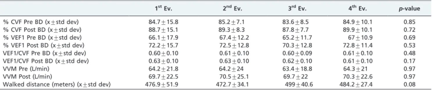
With regard to the 6-minute walking test, we did not notice significant changes in the average distances between the 4 evaluations (Table 1).
Evaluation of complimentary exams
The lung function tests did not reveal significant changes in the average results of the forced vital capacity, forced expiratory volume in 1 second, Tiffeneaux index, and maximal voluntary ventilation between the four evaluations (Table 1); the same was true for the measurements of the peak expiratory flow.
The results of the Pimax and Pemax tests demonstrated significant differences in the Pimax during the different evaluations (p,0.001). After 16 weeks of the exercise program, the average Pimax was 18.1 cmH2O higher than
at the initial evaluation (CI95%= 5.0 cmH20 to 31.3 cmH20,
p= 0.005) and was significantly higher than the average at 4 weeks after discontinuing the exercise program (p= 0.004). The average Pimax values after the initial evaluation and after the interruption of the exercise program were similar (p.0.99) (Table 2).
We also observed significant differences in the Pemax values over the different evaluations (p,0.001). After the 16-week exercise program, the Pemax average was 18.9 cmH2O
higher than at the initial evaluation (CI95%= 3.1 cmH2O to
34.8 cmH2O,p= 0.02) and was significantly higher than that observed at 4 weeks after interruption of the exercise program (p,0.001).
DISCUSSION
In recent years, asthma treatment has been focused on pharmacological protocols designed to control asthma and the inflammatory process of the disease, and other therapeutic approaches to help control asthma have been neglected.17,18 Studies on physical exercises, breathing exercises, and physiotherapeutic approaches have been performed to determine the clinical and physical benefits of these interventions.
patients.19 In our literature review, however, we did not
find any studies that, like ours, evaluated the effect of breathing exercises on the inspiratory and expiratory pressures of older asthmatic patients without using incen-tive devices.
In our study, the breathing exercise program was associated with a positive effect on the clinical presentation of patients, with an average reduction in symptoms and in the use of bronchodilating agents. These observations were based on the clinical evaluations performed by the physician and also on the symptoms recorded in the patient diary. With regard to the perception of asthma control by the patients, the majority of patients considered their asthma control to be good or very good at the end of the exercise program.
No significant changes in the lung function parameters were observed throughout the study, which is consistent with the findings in the literature.
The determination of respiratory muscle contraction force can be estimated by the measuring the maximum pressures, Pimax and Pemax. The Pimax is associated with the index of diaphragmatic strength. In contrast, Pemax is associated with abdominal and intercostal muscle strength. Since the 1960s and 1970s, Pimax and Pemax have been regarded as simple, practical and accurate methods to evaluate the strength of the muscles involved in the breathing process in both healthy individuals and in patients with respiratory or neurologic disorders.
Prior to the start of the exercise program, our patients had Pimax and Pemax values lower than expected. This finding may be explained by weaker respiratory muscles, probably due to the pulmonary hyperinflation that is common in
older asthmatic patients and that places the diaphragm in a position of mechanical disadvantage. These findings are consistent with other reports.20
Significant increases in the Pimax and Pemax values were demonstrated after the 16-week breathing exercise program, which suggest improvements in inspiratory muscle strength and endurance. A systematic review of duly validated and controlled studies on the training of inspiratory muscles while using an inspiratory valve (a threshold-incentive spirometer) has reported an increase in maximum respira-tory pressure.20
An improvement in the quality of life was also seen. It resulted from a reduction in physical limitations, from social and economic improvements, and from an improvement in the global quality of life evaluation. However, patients did not demonstrate cardio-respiratory improvements after completing the 16-week exercise program, which may have been due to the low accuracy of the 6-minute walking test compared to the maximum effort cardio-pulmonary test. In addition, strength and endurance training has little or no effect on aerobic capacity and results in limited cardiovas-cular adaptation.
The changes in muscle strength during the initial 4 weeks were due to neural adaptation. There is evidence that other mechanisms, such as structural and peripheral changes, also take place, and they could have been responsible for the increase in strength after 4 weeks of training.21-23A potential positive long-term effect of the program was not observed in this study. An analysis of the test results at 4 weeks after discontinuation of the exercise program revealed a sig-nificant decrease in the Pimax and Pemax, which may have been due to detraining.
Table 1 -The lung function variables and the results of the 6-minute walking test during the 4 patient evaluations (n = 14).
1stEv. 2ndEv. 3rdEv. 4thEv. p-value
% CVF Pre BD (x¡std dev) 84.7¡15.8 85.2¡7.1 83.6¡8.5 84.9¡10.1 0.85
% CVF Post BD (x¡std dev) 88.7¡15.1 89.3¡8.3 87.8¡7.7 89.9¡10.1 0.72
% VEF1 Pre BD (x¡std dev) 66.1¡17.9 67.4¡12.2 65.2¡11.7 67¡10.9 0.69
% VEF1 Post BD (x¡std dev) 72.2¡15.7 72.5¡12.8 70.3¡12.8 72.8¡11.4 0.53
VEF1/CVF Pre BD (x¡std dev) 0.60¡0.10 0.61¡0.10 0.60¡0.09 0.61¡0.10 0.48
VEF1/CVF Post BD (x¡std dev) 0.63¡0.10 0.63¡0.10 0.62¡0.10 0.61¡0.10 0.17
VVM Pre (L/min) 64.2¡21.8 64.2¡24 63.4¡18.8 64.3¡21 0.97
VVM Post (L/min) 69.7¡22.5 70.5¡25.1 69.7¡22 70.3¡22.6 0.97
Walked distance (meters) (x¡std dev) 476.9¡51.9 472.7¡34.1 499¡40.6 484.2¡27.4 0.08
1stEv.: Initial evaluation (1stevaluation); 2ndEv.: Evaluation at 8 weeks after the start of the exercise program; 3rdEv.: Evaluation at 16 weeks after the
start of the exercise program; 4thEv.: Evaluation at 4 weeks after discontinuing the exercise program. X: Average; ‘‘std dev’’: Standard Deviation.
Table 2 -The measures of respiratory muscle strength assessed during the 4 patient evaluations (women = 8, men = 6, total = 14).
Pimax (cmH2O) (x¡std dev) Pemax (cmH2O) (x¡std dev)
Women Men All Women Men All
1stEvaluation 63.1
¡23.5 75.2+23.5 68.0+23.4 86.6¡23.1 99.2+40.5 92.0+31.0
2ndEvaluation 76.4¡25.0 80.8
+18.5 78.3+21.8 97.3¡17.9 108+39.5 101.9+28.3
3rdEvaluation 82.8¡21.1 91.3
+22.1 86.4+21.2 105.8¡27.8 117.8+35.7 110.9+30.7
4thEvaluation 68.8
¡20.9 66.8+17.7 67.9+18.9 68.6¡15.4 70.7+24.3 69.5+18.9
X: Average; ‘‘std dev’’: Standard Deviation.
1stEvaluation: Initial evaluation; 2ndEvaluation: Evaluation at 8 weeks after the start of the exercise program; 3rdEvaluation: Evaluation at 16 weeks after
the start of the exercise program; 4thEvaluation: Evaluation at 4 weeks after discontinuing the exercise program.
Detraining is defined as the partial or complete loss of anatomical, physiological and performance adaptations due to a reduction in or interruption of a training program. A significant reduction in metabolic and work capacities is already present after one or two weeks of detraining, and a number of training-induced improvements disappear within a few months.24
Our results are consistent with previous reports because reductions in the variables associated with respiratory muscle strength (Pimax and Pemax) and negative impacts on clinical presentation and quality of life were observed at four weeks after discontinuation of the exercise program. We speculate that this reduction in the parameters associated with respiratory muscle strength may have had negative consequences for the clinical presentation and patient quality of life.
Any study of complimentary therapeutic approaches, such as respiratory exercises, has a number of difficulties: establishing and maintaining an adequate control group; the need for intensive interaction between the patients, the professional in charge of the respiratory therapy, and the physician; difficulties in advising control patients; difficul-ties in blinding the patients and researchers; difficuldifficul-ties in ensuring a standard intervention; a lack of financial support because these interventions have neither pharmacological nor commercial application; and the need to ensure treatment compliance by the patient.11,25,26 Although the present study was an open trial with few patients, it showed relevant results.
The breathing exercise program used in the current study was accompanied by improvements in clinical presentation (reductions in respiratory symptoms and in the use of bronchodilating agents), respiratory muscle strength and patient quality of life. The study also showed that the measures of clinical control and quality of life may be directly related to measures of patient physical status.
The use of breathing exercises in the clinical treatment of older adults with asthma may be effective, and the improvements in muscle strength may have been associated with a greater ability to deal with asthma crises. New randomized, double-blind, placebo-controlled studies with larger sample populations are needed, especially for older asthmatic patients. To better understand the physiological mechanisms of these interventions, future studies could examine both the outcomes used in this study and outcomes associated with airway hyper-reactivity and inflammatory markers.
REFERENCES
1. GINA. The Global Initiative for Asthma: www.ginasthma.com (accessed 01/10/2010).
2. The International Study of Asthma and Allergy in Childhood (ISSAC) Steering Committee. Worlwide variation in prevalence of asthma
symptoms: The Internationsl Study of Asthma and Allergy in Childhood (ISAAC). Eur Respir J. 1998;12:315-35, doi: 10.1183/ 09031936.98.12020315.
3. Choy DK, Hui DS, Li ST, Ko FW, Ho S, Woo J, et al. Prevalence of wheeze, bronchial hyper-responsiveness and asthma in the elderly Chinese. Clin Exp Allergy. 2002;32:702-7, doi: 10.1046/j.1365-2222.2002. 01395.x.
4. Jack CIA, Lye M. Asthma in the Elderly Patient. Gerontology 1996;42:61-8.
5. Banerjee DK, Lee GS, Malik SK, Daly S. Underdiagnosis of asthma in the elderly. Br J Dis Chest. 1987;81:23-9, doi: 10.1016/0007-0971(87)90104-5. 6. Braman SS, Kaemmerlen JT, Davis SM. Asthma in the elderly: a
comparison between patients with recently acquired and long-standing disease. Am Rev Respir Dis. 1991;143:336-40.
7. Lee HY, Stretton TB. Asthma in the elderly. Br Med J. 1972;4:93-5, doi: 10. 1136/bmj.4.5832.93.
8. Connolly MJ, Crowley JJ, Charan NB, Nielson CP, Vestal RE. Reduced subjective awareness of bronchoconstriction provoked by methacholine in elderly asthmatic and normal subjects as measured on a simple awareness scale. Thorax. 1992;47:410-3, doi: 10.1136/thx.47.6.410. 9. Braman SS, Hanania NA. Asthma in older adults. Clin Chest Med.
2007;28:685-702, doi: 10.1016/j.ccm.2007.08.007.
10. Bisaccioni C, Aun MV, Cajuela E, Kalil J, Agondi RC, Giavina-Bianchi P. Comorbidities in severe asthma: frequency of rhinitis, nasal polyposis, gastroesophageal reflux disease, vocal cord dysfunction and bronchiec-tasis. Clinics. 2009;64:769-73, doi: 10.1590/S1807-59322009000800010. 11. Decramer M. Effects of hyperinflation on the respiratory muscles. Eur
Respir 1989;2:299-302.
12. Veiga J, Lopes AJ, Jansen JM, Melo PL. Within-breath analysis of respiratory mechanics in asthmatic patients by forced oscillation. Clinics. 2009;64:649-56, doi: 10.1590/S1807-59322009000700008.
13. American College of Sports Medicine. Physical activity programs and behaviour counseling in older adults populations. Med Sci Sports Exerc. 2004;36:1997-2003, doi: 10.1249/01.MSS.0000145451.08166.97.
14. Strunk RC, Mascia AV, Lipkowitz MA, Wolf SI. Rehabilitation of a patient with asthma in the outpatient setting. J Allergy Clin. Immunol. 1991;87:601-11, doi: 10.1016/0091-6749(91)90374-W.
15. Fernandes ALG, Oliveira MA. Avaliac¸a˜o da qualidade de vida na asma. Jornal de Pneumologia. 1997;23:148-52.
16. Miller MR, Hankinson J, Brusasco V, Burgos F, Casaburi, et al. ATS/ERS Task Force. Standardization of spirometry. European Respiratory Journal. 2005;26:319-38.
17. Giavina-Bianchi P, Aun MV, Bisaccioni C, Agondi R, Kalil J. Difficult-to-control asthma management through the use of a specific protocol. Clinics. 2010;65:905-18.
18. Sterk PJ, Fabbri LM, Quanjer PJ, et al. Airway responsiveness: standartised challenge testing with pharmacological, physicial and sensitizing stimuli in adults. Eur Respir J. 1994;6:53-83.
19. Weiner P, Azgad Y, Ganam R, Weiner M. Ispiratory muscle training in patients with bronchial asthma. Chest. 1992;102:1357-61, doi: 10.1378/ chest.102.5.1357.
20. Ram FSP, Wellington SR, Barnes NC. Inspiratory muscle training for asthma (Cochrane Review). In: The Chochrane Library, Issue 3, 2008. Oxford:Update Software.
21. Enoka R. Neural adaptations with chronic physical activity. Journal of Biomechanics. 1997;30:447-55, doi: 10.1016/S0021-9290(96)00170-4. 22. Moritani T, de Vries HA. Neural factors versus hypertrophy in the time
course of muscle strenght gain. American Journal of Physical Medicine. 1979;58:115-30.
23. Sale DG. Neural adaptation to resistance exercise. Medicine and Science in Sports and Exercise 1988;20:S135-S145.
24. Mujika I, Padilla S. Detraining: loss of training-induced physiological and performance adaptations. Part I. Short-term insufficient training stimulus. Sports Med. 2000;30:79-87.
25. Pearson MG. Breathing exercises for asthma: panacea or placebo? Thorax. 2007;62:1033-34, doi: 10.1136/thx.2007.084707.