CLINICAL SCIENCE
T-cell large granular lymphocytic leukemia: treatment
experience with fludarabine
Renata Oliveira Costa,II,IIIMarcelo Bellesso,I,IIDalton Alencar Fischer Chamone,I,IIMilton Artur Ruiz,I Abraha˜o Elias Hallack Neto,IVVera Lucia Aldred,V Juliana PereiraI,II
IHospital das Clı´nicas da Faculdade de Medicina da Universidade de Sa˜o Paulo, Hematology Department, Sa˜o Paulo/SP, Brazil.IIInstituto do Caˆncer do
Estado de Sa˜o Paulo da Faculdade de Medicina da Universidade de Sa˜o Paulo Hematology Department, Sa˜o Paulo/SP, Brazil.IIILusiadas University School of Medicine, Internal Medicine Department, Santos/SP, Brazil.IVFaculdade de Medicina da Universidade Federal de Juiz de Fora, Internal Medicine Department, Juiz de Fora/MG, Brazil.VFaculdade de Medicina da Universidade de Sa˜o Paulo, Pathology Department, Sa˜o Paulo/SP, Brazil.
OBJECTIVES:The aim of this retrospective study was to investigate the results of T-cell large granular lymphocytic leukemia treatment with fludarabine by assessing the complete hematologic response, the complete molecular response, progression-free survival, and overall survival.
METHODS: We evaluated the records of six patients with T-cell large granular lymphocytic leukemia who were treated with fludarabine as a first-, second-, or third-line therapy, at a dose of 40 mg/m2, for three to five days per month and 6 to 8 cycles.
RESULTS: Of the six patients investigated with T-cell large granular lymphocytic leukemia who were treated with fludarabine, five (83.3%) were female, and their median age was 36.5 years (range 18 to 73). The median lymphocyte level was 3.46109/L (0.5 to 8.9). All patients exhibited a monoclonal T-cell receptor gamma gene rearrangement at diagnosis. Two (33.3%) patients received fludarabine as first-line treatment, two (33.3%) for refractory disease, one (16.6%) for relapsed disease after the suspension of methotrexate treatment due to liver toxicity, and one (16.6%) due to dyspesia. A complete hematologic response was achieved in all cases, and a complete molecular response was achieved in five out six cases (83.3%). During a mean follow-up period of 12 months, both the progression-free survival and overall survival rates were 100%.
CONCLUSION:T-cell large granular lymphocytic leukemia demonstrated a high rate of complete hematologic and molecular response to fludarabine, with excellent compliance and tolerability rates. To confirm our results in this rare disease, we believe that fludarabine should be tested in clinical trials as a first-line treatment for T-cell large granular lymphocytic leukemia.
KEYWORDS: Fludarabine; Treatment; Large Granular Lymphocyte Leukemia.
Costa RO, Bellesso M, Chamone DAF, Ruiz MA, Hallack Neto AE, Aldred VL, Pereira J. T-cell large granular lymphocytic leukemia: treatment experience with fludarabine. Clinics. 2012;67(7):745-748.
Received for publication onMarch 10, 2012;First review completed onMarch 16, 2012;Accepted for publication onMarch 18, 2012 E-mail: dr.marcelobellesso@gmail.com
Tel.: 55 11 3061-5544
INTRODUCTION
T-cell large granular lymphocytic (T-LGL) leukemia is characterized by a monoclonal expansion of CD3-positive T-LGL cells, as described in 1975 (1). This rare and indolent disorder represents 2% to 3% of chronic lymphoid leukemia cases, with a median age at diagnosis of 60 years and an equal male to female ratio (2). The diagnostic criterion is a persistent increase in circulating monoclonal large granular lymphocytes lasting at least six months. The neoplastic cells are CD3, CD8 and TCRabin 80% of cases, but they may be
CD4-CD8-, CD4+CD8- or CD4+CD8+in rare variants of the
disorder. The associated natural killer cell antigens (NKa) CD16, CD56 and CD57 are variably expressed; CD57 is the most commonly expressed cell antigen and CD56 is the least commonly expressed cell antigen. A total or partial lack of other T-cell antigens, such as CD2, CD5 and CD7, can be observed (3-4).
T-LGL leukemia is associated with autoimmune disor-ders, such as rheumatoid factor with or without rheumatoid arthritis, Felty’s syndrome, Coombs-positive hemolytic anemia, idiopathic thrombocytopenic purpura, pure red cell aplasia, the presence of positive anti-nuclear antibodies, the presence of anti-neutrophil cytoplasmic antibodies, hypogammaglobulinemia, and polyclonal hypergammaglo-bulinemia. Anemia and neutropenia are frequent (5). At diagnosis, 70% of patients require treatment, but a watch-and-wait approach may be appropriate for asymptomatic patients (2). The most common indications for treatment are Copyrightß2012CLINICS– This is an Open Access article distributed under
the terms of the Creative Commons Attribution Non-Commercial License (http:// creativecommons.org/licenses/by-nc/3.0/) which permits unrestricted non-commercial use, distribution, and reproduction in any medium, provided the original work is properly cited.
No potential conflict of interest was reported.
CLINICS 2012;67(7):745-748 DOI:10.6061/clinics/2012(07)07
cytopenia, recurrent infection and pure red cell aplasia, progressive splenomegaly and B symptoms (5).
There is currently no gold standard treatment for T-LGL leukemia. Most patients are treated with low doses of methotrexate, cyclophosphamide, and cyclosporine-A. As a rule, the treatment should be continuously monitored and, if necessary, adjusted to maintain the desired response. However, treatment compliance is frequently low, with a consequent relapse of the disease. Moreover, side effects are common and may limit the treatment effectiveness (6-7). We therefore changed our approach to treating T-LGL leukemia in an attempt to improve the hematological and molecular response rates and to reduce the treatment duration. Previous studies have shown promising results with fludarabine (8), 29-deoxycoformycin and alemtuzumab in T-LGL leukemia, with a response rate of 40% to 60% when used as second-line therapy (9). Based on our long-term, positive experience with fludarabine in other hematological disorders and in view of the high cost and toxicity of alemtuzumab, we changed our treatment approach to T-LGL leukemia by adopting fludarabine as the drug of choice. Herein, we report our experience using fludarabine in the treatment of six patients with T-LGL leukemia.
PATIENTS AND METHODS
Patients and end points
This study was a retrospective analysis of data obtained by reviewing the medical records of six patients with T-LGL leukemia. They were treated with fludarabine by the same Hematology Service of the Clinical Hospital of Sa˜o Paulo and Cancer Institute (Sa˜o Paulo, Brazil) from January 2007 to January 2009 and February 2009 to October 2010, respectively. The primary and secondary end points were complete hematologic (CHR) and complete molecular (CMR) response and progression-free (PFS) and overall survival (OS), respectively.
Diagnostic criteria for T-LGL leukemia
The diagnostic criterion for T-LGL leukemia was a persistent (at least six months) increase in circulating T-LGL cells, as evaluated by cell morphology and phenotype and confirmed by a monoclonal T-cell receptor (TCR) gamma gene rearrangement. A complete blood cell count, peripheral blood (PB) slide examination, and phenotype determination by flow cytometry were carried out using a standard multiparameter methodology to assess the antigens CD2, CD3, CD4, CD5, CD7, CD8, CD56, CD16, CD58, TCRb, and TCRcd, as previously described (10). The equipment used was a FACSCalibur flow cytometer (Becton Dickinson, San Jose, CA) and the CellQuestPro software. For analysis, the lymphocyte region was delimitated by an electronic gate in the forward side scatter versus side scatter display. Populations of T-LGL cells were defined by the co-expression of the T-cell CD3 antigen and at least one NKa CD57, CD16 or CD56 antigen. The normal range of T-LGL cell numbers assumed in the analysis was 0.1-0.36109/L (11).
T-cell monoclonality assessment
The monoclonal rearrangement of the TCR gamma gene was assessed by polymerase chain reaction (PCR) using primer sequences, as described previously by Shadrach B and Warshawsky I (12). Briefly, each 25-ml PCR reaction contained 200 ng of DNA, 1.5 U of Taq, 1.5 nmol/L of
MgCl2, 0.2 nmol/L of dNTP, and 0.5mmol/L of each primer
(IDT Technologies, Illinois, USA). PCR was performed in a PTC-100 DNA Engine Tetrad (MJ Research, Inc., Waltham, MA) with one cycle at 94
˚
C for three min, followed by 40 cycles at 95˚
C for 60 sec, 61.8˚
C for 30 sec, and 72˚
C for 30 sec, with a final extension of 10 min at 72˚
C and then held at 4˚
C. A 0.1-mL aliquot of the PCR product was mixed with 12mL of deionized formamide and 0.5mL of GeneScan 500 HD Rox size standard (Applied Biosystems, Foster City, CA). Then, the mixture was injected into an ABI Prism 3130 Genetic Analyzer, and the resulting data were analyzed using the GeneMapper V 3.2 software (Applied Biosystems, Foster City, CA). A monoclonality spike was determined by visual examination of the electropherograms (12).Treatment
Patients with T-LGL were treated with fludarabine if they presented with B symptoms, progressive splenomegaly, recurrent infection or anemia. Fludarabine was used as a first-line treatment or salvage therapy. HIV-positive patients were excluded from the study.
Fludarabine was used at a dosage of 40 mg/m2/day path-way oral for three to five days monthly for 6/8 cycles. Granulocyte colony-stimulating factor was given to all patients beginning from the 10thday of each cycle until the end of the
cycle, and it was given for five days if the neutrophil count was less than 1.06109/L because it was not possible to differentiate between neutropenia secondary to the disease or due to drug toxicity. Prophylaxis for Pneumocystis jiroveci with trimetho-prim-sulfamethoxazole was also indicated for all patients.
Assessment of treatment response
The treatment response was evaluated by periodic clinical assessments, complete blood cell counts and phenotype and molecular analysis after cycles four, six and eight. CHR was defined as the complete normalization of blood counts (i.e., hemoglobin.120 g/L, platelets .1506109/L, and neutro-phils.1.56109/L) and the absence of T-LGL cells. Partial hematologic response (PHR) was defined as an improve-ment of more than 50% in the complete blood cell count. CMR was defined as the absence of a monoclonal popula-tion as assessed by PCR (13).
RESULTS
Clinical features
Five (83.3%) patients were female, and the median patient age was 36.5 years (18 to 73) (Table 1). The median lymphocyte level was 3.46109/L (0.5 to 8.9), with a median T-LGL cell number of 2.26109/L (0.4-5.3). The median neutrophil count was 0.356109/L (0.2-0.8). Five (83.3%) patients had anemia, with a median hemoglobin level of 105 g/L (38-143); two patients (33.3%) presented with pure red cell aplasia, and the disease was associated with Felt’s syndrome in two patients (33.3%) (Table 1). Four (66.6%) patients were CD3+CD8+TCRab+, one (16.6%) was CD4+
CD8-TCRab+, and one (16.6%) was CD4-CD8-TCRcd+. Five (83.3%) patients were CD57+CD16+, and none of them were CD56+. All patients were positive for the monoclonal TCR
gamma gene rearrangement.
Two (33.3%) patients received fludarabine as first-line treatment, another two (33.3%) for refractory disease, one (16.6%) for relapse disease after discontinuing methotrexate treatment due to liver toxicity, and one (16.6%) due to T-LGL with Fludarabine
Costa RO et al. CLINICS 2012;67(7):745-748
dyspesia. The most common sign of toxicity was grade 4 neutropenia without infection.
CHR was achieved in all cases, and CMR was achieved in five out of the six patients (83.3%) (Figure 1). No case of hematological relapse occurred within a median follow-up time of 12 months. All patients are still alive. Bone marrow transplantation was not performed after the treatment with fludarabine because T-LGL is an indolent disease and is associated with a high rate of mortality and morbidity.
DISCUSSION
Herein, we describe our experience concerning T-LGL leukemia treatment with fludarabine. We observed a high rate of CHR and CMR, with good compliance and minimal side effects. Because T-LGL leukemia is a rare disease, prospective, randomized studies to evaluate different treat-ment protocols are not feasible. Therefore, the treattreat-ment of T-LGL leukemia is based on the findings of case reports and retrospective studies (14-15). Thus, until 2006, cyclosporine and methotrexate were the only first-line therapy options for T-LGL leukemia. Although effective, maintenance therapy is required to achieve sustained responses with these drugs. However, maintenance therapy results in significant side effects, especially in younger patients (7,14,15). In fact, in our institutions, the median age of T-LGL leukemia patients was less than described in the literature. Therefore, a short time of therapy could certainly produce better results because it avoids adverse events. Although indolent diseases, such as T-LGL leukemia, require a long follow-up period for the treatment to lead to a better overall survival, our experience
showed that with cyclosporine-A and methotrexate, only a minority of patients were able to achieve better results, due to low compliance rates and drug toxicity.
Fludarabine has been used to treat malignant lymphoid disorders since the 1990s (16) and has shown high efficacy in indolent diseases, such as chronic lymphocyte leukemia and non-Hodgkin lymphoma. In 1994, Witzig et al. (17) described a case of T-LGL leukemia with transfusion-dependent anemia that was successfully treated with four cycles of fludarabine after a seven-year history of partial response to several drugs. This patient became transfusion-free, with a partial molecular response lasting more than 15 months. Another study showed CHR in four symptomatic patients treated with fludarabine but with no CMR (8). In another report including 26 patients (14), 19 (73%) were treated with different drugs, either alone or in combination, such as cyclosporine-A, erythropoietin, granulocyte colony-stimulat-ing factor, prednisone, alemtuzumab, pentostatin, fludara-bine, cyclophosphamide and infliximab, in addition to splenectomy and hematopoietic stem cell transplantation. Cyclosporine-A was the most commonly used drug; six patients achieved CHR, but none of them achieved immu-nophenotype CR or CMR. Although the authors reported an absence of response in two patients treated with fludarabine, they had been previously treated at another institution and laboratory data were not available to confirm the diagnosis.
In contrast, the best results concerning CMR in T-LGL leukemia were reported in patients treated with fludarabine, mitoxantrone and dexamethasone (18). Based on this study, we decided to use fludarabine to treat our T-LGL leukemia patients. Our results demonstrated a high rate of CHR and
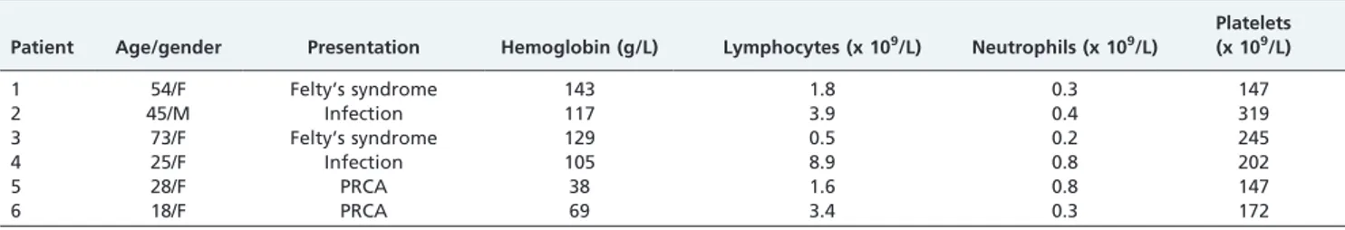
Table 1 -Clinical Characteristics of Patients.
Patient Age/gender Presentation Hemoglobin (g/L) Lymphocytes (x 109/L) Neutrophils (x 109/L)
Platelets (x 109/L)
1 54/F Felty’s syndrome 143 1.8 0.3 147
2 45/M Infection 117 3.9 0.4 319
3 73/F Felty’s syndrome 129 0.5 0.2 245
4 25/F Infection 105 8.9 0.8 202
5 28/F PRCA 38 1.6 0.8 147
6 18/F PRCA 69 3.4 0.3 172
F = Female; M = Male; PRCA = pure red cell aplasia.
Figure 1 -Peripheral blood of patient 4: (A) at diagnosis, showing T-cell receptor gamma gene monoclonal rearrangement; and (B) after eight cycles of fludarabine, showing that the monoclonal cells were completely replaced by polyclonal T-cells, which is characteristic of a complete molecular response (CMR).
CLINICS 2012;67(7):745-748 T-LGL with Fludarabine
Costa RO et al.
CMR, along with excellent treatment compliance and drug tolerance. In accordance with the literature, our main indica-tion for treatment was cytopenia and recurrent infecindica-tion (19). In our series of cases, we found a high rate of CHR and CMR with fludarabine, along with excellent compliance and tolerability. Even though our number of cases was small, we suggest that fludarabine should be further tested as a first-line treatment option for T-LGL to confirm these prelimin-ary data.
AUTHOR CONTRIBUTIONS
Bellesso M and Pereira J were responsible for the data review, manuscript writing and review. Oliveira RC was responsible for the data collection and review, manuscript writing and review. Chamone DAF was responsible for the manuscript review. Ruiz MA was responsible for the manuscript writing. Hallack AE was responsible for the manuscript writing and review. Aldred VL was responsible for the diagnosis review.
REFERENCES
1. Brouet JC, Sasportes M, Flandrin G, Preud’Home JL, Seligman M. Chronic lymphocytic leukaemia of T-cell origin. Immunological and clinical evaluation in eleven patients. Lancet. 1975(7941);2:890-3. 2. Swerdlow SH, Campo E, Harris NL, Jaffe ES, Pileri SA, Stein H, et al.
World Health Organization classification of tumours of Haematopoietic and Lymphoid Tissues. 4thed. International Agency for Research on Cancer Press, Lyon, 2008.
3. Morice WG, Kurt PJ, Leibson PJ, Teferi A, Hanson CA. Demonstration of aberrant T-cell and natural killer cell antigen expression in all cases of granular lymphocytic leukemia. Br J Haematol. 2003;120(6):1026-36, http://dx.doi.org/10.1046/j.1365-2141.2003.04201.x.
4. Lundell R, Hartung L, Hill S, Perkins SL, Bahler DW. T-cell large granular lymphocyte leukemias have multiple phenotypic abnormalities involving pan-T cell antigens and receptors for MHC molecules. Am J Clin Pathol. 2005;124(6):937-46, http://dx.doi.org/10.1309/ PH7X78HF4FW4PRKW.
5. Lamy T, Loughran TP. Clinical features of large granular lymphocyte leukemia. Semin Hematol. 2003;40(3):185-95, http://dx.doi.org/10.1016/ S0037-1963(03)00133-1.
6. Loughran TP, Kidd PG, Starkebaum G. Treatment of large granular lymphocyte leukemia with oral low-dose methotrexate. Blood. 1994; 84(7):2164-70.
7. Sood R, Stewart CC, Aplan PD, Murai H, Ward P, Barcos M, Baer MR. Neutropenia associated with T-cell large granular lymphocyte leukemia: long-term response to cyclosporine therapy despite persistence of abnormal cells. Blood. 1998;91(9):3372-8.
8. Sternberg A, Eagleton H, Pillai N, Leyden K, Turner S, Pearson D, et al. Neutropenia and anaemia associated with T-cell large granular lymphocyte leukaemia responds to fludarabine with minimal toxicity. Br J Haematol. 2003;120(4):699-701, http://dx.doi.org/10.1046/j.1365-2141.2003.04148.x.
9. Fortune AF, Kelly K, Sargent J, O’Brien D, Quinn F, Chadwick N, et al. Large granular lymphocyte leukemia: natural history and response to treatment. Leuk Lymphoma. 2010;51(5):839-45, http://dx.doi.org/ 10.3109/10428191003706947.
10. Stewart CC. Clinical applications of flow cytometry: Immunologic methods for measuring cell membrane and cytoplasmic antigens. Cancer. 1992;15;69(6 Suppl):1543-52, http://dx.doi.org/10.1002/1097-0142(19920315)69:6+,1543::AID-CNCR2820691307.3.0.CO;2-O. 11. O’Malley DP. T-cell large granular leukemia and related proliferations.
Am J Clin Pathol. 2007;127(6):850-9, http://dx.doi.org/10.1309/ A8FHDA0VVRJ05GJP.
12. Shadrach B, Warshawsky I. A comparison of multiplex and monoplex T cell receptor gamma PCR. Diagn Mol Pathol. 2004;13(3):127-34, http:// dx.doi.org/10.1097/01.pdm.0000126419.92931.a3.
13. Dhopapkar M, Chin-Yang, Lust L, Tefferi A, Phyliky R. Clinical spectrum of clonal proliferations of T-large granular lymphocytes: a T-cell clonopathy of undetermined significance. Blood. 1994;84(5): 1620-7.
14. Aribi A, Huh Y, Keating M, Brin S, Ferrajoli A, Faderl S, et al. T-cell large granular lymphocytic (T-LGL) leukemia: Experience in a single institu-tion over 8 years. Leuk Res. 2007;31(7):939-45, http://dx.doi.org/ 10.1016/j.leukres.2006.09.003.
15. Lamy T, Loughran TP. How I treat LGL leukemia. Blood. 2011;117:2764-74, http://dx.doi.org/10.1182/blood-2010-07-296962.
16. Kantarjian HM, Childs C, O’Brien S, Huh Y, Beran M, Schachner J, et al. Efficacy of fludarabine, a new adenine nucleoside analogue in patients with prolymphocytic leukemia and the prolymphocytoid variant of chronic lymphocytic leukemia. Am J Med. 1991;90(2):223-8.
17. Witzig TE, Weitz JJ, Lundberg JH, Tefferi A. Treatment of refractory T-cell chronic lymphocytic leukemia with purine nucleoside analogues. Leuk Lymphoma. 1994;14(1-2):137-9, http://dx.doi.org/10.3109/ 10428199409049659.
18. Tse E, Chan JCW, Pang A, Au W-Y, Leung AYH, Lam CCK, et al. Fludarabine, mitoxantrone and dexamethasone as first-line treatment for T-cell large granular lymphocyte leukemia. Leukemia. 2007;21(10):2225-6, http://dx.doi.org/10.1038/sj.leu.2404767.
19. Bareau B, Rey J, Hamidou M, Donadieu J, Morcet J, Reman O, et al. Analysis of French cohort of patients with large granular lymphocyte leukemia: a report on 229 cases. Haematologica 2010;95(9):1534-41.
T-LGL with Fludarabine
Costa RO et al. CLINICS 2012;67(7):745-748