Evaluation of cost-efectiveness from the funding
body’s point of view of ultrasound-guided central
venous catheter insertion compared with the
conventional technique
INTRODUCTION
he central venous catheter (CVC) is currently regarded as one of the fundamental tools in hospital medical practice. he indications for its use are numerous: administration of vasopressors, hemodynamic monitoring Danilo Teixeira Noritomi1, Rogério Zigaib2, Otavio
T. Ranzani1,3, Vanessa Teich4
1. Grupo de Cuidados Críticos Amil, Intensive Care Unit, Hospital Paulistano - São Paulo (SP), Brazil.
2. Intensive Care Unit, Clinical Emergency Department, Hospital das Clínicas, Faculdade de Medicina, Universidade de São Paulo - São Paulo (SP), Brazil.
3. Department of Pulmonology, Hear Instituto do Coração, Hospital das Clínicas, Faculdade de Medicina, Universidade de São Paulo - São Paulo (SP), Brazil.
4. Insper - Instituto de Ensino e Pesquisa - São Paulo (SP), Brazil.
Objective: To evaluate the cost-efectiveness, from the funding body’s point of view, of real-time ultrasound-guided central venous catheter insertion compared to the traditional method, which is based on the external anatomical landmark technique.
Methods: A theoretical simulation based on international literature data was applied to the Brazilian context, i.e., the Uniied Health System (Sistema
Único de Saúde - SUS). A decision
tree was constructed that showed the two central venous catheter insertion techniques: real-time ultrasonography versus external anatomical landmarks. he probabilities of failure and complications were extracted from a search on the PubMed and Embase databases, and values associated with the procedure and with complications were taken from market research and the Department of Information Technology of the Uniied Health System (DATASUS). Each central venous catheter insertion alternative had a cost that could be calculated by following each of the possible paths on the decision tree. he incremental cost-efectiveness ratio was calculated
Conflicts of interest: None. Submitted on November 2, 2015 Accepted on November 30, 2015
Corresponding author:
Rogério Zigaib
Unidade de Terapia Intensiva do Hospital das Clínicas da Faculdade de Medicina da Universidade de São Paulo - Departamento de Emergências Clínicas
Avenida Enéas de Carvalho Aguiar, 255, 5º andar Zip code: 050403-000 - São Paulo (SP), Brazil E-mail: [email protected]
Responsible editor: Luciano César Pontes de Azevedo
Avaliação de custo-efetividade da passagem de cateter venoso
central guiada por ultrassonograia comparada com a técnica
convencional sob perspectiva da fonte pagadora
ABSTRACT
Keywords: Central venous cateteres/ economics; Ultrasonography/economics; Diagnostic techniques and procedures; Costs and cost analysis; Helath care costs; Uniied Health System/economics by dividing the mean incremental cost of real-time ultrasound compared to the external anatomical landmark technique by the mean incremental beneit, in terms of avoided complications.
Results: When considering the incorporation of real-time ultrasound and the concomitant lower cost due to the reduced number of complications, the decision tree revealed a inal mean cost for the external anatomical landmark technique of 262.27 Brazilian reals (R$) and for real-time ultrasound of R$187.94. he inal incremental cost of the real-time ultrasound-guided technique was -R$74.33 per central venous catheter. he incremental cost-efectiveness ratio was -R$2,494.34 due to the pneumothorax avoided.
Conclusion: Real-time ultrasound-guided central venous catheter insertion was associated with decreased failure and complication rates and hypothetically reduced costs from the view of the funding body, which in this case was the SUS.
when peripheral venipuncture in not possible. Currently, more than 5 million CVC are used in the United States per year.(1) he Department of Information Technology of the Uniied Health System (Departamento de Informática do Sistema Único de Saúde - DATASUS), maintains a national database that contains procedures reimbursed by the SUS, which shows that 103,922 CVC were used in Brazil in 2013.(2) his number may be underestimated, as the database does not account for procedures reimbursed by the supplementary health system.
he use of CVC is not free of complications, either in terms of insertion or maintenance of the device.(3) Traditionally, the devices are inserted using the external anatomical landmark technique (EALT), in which observation and palpation of anatomical landmarks serve as a reference for deciding the best place to make the puncture. However, this technique is subject to error, mainly because of anatomical variations in the population.
Recently, the use of real-time ultrasound-guided (RTUSG) CVC has been incorporated into medical practice(4) his method has become popular over the last decade, and a series of studies have demonstrated its safety and applicability as well as a reduction in complications of CVC insertion.(4,5)
However, the fact that the incorporation of technologies can result in signiicantly increased costs in health care without there necessarily being a proportional improvement in the quality of care ofered to the public must be considered. In part, this discrepancy may be due to the incorporation of technologies that are inefective or too costly. Despite the scientiic sustainability of ultrasound-guided CVC insertion due to its being an efective procedure in reducing complications, systemic incorporation of this technology presents a challenge. Incorporating a new technology that requires signiicant resources can result in a lack of resources for other care activities that are already in place. In practice, the health manager inds little evidence to support his decision within the scientiic literature and is often guided by non-measurable elements, which leads to the possibility of cognitive bias.(6,7) A recent survey showed that the incorporation of health technology in hospitals is rarely based on any cost-efectiveness analysis.(8)
he objective of this study was to evaluate from the perspective of the funding body, in this case the SUS, the cost-efectiveness of incorporating a relatively new clinical practice - RTUSG central venous catheter insertion - compared with the traditional method based on EALT.
METHODS
his study consisted of a theoretical simulation based on data from the international literature applied to the Brazilian context. A decision tree was constructed that presented both alternatives for CVC insertion and that then followed the possible outcomes that could be observed in patients. he proposed model is shown in igure 1.
Figure 1 - Decision tree. CVC - central venous catheter; EALT - external anatomical landmark technique; RTUSG - real-time ultrasound-guided technique; PTX - pneumothorax; HTX - hemothorax.
Likelihood of complications
To construct the described theoretical model, it was necessary to map the probability of each outcome/ complication after attempting to insert the CVC with the use of each technique. To that end, a review was conducted of the scientiic literature to ind the best estimates of the eicacy of the methods that could be used as a basis for the cost-efectiveness simulation in a national context. We conducted a search of the PubMed and Embase databases, from the irst entries in these databases up to August 2013, using theBoolean search method, with the following terms: (“central venous line” OR “central venous line insertion” OR “central venous catheter” OR “central venous access” OR “central line insertion” OR “CVC” OR “IJV” OR “FV”) AND (“ultrasonography” OR “echography” OR “ultrasound” OR “ultrasonic” OR “image guidance” OR “image guided”) AND (“mechanical complication” OR “pneumothorax” OR “cost-efectiveness” OR “cost” OR “length of stay” OR “los” OR “arterial puncture” OR “hematoma” OR “haematoma” OR “hemothorax” OR “haemothorax”). Among the studies found, a systematic review and meta-analysis by Wu et al.(4) met the inclusion criteria for our research, as it was the most recent, had a rigorous methodology and provided the parameters required for building our comparative analysis of the two methods.(9-31) We therefore chose to use the results of that study as a basis for our analysis.
Costs
he following describes the methods used to calculate the costs of the resources associated with CVC insertion and of the treatment of complications. To deine the costs associated with CVC insertion and its complications at a national level, we interviewed a convenience sample of ive qualiied experts in intensive care medicine who had extensive experience in CVC insertion and who worked mostly in Brazilian public hospitals.
- CVC insertion: the cost of CVC insertion was estimated using the corresponding DATASUS
codes,(3) considering the weighted mean
reimbursement amount when inserting double-lumen and single-double-lumen CVCs and short-term hemodialysis catheters in the proportions 80%, 10% and 10%, respectively, according to the expert’s opinion.
- Cost of the ultrasound device: he cost associated with ultrasound is incurred at the time the equipment is purchased by the hospital. he
cost associated with each procedure that uses an ultrasound device was calculated by projecting the expected number of cases of CVC insertion in a hospital. he cost per procedure associated with the ultrasound device is then the result of dividing the cost of the equipment by the total number of tests that this device performs before obsolescence (obsolescence is considered to be after 5 years). For the purpose of this analysis, we considered a hypothetical service with insertion of 325 CVCs per year, keeping in mind that this number can vary greatly among services. he cost was estimated in August 2013 according to market research and consulting public tenders for purchase of the device.
- Cost of protective devices: obtained from market research of device suppliers.
- Cost of complications: the cost of interventions needed to treat complications. Each complication requires a potential set of interventions. he amount of each resource used for the treatment of complications (e.g., probability of thoracotomy for the treatment of pneumothorax) was estimated by consultation with experts. hen, to calculate the cost of a particular type of complication (e.g., pneumothorax), the probability that the patient would require each potential intervention was calculated, and the result was multiplied by the cost of the intervention. Speciically, to calculate the cost of blood products used in hemothorax treatment, the various costs involved were considered, such as blood collection, transfusion tests and transfusion itself - from blood donation to processing and administration of the blood product. he cost of a particular complication type is the sum of each of these values associated with each therapeutic intervention.
Comparison of methods
ICER = ΔC Equation 1
ΔE
ΔC - incremental cost of the RTUSG technique compared to the EALT technique; ΔE = incremental effectiveness of the RTUSG technique compared to the EALT technique, in terms of avoided complications.
To compare methods, a hypothetical base case was analyzed, namely, insertion of 325 CVC per year over a period of 5 years in a service that performs CVC insertion using the EALT technique and another with the same characteristics carrying out the insertions using the RTUSG technique.
Sensitivity analysis
Key parameters were varied in the univariate sensitivity analysis to evaluate the uncertainty efect of these parameters on the results of the analysis. he main parameters varied were the pneumothorax rate associated with the standard technique (0.5 to 2 times the central estimate) and the mean number of CVC inserted per service (0.25 to 4 times the central estimate). Pneumothorax (ICER per avoided pneumothorax) was chosen as the primary outcome in the sensitivity analyses because it is the most common complication with an associated cost.
RESULTS
Efficacy
Table 1 was constructed based on the selected meta-analysis, which conirmed better eicacy of CVC insertion with RTUSG.
Costs
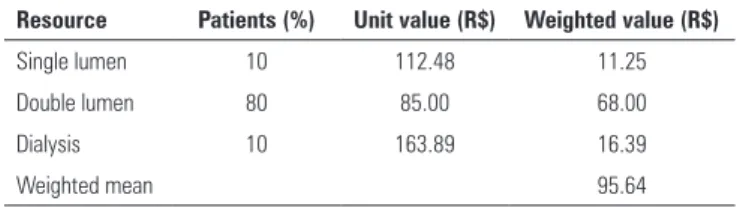
he estimated CVC insertion procedure cost considered three possible diferent types of catheter insertion in the ICU, as shown in table 2. he mean cost per insertion procedure using the EALT technique was R$95.64.
According to market research, the cost of an ultrasound machine is R$45,000.00. he efect of the device value on each procedure was R$87.69. he cost of protective devices was estimated at R$60.00 per CVC inserted. herefore, the additional cost of CVC using the RTUSG method would be R$147.69; this is not taking into consideration that ultrasound reduces complications.
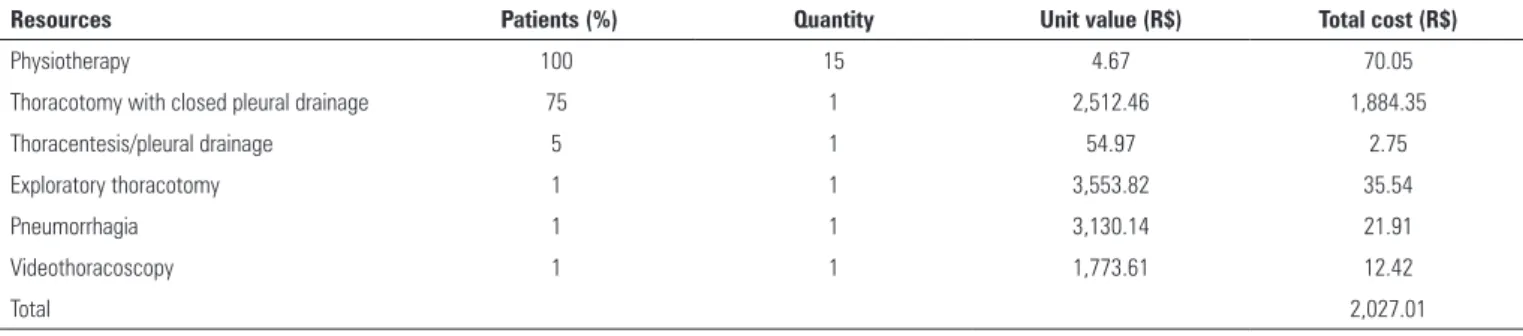
he cost of complications was calculated based on the expert panel opinion and on data from the funding body, SUS. Detailed costs are as shown in tables 3 and 4.
Table 1 - Probability of outcomes and incremental effectiveness of the two methods Outcome EALT (%) RTUSG (%) Incremental
effectiveness %
Failure rate 10.80 1.30 9.50
Pneumothorax rate 3.09 0.11 2.98
Hemothorax rate 2.60 0.00 2.60
Arterial puncture rate 10.80 1.50 9.30
No complications 83.51 98.39 14.88
EALT - external anatomical landmark technique; RTUSG - real-time ultrasound-guided technique.
Table 2 - Weighted cost of central venous catheter insertion
Resource Patients (%) Unit value (R$) Weighted value (R$)
Single lumen 10 112.48 11.25
Double lumen 80 85.00 68.00
Dialysis 10 163.89 16.39
Weighted mean 95.64
We considered the treatment cost of a hematoma due to puncture to be negligible.
Cost-effectiveness analysis
Using the decision tree and considering the incorporation of the new technology and the concomitant cost reduction by reducing complications, the inal mean estimated costs were R$262.27 for the EALT technique and R$187.94 for the RTUSG. he inal incremental cost was therefore -R$74.33. Table 5 shows the cost-efectiveness results based on the additional cost of avoided complications when using the RTUSG technique.
Base case analysis
Table 3 - Mean estimated cost of treatment of a pneumothorax case
Resources Patients (%) Quantity Unit value (R$) Total cost (R$)
Physiotherapy 100 15 4.67 70.05
Thoracotomy with closed pleural drainage 75 1 2,512.46 1,884.35
Thoracentesis/pleural drainage 5 1 54.97 2.75
Exploratory thoracotomy 1 1 3,553.82 35.54
Pneumorrhagia 1 1 3,130.14 21.91
Videothoracoscopy 1 1 1,773.61 12.42
Total 2,027.01
Table 4 - Mean estimated cost of treatment of a hemothorax case
Resources Patients (%) Quantity Unit value (R$) Total cost (R$)
Physiotherapy 100 15 4.67 70.05
Thoracotomy with closed pleural drainage 90 1 2,512.46 2,261.21
Red cell concentrate transfusion 40 2 91.11 72.89
Thoracentesis/pleural drainage 30 1 54.97 16.49
Exploratory thoracotomy 15 1 3,553.82 533.07
Videothoracoscopy 30 1 1,773.61 532.08
Plasma transfusion 20 2 91.11 36.44
Intrathoracic retained clot treatment 10 1 2,582.02 25.82
Platelet transfusion 10 6 91.11 54.67
Total 3,602.73
Table 5 - Cost-effectiveness results
Incremental cost-effectiveness ratio Results (R$)
Incremental cost per case of pneumothorax avoided -2,494.34
Incremental cost per case of hemothorax avoided -2,858.90 Incremental cost per case of hematoma avoided -799.26
ICER - incremental cost-effectiveness ratio.
Figure 2 - Flow chart with percentages attributed to complications in each CVC insertion technique. CVC - central venous catheter; EALT - external anatomical landmark technique; Sensitivity analysis
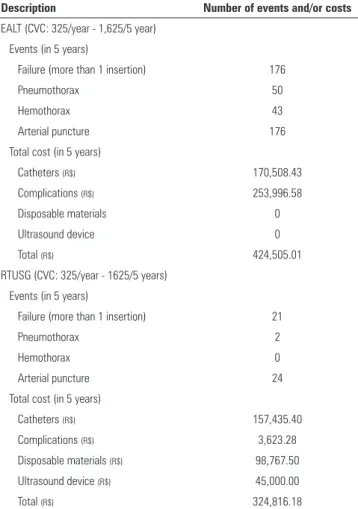
Table 6 - Number of complications and costs associated with each central venous catheter insertion technique
Description Number of events and/or costs
EALT (CVC: 325/year - 1,625/5 year) Events (in 5 years)
Failure (more than 1 insertion) 176
Pneumothorax 50
Hemothorax 43
Arterial puncture 176
Total cost (in 5 years)
Catheters (R$) 170,508.43
Complications (R$) 253,996.58
Disposable materials 0
Ultrasound device 0
Total (R$) 424,505.01
RTUSG (CVC: 325/year - 1625/5 years)
Events (in 5 years)
Failure (more than 1 insertion) 21
Pneumothorax 2
Hemothorax 0
Arterial puncture 24
Total cost (in 5 years)
Catheters (R$) 157,435.40
Complications (R$) 3,623.28
Disposable materials (R$) 98,767.50
Ultrasound device (R$) 45,000.00
Total (R$) 324,816.18
EALT - external anatomical landmark technique; CVC - central venous catheter; RTUSG - real-time ultrasound-guided technique.
DISCUSSION
According to our model, RTUSG CVC insertion is a cost-saving intervention and prevents complications, as shown by the negative ICER in the prevention of complications.
Cost-efective interventions are considered those with increased care costs below a threshold arbitrarily deined as acceptable. When the intervention is able to reduce mortality, there are some suggested thresholds. he World Health Organization (WHO) recommends that an intervention is highly cost-efective if the incremental cost per additional year of life adjusted for quality of life does not exceed the per capita gross domestic product (GDP) of the country in question. An intervention is cost-efective if the ICER is one to three times theper capita GDP; if it exceeds three times the per capita GDP, it is not a cost-efective intervention.(32) his is a concrete element upon
which to guide the administrator’s decision, given that the Brazilian GDP per capita in 2013 was approximately US$11,700.00 (close to R$25,000.00).(33) However, studies comparing the RTUSG technique to the EALT technique do not consider the possibility of a change in mortality with the acquisition of the new technology.
We can state with some certainty that the new technology is cost-saving for a number of reasons. In terms of cost increase, the major determinants of cost tend to become progressively less than those estimated in the base case, as there is a downward trend in device and disposable material costs over the years, given the normal technological evolution in this area. Furthermore, we did not consider the possibility of sharing equipment, which minimizes the cost of the intervention, rendering it even more cost-saving. Still on that side of the equation, the device usage rate in the baseline scenario was quite low, and the obsolescence interval was relatively short. Regarding the costs saved related to complications, values were determined primarily by inarguably necessary components in most cases (such as thoracostomy with pleural drainage).
In our sensitivity analysis, if the number of CVC inserted by RTUSG was below 87 per year, the number of complications would still be much smaller, but costs would not be saved. his situation is usually found when incorporating new health technologies. However, if the annual number of CVC insertions per center were lower, it would be expected that the chance of complications associated with CVC insertion would be higher due to the lower volume of insertion and training. herefore, although we did not simulate this scenario (for example, fewer CVC inserted annually, by center, leading to an increased risk of complications), we can speculate that the use of RTUSG would be efective and cost-saving even in scenarios with low use. Moreover, the sensitivity analysis related to pneumothorax complications always showed a negative ICER.
consider the weaknesses of the analysis, some of which have already been highlighted, and factors that were not taken into account. One of these factors is the immediate impact on the budget. Although the intervention may save resources over time, the funding body must assume a cost that takes place in the present, and the manager should consider whether he is able to pay such an amount immediately. Another point to consider is the usefulness of an ultrasound machine in the intensive care setting for other interventions, such as hemodynamic and cardiovascular evaluations and procedures such as paracentesis, pericardiocentesis and chest puncture. he use of the device can improve and add safety to patient care;(4,5) this is another element not measured in the study that the manager needs to consider.
his study has some limitations. he occurrence rates of events occurring with the EALT and RTUSG techniques were drawn from an international meta-analysis that included no experience from our environment. However, because CVC insertion is a standard technique across the world, we do not believe that Brazilian rates would be much diferent from those observed in the meta-analysis. In addition, the meta-analysis included only randomized studies, which may underestimate complication rates because such studies usually have more controlled samples
than a hospital or clinic will encounter. However, these randomized studies included physicians with and without experience, which may have diluted any efect caused by the better results observed in randomized studies. he cost estimate associated with each complication involved some assumptions, such as the likelihood of the need for each therapeutic intervention. hese probabilities were estimated using the experience of an expert panel and are subject to error. However, we consider the values to be relatively conservative within the consensus range. Furthermore, all directly calculated costs (cost of materials, equipment, etc.) can vary greatly over time and afect the analysis presented at this time. Finally, the economic analysis was performed with data extracted from DATASUS and is based on the SUS’s perspective. It should therefore be interpreted in this context, without the possibility of direct extrapolation of these results to other settings.
CONCLUSION
Real-time ultrasound-guided central venous catheter insertion was associated with decreased failure and complication rates and hypothetically reduced costs from the point of view of the funding body, which in this case was the Brazilian SUS.
Objetivo: Avaliar o custo-efetividade da inserção de cateter venoso central guiada por ultrassonograia em tempo real, em comparação com a técnica tradicional, que é baseada na técnica de reparos anatômicos externos, sob a perspectiva da fonte pagadora.
Métodos: Uma simulação teórica, baseada em dados de literatura internacional foi aplicada ao contexto brasileiro, ou seja, ao Sistema Único de Saúde (SUS). Foi estruturada uma árvore de decisão, que apresentava as duas técnicas para inserção de cateter venoso central: ultrassonograia em tempo real versus reparos anatômicos externos. As probabilidades de falha e complicações foram extraídas de uma busca nas bases PubMed e Embase, e os valores associados ao procedimento e às complicações foram extraídos de pesquisa de mercado e do Departamento de Informática do Sistema Único de Saúde (DATASUS). Cada alternativa de passagem do cateter venoso central teve um custo calculado por meio do seguimento de cada um dos possíveis caminhos da árvore de decisão. A razão de custo-efetividade incremental foi calculada considerando-se a
divisão do custo incremental médio da técnica de ultrassonograia em tempo real comparada à técnica de reparos anatômicos externos pelo benefício incremental médio, em termos de complicações evitadas.
Resultados: O custo inal médio avaliado pela árvore de decisão, considerando a incorporação da ultrassonograia em tempo real e a redução de custo por diminuição de complicações, para a técnica de reparos anatômicos externos foi de R$262,27 e, para ultrassonograia em tempo real, de R$187,94. O custo incremental inal foi de -R$74,33 por cateter venoso central. A razão de custo-efetividade incremental foi -R$2.494,34 por pneumotórax evitado.
Conclusão: A inserção de cateter venoso central com auxílio de ultrassonograia em tempo real esteve associada à diminuição da taxa de falhas e complicações, além de hipoteticamente reduzir custos na perspectiva da fonte pagadora, no caso o SUS.
RESUMO
REFERENCES
1. Raad I. Intravascular-catheter-related infections. Lancet. 1998; 351(9106):893-8. Review.
2. Brasil. Ministério da Saúde. DataSUS. SIGTAP - Sistema de Gerenciamento da Tabela de Procedimentos, Medicamentos e OPM do SUS. [citado 2015 Out 5]. Disponível em: http://sigtap.datasus.gov.br/tabela-unificada/app/ sec/inicio.jsp
3. Mcgee DC, Gould MK. Preventing complications of central venous catheterization. N Engl J Med. 2003;348(12):1123-33.
4. Wu SY, Ling Q, Cao LH, Wang J, Xu MX, Zeng WA. Real-time two-dimensional ultrasound guidance for central venous cannulation: a meta-analysis. Anesthesiology. 2013;118(2):361-75.
5. Parienti JJ, Mongardon N, Mégarbane B, Mira JP, Kalfon P, Gros A, Marqué S, Thuong M, Pottier V, Ramakers M, Savary B, Seguin A, Valette X, Terzi N, Sauneuf B, Cattoir V, Mermel LA, du Cheyron D; 3SITES Study Group. Intravascular complications of central venous catheterization by insertion site. N Engl J Med. 2015;373(13):1220-9.
6. Bero LA, Grilli R, Grimshaw JM, Harvey E, Oxman AD, Thomson MA. Closing the gap between research and practice: an overview of systematic reviews of interventions to promote the implementation of research findings. The Cochrane Effective Practice and Organization of Care Review Group. BMJ. 1998;317(7156):465-8.
7. Cabana MD, Rand CS, Powe NR, Wu AW, Wilson MH, Abboud PA, et al. Why don’t physicians follow clinical practice guidelines? A framework for improvement. JAMA. 1999;282(15):1458-65.
8. Weingart SN. Acquiring advanced technology. Decision-making strategies at twelve medical centers. Int J Technol Assess Health Care.1993;9(4):530-8. 9. Troianos CA, Hartmman GS, Glas KE, Skubas NJ, Eberhardt RT, Walke
JD, Reeves ST; Councils Intraoperative Echocardiography and Vascular Ultrasound of the American Society of Echocardiography. Guidelines for performing ultrasound guided vascular cannulation: recommendations of the American Society of Echocardiography and the Society of Cardiovascular Anesthesiologists. J Am Soc Echocardiogr. 2011;24(12):1291-318. 10. Trerotola SO, Johnson MS, Harris VJ, Shah H, Ambrosius WT, McKusky
MA, et al. Outcome of tunneled hemodialysis catheters placed via the right internal jugular vein by interventional radiologists. Radiology. 1997;203(2):489-95.
11. Docktor BL, Sadler DJ, Gray RR, Saliken JC, So CB. Radiologic placement of tunneled central catheters: rates of success and of immediate complications in a large series. AJR Am J Roentgenol. 1999;173(2):457-60.
12. Mallory DL, McGee WT, Shawker TH, Brenner M, Bailey KR, Evans RG, et al. Ultrasound guidance improves the success rate of internal jugular vein cannulation. A prospective, randomized trial. Chest. 1990;98(1):157-60. 13. Soyer P, Lacheheb D, Levesque M. High-resolution sonographic guidance
for transjugular liver biopsy. Abdom Imaging. 1993;18(4):360-2. 14. Gualtieri E, Deppe SA, Sipperly ME, Thompson DR. Subclavian venous
catheterization: greater success rate for less experienced operators using ultrasound guidance. Crit Care Med. 1995;23(4):692-7.
15. Hilty WM, Hudson PA, Levitt MA, Hall JB. Real-time ultrasound- guided femoral vein catheterization during cardiopulmonary resuscitation. Ann Emerg Med. 1997;29(3):331-6; discussion 337.
16. Slama M, Novara A, Safavian A, Ossart M, Safar M, Fagon JY. Improvement of internal jugular vein cannulation using an ultrasound-guided technique. Intensive Care Med. 1997;23(8):916-9.
17. Teichgräber UK, Benter T, Gebel M, Manns MP. A sonographically guided technique for central venous access. AJR Am J Roentgenol. 1997;169(3):731-8.
18. Sulek CA, Blas ML, Lobato EB. A randomized study of left versus right internal jugular vein cannulation in adults. J Clin Anesth. 2000;12(2):142-5. 19. Cajozzo M, Quintini G, Cocchiera G, Greco G, Vaglica R, Pezzano G, et al.
Comparison of central venous catheterization with and without ultrasound guide. Transfus Apher Sci. 2004;31(3):199-202.
20. Bansal R, Agarwal SK, Tiwari SC, Dash SC. A prospective randomized study to compare ultrasound-guided with nonultrasound- guided double lumen internal jugular catheter insertion as a temporary hemodialysis access. Ren Fail. 2005;27(5):561-4.
21. Milling TJ Jr, Rose J, Briggs WM, Birkhahn R, Gaeta TJ, Bove JJ, et al. Randomized, controlled clinical trial of point-of-care limited ultrasonography assistance of central venous cannulation: the Third Sonography Outcomes Assessment Program (SOAP-3) Trial. Crit Care Med. 2005;33(8):1764-9. 22. Karakitsos D, Labropoulos N, De Groot E, Patrianakos AP, Kouraklis G,
Poularas J, et al. Real-time ultrasound-guided catheterization of the internal jugular vein: a prospective comparison with the landmark technique in critical care patients. Crit Care. 2006;10(6):R162.
23. Leung J, Duffy M, Finckh A. Real-time ultrasonographically-guided internal jugular vein catheterization in the emergency department increases success rates and reduces complications: a randomized, prospective study. Ann Emerg Med. 2006;48(5):540-7.
24. Agarwal A, Singh DK, Singh AP. Ultrasonography: a novel approach to central venous cannulation. Indian J Crit Care Med. 2009;13(4):213-6. 25. Palepu GB, Deven J, Subrahmanyam M, Mohan S. Impact of
ultrasonography on central venous catheter insertion in intensive care. Indian J Radiol Imaging. 2009;19(3):191-8.
26. Turker G, Kaya FN, Gurbet A, Aksu H, Erdogan C, Atlas A. Internal jugular vein cannulation: an ultrasound-guided technique versus a landmark-guided technique. Clinics (Sao Paulo). 2009;64(10):989-92.
27. Aouad MT, Kanazi GE, Abdallah FW, Moukaddem FH, Turbay MJ, Obeid MY, et al. Femoral vein cannulation performed by residents: a comparison between ultrasound-guided and landmark technique in infants and children undergoing cardiac surgery. Anesth Analg. 2010;111(3):724-8.
28. Prabhu MV, Juneja D, Gopal PB, Sathyanarayanan M, Subhramanyam S, Gandhe S, et al. Ultrasound-guided femoral dialysis access placement: a single-center randomized trial. Clin J Am Soc Nephrol. 2010;5(2):235-9. 29. Fragou M, Gravvanis A, Dimitriou V, Papalois A, Kouraklis G, Karabinis A,
et al. Real-time ultrasound-guided subclavian vein cannulation versus the landmark method in critical care patients: a prospective randomized study. Crit Care Med. 2011;39(7):1607-12.
30. Shrestha BR, Gautam B. Ultrasound versus the landmark technique: a prospective randomized comparative study of internal jugular vein cannulation in an intensive care unit. JNMA J Nepal Med Assoc. 2011;51(182):56-61.
31. Zhang YL, Mi WD, Yu DJ, Fu Q, Feng XX. [Application of ultrasonic surface location for internal jugular vein catheterization via central approach]. Zhongguo Yi Xue Ke Xue Yuan Xue Bao. 2011;33(5):479-84. Chinese. 32. Johns B, Baltussen R, Hutubessy R. Programme costs in the economic
evaluation of health interventions. In: World Health Organization. Making choices in health: WHO guide to cost-effectiveness analysis. Geneva: Worl Health Organization; 2003. p. 177-93.