Rev Bras Ter Intensiva. 2016;28(1):5-7
Lung ultrasound: a useful tool in the weaning
process?
COMMENTARY
INTRODUCTION
he incidence of pulmonary complications related to mechanical ventilation is an important issue among critically ill patients. Reducing the duration of respiratory support is essential for minimizing these complications. he extubation of a patient marks the end of the weaning process. Unfortunately, even after a successful spontaneous breathing trial (SBT), approximately 30% of patients develop respiratory distress within 48 hours of extubation; this results in extubation failure and requires either therapeutic non-invasive ventilation or reintubation.(1) he loss of pulmonary aeration following extubation is a hallmark of extubation failure, leading to impaired gas exchange, prolonged mechanical
ventilation, and increased morbidity and mortality.(2) he pathophysiology is
multifactorial.
he amount of lung aeration loss can be quantiied via lung ultrasound during diferent clinical conditions including the weaning process. It is a non-invasive and radiation-free procedure, which can be performed quickly at the bedside and enables a dynamic assessment of lung aeration changes depending on ventilation conditions, as opposed to a chest x-ray. For many years, lungs were not considered accessible by ultrasound because air does not allow for the transmission of ultrasound waves. However, the artifacts produced at the interface between the lungs and luids, for example, can be easily identiied by lung ultrasound.
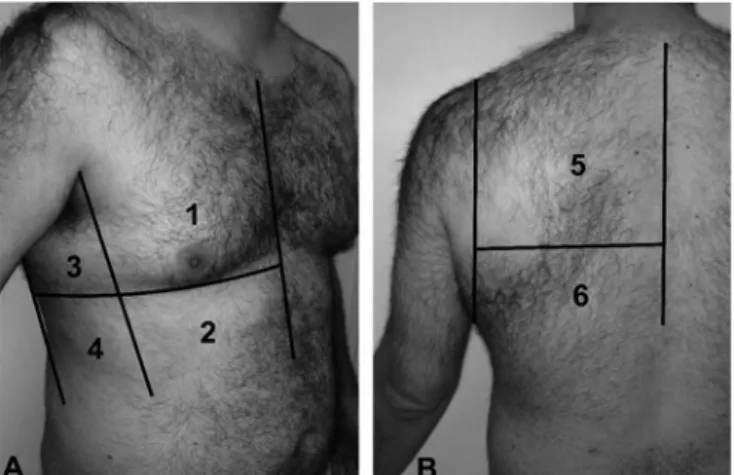
Lung aeration loss can be estimated using by a validated score called the
Lung Ultrasound Score (LUS). As previously recommended,(3-5) all of the
intercostal spaces of the anterior, lateral and posterior regions of both lungs (6 regions per side) are evaluated (Figure 1). For each region, the worst ultrasound pattern isconsidered to be representative of the entire region. Normal aeration is represented by the presence of lung sliding and horizontal A lines, or less than 3 vertical B lines; a score of 0 is assigned to a lung region if all of the intercostal spaces show normal aeration. A moderate loss of aeration is characterized by multiple regularly or irregularly spaced B lines that originate from pleural line or from small juxta-pleural consolidations; a score of 1 is assigned to a lung region if all of the intercostal spaces show a moderate loss of aeration. Severe loss of aeration is characterized by the presence of coalescent B lines in several intercostal spaces, occupying the whole intercostal space; a score of 2 is assigned to the examined region. Complete loss of lung aeration, as observed in lung consolidation, is characterized by tissue echogenicity with static or dynamic air bronchograms; a score of 3 is assigned to the examined region. he scores of Fabiola Prior Caltabeloti1, Jean-Jacques Rouby2
1. Department of Anesthesiology, Hospital das Clínicas, Faculdade de Medicina, Universidade de São Paulo - São Paulo (SP), Brazil.
2. Departament of Anesthesiology and Critical Care Medicine, Pitié-Salpêtrière Hospital, University Pierre and Marie Curie of Paris 6, Paris, France.
Conflicts of interest: None.
Submitted on December 3, 2015 Accepted on December 13, 2015
Corresponding author: Fabiola Prior Caltabeloti
Departamento de Anestesiologia do
Hospital das Clínicas da Faculdade de Medicina da Universidade de São Paulo
Avenida Dr. Eneas de Carvalho Aguiar, 255 Zip code: 05403-000 - São Paulo (SP), Brazil E-mail: [email protected]
Responsible editor: Jorge Ibrain Figueira Salluh
Ultrassonograia pulmonar: uma ferramenta útil no processo de
desmame?
6 Caltabeloti FP, Rouby JJ
Rev Bras Ter Intensiva. 2016;28(1):5-7
the 12 examined regions are summed to calculate the LUS score, which ranges between 0 and 36. Video iles and detailed ultrasound patterns that characterize the diferent stages of aeration can be freely downloaded by visiting http://www.reapitie-univparis6.aphp.fr and clicking on the “Basic skills in lung ultrasound” link.
Figure 1 - Lung ultrasound score.
LUNG ULTRASOUND IN GENERAL CLINICAL PRACTICE
Alveolar recruitment resulting from the administration of positive end-expiratory pressure in patients with acute respiratory distress syndrome (ARDS)(4) and after recovery from ventilator-associated pneumonia during antibiotic treatment can be successfully assessed by lung ultrasound.(5) he evolution of ARDS can also be monitored using lung ultrasound.(6)
Lung ultrasonography has also been validated as a sensitive tool for assessing the risk-beneit ratio of luid loading in patients with septic shock and ARDS. While hemodynamic parameters and oxygenation improved in the study sample, the patients’ LUSs increased, indicating aeration loss. herefore, the use of lung untrasonography may prevent luid overload.(7)
Recently, Soummer et al.(8) showed that a LUS < 13 at the end of a SBT is predictive of extubation success. On the other hand, a LUS > 17 is highly predictive of postextubation distress and extubation failure. Lung derecruitment during the SBT in patients who later experienced extubation failure mainly comprised partial loss of lung aeration rather than new consolidation. his inding suggests that prophylactic non-invasive ventilation (NIV) can prevent derecruitment. A recent multicenter randomized controlled trial demonstrated that high low
nasal oxygen (HFNO) is as eicient as NIV in preventing reintubation in cardiac patients with severe postoperative
respiratory failure.(9) Another multicenter randomized
controlled trial reported that compared to a combination of NIV and HFNO, continuous HFNO is more efective at preventing intubation in severely hypoxemic patients
who were admitted to the intensive care unit (ICU) for
acute respiratory distress caused by community acquired
pneumonia.(10) his positive efect has been shown to be
associated with a reduction in mortality.(11) Interestingly, a randomized monocenter study found that HFNO is much more efective than conventional oxygen therapy in preventing extubation failure during the weaning
process.(12) In addition, it appears to be much more
comfortable for patients.
IMPLEMENTATION OF LUNG ULTRASOUND IN THE WEANING PROCESS
Based on these diferent studies, we designed a multicenter randomized controlled trial, the WEANLUS Brazil Study, to assess whether the continuous administration of HFNO prevents extubation failure in patients categorized as at-risk based on a LUS > 13 at the end of a successful SBT. he trial is now underway and will include 640 patients who were older than 18 years and
mechanically ventilated for more than 48 hours. Patients
will be divided in two groups after extubation: the control group and intervention group. A LUS will be calculated for each patient at the end of their successful SBT. In the control group, the physician in charge of the patient will be blinded to the LUS, and all of the patients will receive conventional oxygen therapy after extubation. NIV will be administered to the patients with well-deined criteria of severe respiratory failure, and they will be classiied as experiencing “extubation failure”. In the intervention group, the physician in charge of the patient will not be blinded to the LUS. HFNO will be administered only to the patients at a high risk of extubation failure (i.e., patients with a LUS > 13 at the end of their SBT). he goal of the study is to show a 30% decrease in the incidence of extubation failure, an increase in the number of days without mechanical ventilation (invasive and non-invasive) from randomization, a decrease in the length of stay in the ICU and a reduction in mortality at D30 and D90 in the high-risk group (LUS >13). Furthermore, patient comfort under each technique of oxygenation
(conventional O2 and HFNO) will be assessed by assigning
Lung ultrasound 7
Rev Bras Ter Intensiva. 2016;28(1):5-7 REFERENCES
1. Boles JM, Bion J, Connors A, Herridge M, Marsh B, Melot C, et al. Weaning from mechanical ventilation. Eur Resp J. 2007;29(5):1033-56. 2. Peñuelas O, Frutos-Vivar F, Fernández C, Anzueto A, Epstein SK, Apezteguía C,
González M, Nin N, Raymondos K, Tomicic V, Desmery P, Arabi Y, Pelosi P, Kuiper M, Jibaja M, Matamis D, Ferguson ND, Esteban A; Ventila Group. Characteristics and outcomes of ventilated patients according to time to liberation from mechanical ventilation. Am J Resp Crit Care Med. 2011;184(4):430-7. 3. Bouhemad B, Zhang M, Lu Q, Rouby JJ. Clinical review: Bedside lung
ultrasound in critical care practice. Crit Care. 2007;11(1):205.
4. Bouhemad B, Brisson H, Le-Guen M, Arbelot C, Lu Q, Rouby JJ. Bedside ultrasound assessment of positive end-expiratory pressure-induced lung recruitment. Am J Respir Crit Care Med. 2011;183(3):341-7.
5. Bouhemad B, Liu ZH, Arbelot C, Zhang M, Ferarri F, Le-Guen M, et al. Ultrasound assessment of antibiotic-induced pulmonary reaeration in ventilator-associated pneumonia. Crit Care Med. 2010;38(1):84-92. 6. Arbelot C, Ferrari F, Bouhemad B, Rouby JJ. Lung ultrasound in acute
respiratory distress syndrome and acute lung injury. Curr Opin Crit Care. 2008;14(1):70-4.
7. Caltabeloti F, Monsel A, Arbelot C, Brisson H, Lu Q, Gu WJ, et al. Early fluid loading in acute respiratory distress syndrome with septic shock deteriorates lung aeration without impairing arterial oxygenation: a lung ultrasound observational study. Crit Care. 2014;18(3):R91.
8. Soummer A, Perbet S, Brisson H, Arbelot C, Constantin JM, Lu Q, Rouby JJ; Lung Ultrasound Study Group. Ultrasound assessment of lung aeration loss during a successful weaning trial predicts postextubation distress. Crit Care Med. 2012;40(7):2064-72.
9. Stéphan F, Barrucand B, Petit P, Rézaiguia-Delclaux S, Médard A, Delannoy B, Cosserant B, Flicoteaux G, Imbert A, Pilorge C, Bérard L; BiPOP Study Group. High-flow nasal oxygen vs noninvasive positive airway pressure in hypoxemic patients after cardiothoracic surgery: a randomized clinical trial. JAMA. 2015;313(23):2331-9.
10. Frat JP, Thille AW, Mercat A, Girault C, Ragot S, Perbet S, Prat G, Boulain T, Morawiec E, Cottereau A, Devaquet J, Nseir S, Razazi K, Mira JP, Argaud L, Chakarian JC, Ricard JD, Wittebole X, Chevalier S, Herbland A, Fartoukh M, Constantin JM, Tonnelier JM, Pierrot M, Mathonnet A, Béduneau G, Delétage-Métreau C, Richard JC, Brochard L, Robert R; FLORALI Study Group; REVA Network. High-flow oxygen through nasal cannula in acute hypoxemic respiratory failure. N Engl J Med. 2015;372(23):2185-96.
11. Matthay MA. Saving lives with high-flow nasal oxygen. N Engl J Med. 2015;372(23):2225-6.