1. Programa de Pós-Graduação em Ciências Biológicas, São José dos Cam-pos, SP.
2. Laboratório de Processamento de Sinais Biológicos, IP&D / UniVap, São José dos Campos, SP.
3. Laboratório de Farmacologia e Fototerapia da Inflamação. Departamen-to de Farmacologia, InstituDepartamen-to de Ciências Biomédicas, Universidade de São Paulo, São Paulo, SP.
4. Laboratório de Histologia e Neuroanatomia, Faculdade de Ciências da Saúde, Universidade Cruzeiro do Sul – UNICSUL, São Miguel Paulista, SP.
5. Grupo de Engenharia Biomédica – GENEBIO, Centro de Pesquisa e De-senvolvimento em Engenharia Elétrica, Universidade Federal de Minas Gerais, Belo Horizonte, MG.
Approved in 15/1/07.
Correspondence to: Rodrigo Alvaro Brandão Lopes Martins, Laboratório de Farmacologia e Fototerapia da Inflamação, Departamento de Farmaco-logia, Instituto de Ciências Biomédicas, Universidade de São Paulo, Av. Lineu Prestes, 1.524 – 05508-900 – São Paulo, SP, Brazil.
E-mail: [email protected]
Characterization of heart rate variability and baroreflex
sensitivity in sedentary individuals and male athletes
Leandro Yukio A. Kawaguchi1, Aline C.P. Nascimento2, Márcio S. Lima3, Lúcio Frigo4,Alderico Rodrigues de Paula Júnior2, Carlos Júlio Tierra-Criollo5 and Rodrigo Alvaro Brandão Lopes-Martins3
O
RIGINALA
RTICLEKeywords: Heart rate variability. Autonomous nervous system. Athletes. ENGLISH VERSION
ABSTRACT
Introduction: The capacity to vary the heart rate represents
im-portant physiologic role in the daily life. The variations of the RR intervals is dependent of biological modulators as the autonomic nervous system. Those variations constitute the heart rate vari-ability (HRV). Methods: 10 athletes (Atl) and 10 sedentary (Sed) male individuals (20-35 age) were submitted to digital electrocar-diography, in rest, before, during and after the maneuver. The valu-es of RR were analyzed (software Matlab 6.1), in the time domain.
Results: Both Sed and Atl presented mean heart rate of 73.5 bpm
± 2,5 and 51 bpm ± 2,4, respectively. Related to the RR intervals, the group of Sed presented average of 826.58 bad ± 5.3 and the group Atl, 1189.18 ± 6.9. The return time of sympathetic system after the maneuver was 72 ± 12 s (Sed) 37 ± 6 s (Atl). The return time of parasympathetic system was 80 ± 11 s (Sed) and 40 ± 8 s (Atl). The pNN50 was of 10 ± 3,3 (Sed) and 42,10 ± 6,9 (Atl). The value of RR variation above the average of the whole sign was 343 ± 40 (Sed) and 175 ± 39 (Atl). The RR variation below the average of the whole sign was 281 ± 27 (Sed) and 425 ± 26 (Atl).
Conclu-sions: The analysis of HRV associated to the Valsalva maneuver
can represent a simple, but important tool, for possible inferences on physical aptitude.
INTRODUCTION
The portion of the nervous system which controls the visceral functions of the body is called autonomic or autonomous nervous system (ANS), which means to regulate itself. The autonomic ad-justments are not usually accessible to conscience; for that rea-son, this system is frequently called involuntary or neurovegeta-tive motor system. This system tonically and reflexely influences blood pressure, peripheral resistance, cardiac frequency and cardi-ac debt(1-3).
The heart is a central organ in the maintenance of homeostasis and in order to reach it, it receives autonomic influences. Within this context, one of its main characteristics consists of constant alteration of its beating frequency. The information reaches the central nervous system (nucleus tractus solitarius) through mar-row and vagal afferent ways; is modulated and returns to the heart through fast vagal efferent fibers (which can occur in the first sub-sequent beat) and slow sympathic efferent ones (an interval of up to 20 seconds can occur). The resulting effect of these autonomic influences is the variability beat after beat of the instantaneous heart rate(4-9).
The RR intervals variations are dependent on biological modula-tors such as the ANS, through the activity of the sympathic and parasympathetic systems. These variations constitute the heart rate variability (HRV), where the aim is to measure the variation between each successive sinusal beat. Such technique has been used as a non-invasive method of evaluation of the neural control over the heart. The clinical relevance of the HRV was first appreci-ated in 1965 by Hon and Lee(10) in the fetal monitoring. In 1977
Wolf et al.(11) were the first ones to demonstrate association
be-tween increase in the risk for post-infarct mortality with decrease of HRV. Actually, the capacity to vary the cardiac beat rate has the physiological meaning of adapting the Cardiovascular System, moment after moment, to the several daily situations, from sleep to strenuous physical activity.
The study of the HRV has allowed recognizing and characteriz-ing some situations in which diseases affect the autonomic con-trol. Some authors have demonstrated that the decrease of the HRV is related to a higher index of cardiovascular morbidity and mortality implying in the presence of individual physiological dys-function(4,6,9,12).
Bioengineering and biological signals processing have allowed countless possibilities of non-invasive diagnosis procedures, es-pecially in the cardiovascular field. Any periodic variable can be analyzed in relation to the rate in which the event occurs. Many of the biological signals are defined as ‘almost periodic’, that is, they vary in a repetitive way at times ‘almost regular’(7).
The autonomic tonus may be evaluated through the HRV during short periods of electrocardiograph monitoring, between 60 sec-onds and 30 minutes, or by prolonged periods, usually through long duration electrocardiogram, 24 hours (Holter system)(13).
According to some authors(4,8,13), the analysis can be performed
in two ways: in the time dominium or in the rate dominium (or the spectral power of the heart frequency): the analysis of the time dominium globally reflects the autonomic activity, that is, whether it presents any alteration or not. The evaluated variables are:
ob-tained at every 5 minutes; d) RMSSD, which represents the square root of the mean of the squares of the differences between suc-cessive normal RR intervals; e) SDNN index – represents the mean of all the standard deviations of the normal R-R intervals obtained at every 5 minutes; f) pNN50 – percentage concerning the total of normal RRs, of the normal RRs which, concerning the previous RRs, have a difference higher than 50 ms.
The analysis of the spectral power allows the quantitative and qualitative, individualized and simultaneous characterization, in absolute and relative terms of the cardiac sympathic and parasym-pathetic activities, through the frequency of the waves and their respective physiological origins: a) very low rate component – (VLF, Very Low Frequency) – (0.015 to 0.04 Hz) – mediated by the ther-moregulation and the renin-angiotensin-aldosterone system; b) low frequency component – (LF of Low Frequency) – (0.04 to 0.15 Hz) – mediated by the baroreceptor reflex, with mixed influences of the sympathic and parasympathetic); c) high frequency component – (HF of High Frequency) – (0.15 to 0.40 Hz) – indicator of expressed vagal tonus to parasympathetic influence over the sinusal node and respiratory frequency.
The analysis of the autonomic activity is based on the evaluation of alterations in the cardiac frequency evoked by the stimulation of cardiovascular reflexes. One of the ways to investigate the cardio-vascular reflexes is the Valsalva Maneuver(9).
This maneuver consists of the act to perform an expiration ef-fort with the glottis closed. This procedure was described in 1704 as a method for pus expelling from the medium ear by the effort with mouth and nose closed. The forced exhaling against a closed glottis commonly occurs during weight lifting and during other ac-tivities which demand fast and maximum application of strength for a short period. The intrathoracic pressure during the maneuver may reach over 150 mm Hg above air pressure. Such pressure is transmitted through the thin walls of the veins which cross the thoracic region. As the venous blood is submitted to a relatively low pressure, these veins are squeezed and the blood flow which returns to the heart suffers a significant reduction(14-15).
The normal response to the valsalva maneuver consists of 4 phases. Phase I is associated with a transitory increase in the sys-temic blood pressure as the effort initiates. Phase II is followed by a perceptive decrease in the systemic venous return, blood pres-sure, pulse pressure and reflex tachycardia promptly detectable. Phase III initiates with the end of the effort and is associated with a transitory and sudden decrease in the blood pressure as well as in the systemic venous return. Finally, phase IV is characterized by a recovery of the systemic arterial pressure and relatively evident reflex bradycardia(16).
According to the author(17), the selection of non-invasive
auto-nomic tests such as: controlled breathing, active postural alteration and valsalva maneuver, were sensitive enough to exemplify the improvement in the cardiovascular aptitude of individuals who par-ticipated in the study.
On the other hand, some authors report benefits of moderate endurance exercise in the parasympathetic system activity, reduc-tion of the sympathic tonus at rest, as well as influence on the baroreflex sensitivity. Differences concerning age and gender have also been mentioned(18-21). High level athletes present heart rate at
rest lower than sedentary individuals from the same age group. Physical exercise induces to a blockage of the parasympathetic activity, as well as the sympathic stimulation in the oblong bone marrow, similarly to the classical reflex of fight and run(22).
In this investigation, we tried to characterize the heart rate vari-ability through processing instruments of biological signals in healthy individuals, athletes and sedentary individuals, in the valsalva test, usually applied for measurement of the baroreflex sensitivity. We try through the parameters analyzed in the valsalva maneuver, to investigate the possible differences found between athletes and sedentary individuals concerning the baroreflex sensitivity,
infer-ring over the use of this classic test as predictor for physical apti-tude.
METHODS
Data collection was conducted in the laboratory of Cardiopul-monary Rehabilitation of the Vale do Paraíba University – São José dos Campos – SP.
20 subjects, age range of 20-35 years, participated in the study. They were divided in two groups: group I (10 athletes (Running Teams from São José dos Campos, participants of the Regional and State Games in long distance events) and group II, (10 seden-tary individuals according to IPAQ (annex 1)...
The inclusion criteria were: healthy individuals with no cardio-vascular pathology; renal and brain history or risk factors such as hypertension, smoking, diabetes, obesity and electrocardiogram of 12 derivations within the normality boundaries.
Individuals who presented atrial fibrillation, sinusal dysfunction, atrioventricular conduction disturbs, artificial pacemaker or ectopic beating were excluded.
All volunteers were informed about the research’s protocol, and after having signed a free and clarified consent form, were submit-ted to the following procedures:
Experimental procedure:
a) anamnesis
b) Initially the electrodes were placed on the torax (care was taken with the skin preparation; 70% alcohol asepsis and 360 sand-paper), in the following regions: root of the right and left upper limbs and right and left supra-iliac region, for acquiring the cardiac depolarization in the DI, DII, DIII, aVR, aVL and aVF derivations, being the DII derivation used for having the R-R intervals.
In the beginning of the protocol the individual remained seating on a steady chair for 5 minutes with relaxed upper limbs; lower limbs at 90 degrees of hip and knee flexion; natural breathing; at room temperature between 22 to 25oC with relative humidity
be-tween 50 to 60%. From that time on, the registration of the RR intervals for 15 continuous minutes was initiated. At the 7th minute the beginning of the valsalva maneuver was asked; after maximal inspiration (maximal inspiratory capacity) the subject kept an expi-ratory pressure of 50 cmH2O for 15 seconds. The manuvacuome-ter was placed in front of his eyes, being held by the author of the research.
c) After the values of the RR intervals had been obtained, these data were treated with the aid of the Matlab 6.1 software, in the time dominium. The association of digital filters with the calcula-tion of spectral energy was used for the processing of the signal. The program developed in order to process the signals of the HRV was implemented through the following steps: signal → temporal correction → removal of the DC level → interpolation → resam-pling → bandpass filter → energy calculation → processed exit signal.
Firstly, the time scale correction and the removal of the DC level of the signal were performed due to the interest in the analysis of the HRV. The signal reconstruction with the same rate of the sam-ple was performed by the use of the cubic spline interpolation func-tion. Afterwards, the signal was reassembled at 4 Hz. A Butter-worth bandpass filter of order 2 with cutting frequencies of 0.04 Hz and 0.15 Hz was used in order to separate the frequencies concerned with the sympathic activity, and for the parasympathet-ic activity a Butterworth bandpass of order 2 with cutting frequen-cies of 0.15 Hz and 0.4 Hz was also used, generating the xa and xb signals respectively. In order to obtain a linear phase, both signals were direct and reversely filtered.
He could choose to use windows with overlaps or not, as repre-sented in figure 1.
Figure 1 – Designing of intervals in the time scale (s) with n = 12 seconds and m = 15 seconds, with a 3-second overlap
For each time interval the energy for the two xa and xb, signals was calculated, using equation 1, generating the Ena and Enb tints.
En = n + m
En = ∑ x(i)2 (1)
En = i = n
where, En is the x(i) energy signal in the interval from n up to n + m [14].
After the calculation of the xa and xb signals energy, the program graphically presents these outcomes concerning time, enabling the operator to define the beginning of the valsalva maneuver. The program also calculated the ratio between Ena and Enb concerning time.
Statistical analysis
In order to compare the means of the studied groups, the anal-ysis of variance – ANOVA was used, followed by the t student test for non-paired samples. The p values < or = 0.05 were considered significant.
RESULTS
Characterization of the athletes and sedentary groups
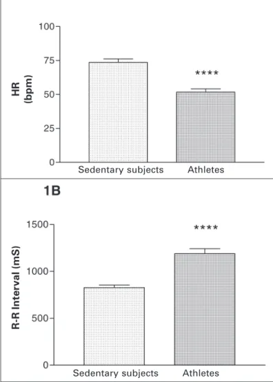
After the data analysis of the group of athletes and sedentary subjects through observation of the heart rate and the R-R inter-vals at real time, we can observe that the group of sedentary sub-jects presents heart rate higher than the group of athletes, with mean heart rate equal to 73.59 bpm ± 2.5 and 51.01 bpm ± 2.4, respectively as observed in graphic 1A. The R-R intervals are rep-resented in graphic 1B, where we observe that the group of sed-entary subjects presents mean of R-R intervals equal to 826.58 ms ± 5.3 and the group pf athletes presents mean of 1189.18 ± 6.9. Such values confirm a low heart rate of the athletes, characterizing hence the groups.
Time of sympathic and parasympathetic return after valsalva maneuver
It was possible to evaluate the time in which the autonomous nervous system takes to stabilize the cardiovascular system after stress through the valsalva maneuver. In graphic 2A we can ob-serve the return time of the sympathic system after the valsalva maneuver. The group of sedentary subjects presents mean time of 72 ± 12 s and for the group of athletes the mean time was of 37 ± 6 s. Comparing the two groups we obtain significant differences with p = 0.02. In graphic 2B, the return time of the parassympa-thetic system after the valsalva maneuver is represented. It is ob-served that the group of sedentary subjects presents mean return time of 80 ± 11 s and the group of athletes of 40 ± 8 s, presenting hence statistic differences with p = 0.01.
Analysis of the heart rate variability in the time dominium
The heart rate variability can be calculated through the R-R inter-vals. In graphic 3 we can observe one of the mean for evaluation of the variability in the time dominium, the pNN50. It can be seen that the mean of the values found for the group of sedentary indi-viduals was of 10 ± 3.3 and for the group of athletes was of 42.10 ± 6.9. When the groups are compared, we observe statistic differ-ences with p = 0.0007.
R-R interval above and below the R-R mean variation of the entire signal
In graphic 4A the mean value of the R-R intervals variation above the mean of the entire signal can be observed. In the group of sedentary individuals this value was 343 ± 40 ms and in the group of athletes it was 175 ± 39 ms. These values when submitted to statistic treatment presented p = 0.0084. The variation of the inter-vals above the mean of the signal expresses the bradycardia gen-erated in the cardiovascular system which is induced by the valsal-va maneuver. Graphic 4B demonstrates the mean valsal-value of the variation in the R-R intervals below the mean of the entire signal. In the group of sedentary subjects we found mean value of 281 ± 27 ms. In the group of athletes we found mean value of 425 ± 26 ms. When the groups were compared, we found p = 0.0012. The variation of the intervals below the mean of the signal expresses the tachycardia generated in the cardiovascular system induced by the valsalva maneuver.
Graphic 1
HR
(bpm)
Sedentary subjects Athletes
R-R Inter
val (mS)
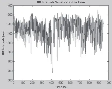
Figures 2 and 3 represent the heart rate variability during the entire experiment for sedentary individuals and athletes respec-tively, which aids in the understanding of the data obtained men-tioned above in graphic 4A and B.
DISCUSSION
As mentioned before, the RR intervals variations, beat after beat, depend on the modulation of the Autonomous Nervous System and received the name of ‘heart rate variability’ (HRV), represent-ing the measurement of the variation between each successive sinusal beating(8). The ability to vary the heart rate in relation to Graphic 2
Sympathic Return
T
ime (s)
Parasympathetic Return
T
ime (s)
Graphic 3
Graphic 4 Above the mean R-R Inter
val V
ariation
p = 0,0084
Below the mean R-R Inter
val V
ariation
p = 0,0012
Figure 2 – R-R intervals variation in the sedentary individual time Variação dos intervalos R-R no tempo
Tempo(s)
Intervalos R-R (ms)
LADO ESQUERDO: RR Intervals (ms) ACIMA: RR Intervals Variation in the Time ABAIXO: Time (s)
external stimuli seems to play an important physiological role in daily life, even in simple situations of postural change, but espe-cially in situations of more intense physical effort, such as sportive activity. Moreover, cardiovascular events or even the natural evo-lution of age, seem to corroborate to the loss or reduction of the ability to vary heart rate. Wolf et al.(11) were the first ones to
dem-onstrate association between increase of post-infarct risk of mor-tality with reduction of HRV. In this investigation, the authors ob-served that 73 out of 176 patients admitted in the Coronary Unit with acute myocardial infarct presented sinusal arrythmia, and con-sequently, higher variability in the R-R interval. These patients pre-sented lower mortality rate. More recently, other authors confirmed this initial hypothesis demonstrating that the decrease of HRV is related to a higher cardiovascular morbidity and mortality in-dex(6,4,9,12).
Sedentary subjects Athletes
Sedentary subjects Athletes
Sedentary subjects Athletes
Sedentary subjects Athletes
In this investigation we analyzed the heart rate variability asso-ciated with the valsalva maneuver with the aim to characterize the response to the maneuver in sedentary individuals as well as ath-letes. Within this context, we intend to identify parameters which are able to aid in the performance of inferences concerning the aptitude of the cardiovascular system, in a fast and simplified way. As already mentioned in the introduction, moderate endurance exercise may significantly influence the activity of the Autonomous Nervous System, as well as of the baroreflex sensitivity(18-21).
Our outcomes corroborate those already demonstrated in the literature, demonstrating that athletes present lower heart rate at rest, than sedentary individuals in the same age group. The mean values observed for heart rate at rest (graphic 1A) were significant-ly lower (HR) in athletes, when compared with the sedentary indi-viduals. Concerning the RR values, the athletes presented RR val-ues significantly higher in comparison to the sedentary subjects, representing a longer interval between each beat. According to some authors, physical training induces to a higher parasympa-thetic activity at rest, explaining the results obtained in this study(22).
Another variable analyzed in this study was the recovery time of the cardiovascular system after sympathic and parasympathetic stimulation of the valsalva maneuver, the HRV basal conditions. The aim of this analysis was to verify through the HRV analysis whether the better physical capacity of the individuals could alter the recovery time of the cardiovascular system or the baroreflex sensitivity.
As can be observed in graphics 2A and 2B, athletes presented in both cases HRV recovery times after valsalva maneuver signifi-cantly lower than sedentary individuals. These outcomes suggest that the baroreflex sensitivity is variable which effectively suffers influences from physical training, being able to accurately differen-tiate two conditions of physical capacity.
As more specific measurement of the HRV, the pNN50 variable was also calculated with the aim to characterize the two studied groups. As expected, the HRV of athletes was significantly higher than in sedentary individuals (graphic 3), also confirming previous findings from the literature and indicating that physical training is able to increase the physiological capacity of the cardiovascular system in varying heart rate, and consequently, better adapting to alterations or stimuli from the external environment. The results here obtained corroborate those previously observed by Levy et al.(18), who stated the increase of the parasympathetic tonus at rest
after 6 months of aerobic training.
The RR intervals variation was calculated during the valsalva maneuver above and below the mean established for all the RR
obtained with the aim to better characterize and differentiate the studied groups concerning the HRV as well as the baroreflex sen-sitivity. As one can see in graphic 4A, the RR intervals variation above the mean, that is, increase of the RR intervals, was signifi-cantly lower for the athletes subjects compared with the seden-tary ones. This finding probably occurred due to the fact that ath-letes already presented RR intervals values significantly higher than the sedentary subjects, in the period before the maneuver, accord-ing to what was previously demonstrated (graphic 1B). Our results corroborate those found by Middleton and De Vito(21) who observed
parasympathetic effect concerned with the baroreflex sensitivity, lower for trained individuals when compared with sedentary ones. On the other hand, the RR intervals variation below the mean, that is, reduction of the RR intervals, was significantly higher for the athletes. Such finding shows that physical training can offer higher capacity of the sympathic system to induce increases of heart rate, reaching higher values when compared with sedentary subjects, in the same period of time and under stimuli of the same intensity. These results are according to those observed by Iella-mo et al.(20), who say that physical training increases the sympathic
response capacity, especially at training peaks.
Within this context, we may suggest that the results obtained in this study using the valsalva maneuver seem to correlate with those reported in the literature where the HRV was analyzed in effort situations. Thus, the present work may be considered the first one to suggest that the classic test of valsalva maneuver will possible be applied as an indicator of physical capacity in different groups of individuals. Further studies are still needed in order to better characterize as well confirm such hypothesis.
All the authors declared there is not any potential conflict of inter-ests regarding this article.
REFERENCES
1. Kandel ER, Schwarts JH, Jessell TM. Principles of neural science. 4th ed. McGraw Hill, 2000.
2. Johnson LR. Fundamentos de fisiologia medica. 2a ed. Rio de Janeiro:
Guanaba-ra Koogan, 2000.
3. Guyton AC, Hall JE. Tratado de fisiologia médica. 10a ed. Rio de Janeiro:
Guana-bara Koogan, 2002.
4. Longo A, Ferreira D, Correia JC. Variabilidade da freqüência cardíaca. Rev Port Cardiol. 1995;14:241-62.
5. Ribeiro FT, Cunha A, Lourenço GCD, Marães VRFS, Catai AM, Gallo Jr L, et al. Estudo da variabilidade da freqüência cardíaca em dois voluntários de meia-ida-de, um coronariopata e outro saudável – Relato de caso. Rev Soc Cardiol Estado de São Paulo. 2000;10:1-10.
6. Campelo M, Coutinho J, Fernandes P, Maciel MJ, Gonçalves FR, Gomes MC. Variabilidade da freqüência cardíaca: uma perspectiva. Rev Port Cardiol. 1992; 11:723-32.
7. Ribeiro MP, Brum JM, Ferrario CM. Análise espectral da freqüência cardíaca. Conceitos básicos e aplicação clínica. Arq Bras Cardiol. 1992;59:141-9.
8 Task Force of the European Society of Cardiology and the North American Soci-ety of Pacing and Electrophysiology. Heart rate variability: standards of mea-surement, physiological interpretation and clinical use. Circulation. 1996;93:1043-65.
9. Pumprla J, Howorka K, Groves D, Chester M, Nolan J. Functional assessment of heart rate variability: physiological basis and practical applications. Int J Cardi-ol. 2002;84:1-14.
10. Hon EH, Lee ST. Electronic evaluations of the fetal heart rate patterns preceding fetal death, further observation. Am J Obstet Gynec. 1965;87:814-26.
11. Wolf MM, Varigos GA, Hunt D, Sloman JG. Sinus arrhythmia in acute myocardial infarction. Med J Aust. 1978;2:52-3.
12. Alonso DO, Forjaz CLM, Rezende LO, Braga AMFW, Barretto ACP, Negrão CE, et al. Comportamento da freqüência cardíaca e da sua variabilidade durante dife-rentes fases do exercício físico progressivo máximo. Arq Bras Cardiol. 1998;71:787-92.
13. Grupi CJ, Moffa PJ, Sanches PCR, Barbosa SA, Bellotti GMV, Pileggi FJC. Varia-bilidade da freqüência cardíaca: significado e aplicação clínica. Rev Assoc Med Brasil. 1994;40:129-36.
Figure 3 – R-R intervals variation in athlete individual time RR Intervals Variation in the Time
Time (s)
14. Fox ML, Keteyian SJ. Bases fisiológicas do exercício e do esporte. Rio de Janei-ro: Guanabara Koogan, 2000.
15. McArdle WD, Katch FI, Katch VL. Fisiologia do exercício, energia, nutrição e desempenho humano. Rio de Janeiro: Guanabara Koogan, 1998.
16. Castro CLB, Nóbrega ACL, Araújo CGS. Testes autonômicos cardiovasculares, uma revisão crítica, parte I. Arq Bras Cardiol. 1992;59:175-85.
17. Guo XH, Yi G, Batchavarov V, Gallagher MM, Malik M. Effect of moderate phys-ical exercise on noninvasive cardiac autonomic test in healthy volunteers. Int J Cardiol. 1999;69:155-68.
18. Levy WC, Cerqueira MD, Harp GD, Johannessen KA, Abrass IB, Schwartz MS, et al. Effect of endurance exercise training on heart rate variability at rest in healthy young and older men. Am J Cardiol. 1998;82:1236-41.
19. Pichot V, Roche F, Gaspoz JM, Enjolras F, Antoniadis A, Minini P, et al. Relation between heart rate variability and training load in middle-distance runners. Med Sci Sports Exerc. 2000;32:1729-36.
20. Iellamo F, Legramante JM, Pigozzi F, Spataro A, Norbiato G, Lucini D, et al. Con-version from vagal to sympathetic predominance with strenuous training in high-performance world class athletes. Circulation. 2002;105(23):2719-24.
21. Middleton N, De Vito G. Cardiovascular autonomic control in endurance-trained and sedentary young women. Clin Physiol Funct Imaging. 2005;25:83-9.
22. Aubert AE, Seps B, Beckers F. Heart rate variability in athletes. Sports Med. 2003; 33:889-919.
ANEXO 1
Questionário Internacional de Atividade Física – versão curta
Nome: ____________________________________________________________________
Data: ______ / ______ / ______ Idade: ______ Sexo: F ( ) M ( )
Nós estamos interessados em saber que tipos de atividade física as pessoas fazem como parte do seu dia a dia. Este projeto faz parte de um grande estudo que está sendo feito em diferentes países ao redor do mundo. Suas respostas nos ajudarão a entender que tão ativos nós somos em relação a pessoas de outros países. As perguntas estão relacionadas ao tempo que você gasta fazendo atividade física na
ÚLTIMA semana. As perguntas incluem as atividades que você faz no trabalho, para ir de um lugar a outro, por lazer, por esporte, por exercício ou como parte das suas atividades em casa ou no jardim. Suas respostas são MUITO importantes. Por favor responda cada questão mesmo que considere que não seja ativo. Obrigado pela sua participação!
Para responder às questões lembre que:
¾ atividades físicas VIGOROSAS são aquelas que precisam de um grande esforço físico e que fazem respirar MUITO mais forte que o normal
¾ atividades físicas MODERADAS são aquelas que precisam de algum esforço físi-co e que fazem respirar UM POUCO mais forte que o normal
Para responder às perguntas pense somente nas atividades que você realiza por pelo menos 10 minutos contínuos de cada vez:
1a. Em quantos dias da última semana você caminhou por pelo menos 10 minutos contínuos em casa ou no trabalho, como forma de transporte para ir de um lugar para outro, por lazer, por prazer ou como forma de exercício?
dias ______por SEMANA( ) Nenhum
1b. Nos dias em que você caminhou por pelo menos 10 minutos contínuos quanto tempo no total você gastou caminhando por dia?
horas: ______ Minutos: ______
2a. Em quantos dias da última semana você realizou atividades MODERADAS por pelo menos 10 minutos contínuos, como por exemplo pedalar leve na bicicleta, na-dar, dançar, fazer ginástica aeróbica leve, jogar vôlei recreativo, carregar pesos le-ves, fazer serviços domésticos na casa, no quintal ou no jardim como varrer, aspirar, cuidar do jardim, ou qualquer atividade que fez aumentar moderadamente sua res-piração ou batimentos do coração (POR FAVOR NÃO INCLUA CAMINHADA)
dias ______por SEMANA( ) Nenhum
2b. Nos dias em que você fez essas atividades moderadas por pelo menos 10 mi-nutos contínuos, quanto tempo no total você gastou fazendo essas atividades por dia?
horas: ______ Minutos: ______
3a. Em quantos dias da última semana você realizou atividades VIGOROSAS por pelo menos 10 minutos contínuos, como por exemplo correr, fazer ginástica aeróbi-ca, jogar futebol, pedalar rápido na bicicleta, jogar basquete, fazer serviços domésti-cos pesados em casa, no quintal ou cavoucar no jardim, carregar pesos elevados ou qualquer atividade que fez aumentar MUITO sua respiração ou batimentos do cora-ção.
dias ______por SEMANA( ) Nenhum
3b. Nos dias em que você fez essas atividades vigorosas por pelo menos 10 minu-tos contínuos quanto tempo no total você gastou fazendo essas atividades por dia? horas: ______ Minutos: ______
PERGUNTA SOMENTE PARA O ESTADO DE SÃO PAULO
5. Você já ouviu falar do Programa Agita São Paulo?