ISSN/$–see front matter © 2013 Sociedade Brasileira de Ortopedia e Traumatologia. Published by Elsevier Editora Ltda. All rights reserved. www.rbo.org.br/
Original Article
Positioning of the acetabular component in cemented
prostheses – radiographic calculation
Pedro José Labronici,
1*Ramon Louro Motta,
2Bruno Bandeira Esteves,
2José Sergio Franco,
3Rolix Hoffmann,
4Luiz Aurélio Costa Ferreira,
5Marcos Giordano,
6Sergio Delmonte Alves
7 1PhD in Medicine from the Universidade Federal de São Paulo – Escola Paulista de Medicina. Head of the “Prof. Dr. Donato D’Ângelo”Orthopedics and Traumatology Service, Hospital Santa Teresa, Petrópolis, Rio de Janeiro, Brazil.
2Resident Physician in Orthopedics and Traumatology, “Prof. Dr. Donato D’Ângelo” Orthopedics and Traumatology Service, Hospital Santa
Teresa, Petrópolis, Rio de Janeiro, Brazil.
3PhD. Head of Department and Associate Professor of the Department of Orthopedics and Traumatology, School of Medicine, universidade
Federal do Rio de Janeiro (UFRJ), Rio de Janeiro, Rio de Janeiro, Brazil.
4Physician in the “Prof. Dr. Donato D’Ângelo” Orthopedics and Traumatology Service, Hospital Santa Teresa, Petrópolis, Rio de Janeiro, Brazil.
5Resident Physician (R4) in the Hip Group, “Prof. Dr. Donato D’Ângelo” Orthopedics and Traumatology Service, Hospital Santa Teresa,
Petrópolis, Rio de Janeiro, Brazil.
6Head of the Orthopedics and Traumatology Service, Galeão Air Force Hospital, Rio de Janeiro; MSc in Medicine Focusing on Orthopedics
and Traumatology, Petrópolis, Rio de Janeiro, Brazil.
7Physician Responsible for the Hip Group, “Prof. Dr. Donato D’Ângelo” Orthopedics and Traumatology Service, Hospital Santa Teresa,
Petrópolis, Rio de Janeiro, Brazil.
Work performed at the “Prof. Dr. Donato D’Ângelo” Orthopedics and Traumatology Service, Hospital Santa Teresa, Petrópolis, Rio de Janeiro, and at the School of Medicine of Petrópolis, Petrópolis, Rio de Janeiro, Brazil.
*Corresponding author at: Av. Roberto Silveira, 187/601, Petropolis, RJ, Brazil. CEP: 25685-040. Phone: (+55 24) 2242 5571.
E-mail: [email protected]
A RT I C L E I N F O Article history:
Received January 18 2012 Approved April 27 2012
Keywords:
Mycobacteria, atypical Keratitis
Corneal transplantation
a b s t r a c t
Objective: to assess the reliability of the inclination angle and anteversion of acetabular cup component in patients with idiopatic osteoarthritis of the hip, aseptic necrosis and hip neck fracture using trigonometric formula and plain radiographs. Methods: 66 patients underwent cemented total arthroplasty of 72 hips. The inclination of acetabular component was measured using plain radiograph. The acetabular component anteversion was measured using trigonometric formula. Results: it was observed that, in the osteoarthritic hips, hip neck fracture and aseptic necrosis, the degree of agreement was highly significant (p < 0.0001), in the measurements of anteversion and inclination angles, among the three assessments, from intra as well as inter-observers. All the agreement pairs were of excellent degree (ICC > 0.80). Conclusion: using plain radiographs and trigonometric formula, the method resulted to be highly accurate and reliable. Besides being easy to be calculated. No significant variation was found in the anteversion and inclination angles when compared with osteoarthritis of the hip, aseptic necrosis and hip neck fracture.
© 2013 Sociedade Brasileira de Ortopedia e Traumatologia. Published by Elsevier Editora Ltda. All rights reserved.
Introduction
The positions of the components in total hip arthroplasty in relation to the femur and pelvis is important with regard to the prognosis for the surgery.1 The inclination and anteversion of
the acetabular component were defined by Murray,2 in relation
to three different perspectives: radiographic, surgical and anatomical. The present study analyzed only the radiographic angle, which is the inclination between the longitudinal and acetabular axes that is projected onto the coronal plane. Several studies in the literature have demonstrated the importance of achieving appropriate inclination and anteversion, and of making measurements on these.2-10 A variety of mathematical,
trigonometric and fluoroscopic methods have been described for determining the position of the acetabular component on conventional radiographs.2,3,7,9,11,12 Lewinnek et al.13 proposed
that the ideal radiographic image would be an acetabular component with anteversion of 15° (SD 10°) and abduction of 40° (SD 10°) with the aim of preventing impact and dislocation. The aim of the present study was to measure the reliability of the inclination and anteversion angles of the acetabular component in patients with idiopathic hip osteoarthrosis, aseptic necrosis and femoral neck fracture who underwent cemented total hip arthroplasty, through using a trigonometric formula for measuring the anteversion and through using direct measurement of the acetabular inclination angle on conventional radiographs.
Methods
Between March 2009 and January 2011, 66 patients were treated with total hip arthroplasty, among which there were 12 bilateral and 60 unilateral cases, thus totaling 72 hips. Forty-eight patients of mean age 67.6 years presented hip osteoarthrosis. Sixteen patients of mean age 72.7 years presented femoral neck fractures, and eight patients of mean age 52.5 years presented aseptic necrosis. All the patients were treated with cemented total arthroplasty, both for the acetabular and for the femoral component, with use of the Hardinge direct lateral access.
The inclusion factors were that these should be patients presenting idiopathic hip osteoarthrosis, aseptic necrosis and femoral neck fractures who were treated with total hip arthroplasty using a cemented acetabular component that had a circumferential metal rim around the entire edge of the ace-tabular polyethylene (BaumerR). The exclusion factors were that these should not be patients who presented acetabu-lar revision components, hip dysplasia, previous acetabuacetabu-lar fractures or osteometabolic diseases.
The first postoperative radiograph was selected and the position of the acetabular component was measured in accordance with Murray’s technique.2 All the patients were
positioned in dorsal decubitus with the radius centered over the public symphysis, to show both hips (foramen obturatum the same on both sides) and including the proximal third of the femur. The inclination of the acetabular component was
measured using the angle between a line joining the ischial tuberosities and a line crossing the long axis of the acetabular component, determined by means of the axis of the major diameter that is formed by the projection of the metal rim on the radiograph (Fig. 1). The anteversion of the acetabular component was measured using Pradhan’s technique.11 A point
M was marked at one-fifth of the distance along the maximum length of the diameter (D) of the ellipse projected on the ring of the acetabular dome (Fig 2). The perpendicular distance (p) was measured from the point M to the rim. Thus, the formula was:
Planar anteversion = arc sin* (p/0.4D)
*arc sin = trigonometric function involving operations with radian degrees.
Fig. 1 - Measurement of the inclination angle of the acetabular component. (a) – line tangential to the ischial tuberosities; (b) – line through the axis of the major diameter formed by the projection of the metal rim on the radiograph; (c) – acetabular inclination angle.
To analyze the inter and intra-observer reproducibility, each acetabulum was measured in random order by three orthopedic surgeons at different times while the result was kept concealed. Table 1 provides a general description of the sample of 72 hips that were studied. Tables 2 and 3 show the mean ± standard deviation, minimum and maximum for the anteversion and inclination angles, respectively, for each observer (Observer 1, Observer 2 and Observer 3) and the first and second measurements.
Methodology
The statistical analysis comprised the intraclass correlation coefficient (ICC) for evaluating the intra and inter-observer agreement of the measurements of the anteversion and inclination angles, and one-way ANOVA14 to investigate
whether there was any significant difference in angles between the three types of etiology. The criterion for determining significance was taken to be the level of 5%. The statistical analysis was processed using the SPSS version 17.0 statistical software.
Result
The intra and inter-observer reliability was assessed using the intraclass correlation coefficient (ICC), which ascertained whether there was any significant agreement in the measurements on the anteversion angle (AA) and inclination angle (AI) between the three evaluators (Obs 1, Obs 2 and Obs 3).
It is known that the closer the ICC is to one, the stronger (or more perfect) the agreement is between the observers. In this case, the observers would be similar regarding numerical (quantitative) values. On the other hand, the closer to zero (0) the ICC is, the greater the disagreement is, i.e. the values are not reproduced and the differences are not random.
Through a variety of studies and simulations, it can be said that:
ICC ≤ 0.20 → no agreement 0.20 < ICC ≤ 0.40 → weak agreement 0.40 < ICC ≤ 0.60 → moderate agreement 0.60 < ICC ≤ 0.80 → good agreement
ICC > 0.80 ≤ very good agreement (excellent)
When strong agreement (ICC > 0.80) predominates in a study, the 95% confidence interval (95% CI) of the ICC is used as a differential, i.e. a narrow interval expresses greater precision. On the other hand, a wide interval expresses low precision, which is thus less reliable.
Tables 4 to 6 show the ICC with its respective 95% CI and the descriptive level (p value) for each pair of observers. The anteversion and inclination angles were studied for the whole sample (n = 72) and separately according to the pathological condition: arthrosis (n = 48), hip fracture (n = 16) and aseptic necrosis (n = 8), respectively.
Among the 74 hips studied, there was highly significant intra and inter-observer agreement (p < 0.0001) in the measurements of the anteversion and inclination angles between the three evaluators. All the pairs of agreement were of excellent degree (ICC > 0.80).
It was observed that, in relation to the hips with arthrosis, there was highly significant intra and inter-observer agreement (p < 0.0001) in the measurements on the anteversion and inclination angles between the three evaluators. All the pairs of agreement were of excellent degree (ICC > 0.80).
Among the hips with fractures, there was highly significant intra and inter-observer agreement (p < 0.0001) in the measurements of the anteversion and inclination
Variable Category n %
Age (years) 67.1 ± 14.2 (30 - 91)
Etiology
Arthrosis 48 66.7
Hip fracture 16 22.2
Aseptic necrosis 8 11.1
Side Right 38 52.8
Left 34 47.2
Source: Hospital Santa Teresa.
SD: standard deviation; age is expressed as the mean ± SD (minimum - maximum).
Table 1 - General description of the sample.
Observer Measurement Mean ± SD Minimum Maximum
Observer 1 AA 1 13.2 ± 7.4 0 34.9
AA 2 13.0 ± 7.4 2.7 32.6
Observer 2 AA 1 13.2 ± 7.5 0 33.3
AA 2 12.2 ± 7.0 0 29.2
Observer 3 AA 1 14.2 ± 7.4 2.6 31.9
11.4 ± 6.6 0 30.0
Source: Hospital Santa Teresa.
AA1 – Anteversion angle of the first measurement; AA2 – Anteversion angle of the second measurement. Table 2 - General description of the anteversion angle (degrees).
Observer Measurement Mean ± SD Minimum Maximum
Observer 1 AI 1 42.6 ± 9.0 22 64
AI 2 42.8 ± 9.1 24 62
Observer 2 AI 1 42.6 ± 8.6 24 62
AI 2 43.8 ± 8.2 24 64
Observer 3 AI 1 42.5 ± 8.8 22 64
AI 2 42.3 ± 8.7 22 60
Source: Hospital Santa Teresa.
Measurement Analysis Observers ICC 95% CI p value
Anteversion angle (AA)
Intra-observer
Obs1 x Obs1 0.951 0.92 - 0.97 < 0.0001 Obs2 x Obs2 0.842 0.76 - 0.90 < 0.0001 Obs3 x Obs3 0.860 0.79 - 0.91 < 0.0001 Inter-observer
(measurement 1)
Obs1 x Obs2 0.932 0.89 - 0.96 < 0.0001 Obs1 x Obs3 0.944 0.91 - 0.97 < 0.0001 Obs2 x Obs3 0.917 0.87 - 0.95 < 0.0001 Inter-observer
(measurement 2)
Obs1 x Obs2 0.901 0.85 - 0.94 < 0.0001 Obs1 x Obs3 0.919 0.88 - 0.95 < 0.0001 0.900 0.85 - 0.94 < 0.0001 Inclination angle (AI)
Intra-observer
Obs1 x Obs1 0.956 0.93 - 0.97 < 0.0001 Obs2 x Obs2 0.917 0.87 - 0.95 < 0.0001 Obs3 x Obs3 0.942 0.91 - 0.96 < 0.0001 Inter-observer
(measurement 1)
Obs1 x Obs2 0.942 0.91 - 0.96 < 0.0001 Obs1 x Obs3 0.958 0.93 - 0.97 < 0.0001 Obs2 x Obs3 0.936 0.90 - 0.96 < 0.0001 Inter-observer
(measurement 2)
Obs1 x Obs2 0.921 0.88 - 0.95 < 0.0001 Obs1 x Obs3 0.946 0.92 - 0.97 < 0.0001 0.911 0.86 - 0.94 < 0.0001 Source: Hospital Santa Teresa.
ICC: intraclass correlation coefficient; 95% CI: 95% confidence interval for the ICC; Obs: Observer.
Table 4 - Agreement analysis for the anteversion and inclination angles, for the total sample (n = 72).
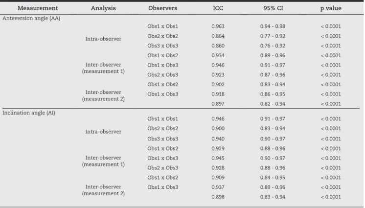
Measurement Analysis Observers ICC 95% CI p value
Anteversion angle (AA)
Intra-observer
Obs1 x Obs1 0.963 0.94 - 0.98 < 0.0001
Obs2 x Obs2 0.864 0.77 - 0.92 < 0.0001
Obs3 x Obs3 0.860 0.76 - 0.92 < 0.0001
Inter-observer (measurement 1)
Obs1 x Obs2 0.934 0.89 - 0.96 < 0.0001
Obs1 x Obs3 0.946 0.91 - 0.97 < 0.0001
Obs2 x Obs3 0.923 0.87 - 0.96 < 0.0001
Inter-observer (measurement 2)
Obs1 x Obs2 0.902 0.83 - 0.94 < 0.0001
Obs1 x Obs3 0.918 0.86 - 0.95 < 0.0001
0.897 0.82 - 0.94 < 0.0001 Inclination angle (AI)
Intra-observer
Obs1 x Obs1 0.946 0.91 - 0.97 < 0.0001
Obs2 x Obs2 0.900 0.83 - 0.94 < 0.0001
Obs3 x Obs3 0.940 0.90 - 0.97 < 0.0001
Inter-observer (measurement 1)
Obs1 x Obs2 0.929 0.88 - 0.96 < 0.0001
Obs1 x Obs3 0.945 0.90 - 0.97 < 0.0001
Obs2 x Obs3 0.928 0.88 - 0.96 < 0.0001
Inter-observer (measurement 2)
Obs1 x Obs2 0.909 0.84 - 0.95 < 0.0001
Obs1 x Obs3 0.937 0.89 - 0.96 < 0.0001
0.898 0.83 - 0.94 < 0.0001 Source: Hospital Santa Teresa. ICC: intraclass correlation coefficient; 95% CI: 95% confidence interval for the ICC; Obs: Observer.
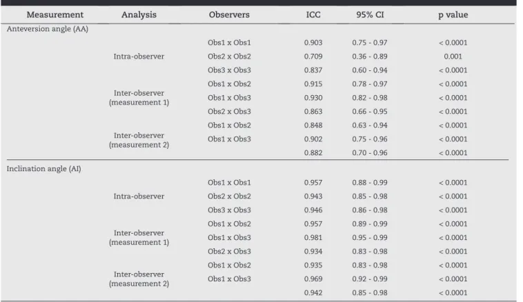
Measurement Analysis Observers ICC 95% CI p value
Anteversion angle (AA)
Intra-observer
Obs1 x Obs1 0.903 0.75 - 0.97 < 0.0001
Obs2 x Obs2 0.709 0.36 - 0.89 0.001
Obs3 x Obs3 0.837 0.60 - 0.94 < 0.0001 Inter-observer
(measurement 1)
Obs1 x Obs2 0.915 0.78 - 0.97 < 0.0001 Obs1 x Obs3 0.930 0.82 - 0.98 < 0.0001 Obs2 x Obs3 0.863 0.66 - 0.95 < 0.0001 Inter-observer
(measurement 2)
Obs1 x Obs2 0.848 0.63 - 0.94 < 0.0001 Obs1 x Obs3 0.902 0.75 - 0.96 < 0.0001 0.882 0.70 - 0.96 < 0.0001 Inclination angle (AI)
Intra-observer
Obs1 x Obs1 0.957 0.88 - 0.99 < 0.0001 Obs2 x Obs2 0.943 0.85 - 0.98 < 0.0001 Obs3 x Obs3 0.946 0.86 - 0.98 < 0.0001 Inter-observer
(measurement 1)
Obs1 x Obs2 0.957 0.89 - 0.99 < 0.0001 Obs1 x Obs3 0.981 0.95 - 0.99 < 0.0001 Obs2 x Obs3 0.934 0.83 - 0.98 < 0.0001 Inter-observer
(measurement 2)
Obs1 x Obs2 0.935 0.83 - 0.98 < 0.0001 Obs1 x Obs3 0.969 0.92 - 0.99 < 0.0001 0.942 0.85 - 0.98 < 0.0001 Source: Hospital Santa Teresa. ICC: intraclass correlation coefficient; 95% CI: 95% confidence interval for the ICC; Obs: Observer. Table 6 - Agreement analysis for the anteversion and inclination angles in the fractured hips (n = 16).
angles between the three evaluators, except for the intra-observer agreement of Observer 2 for the anteversion angle (ICC = 0.709; p = 0.001). All the pairs of agreement were of excellent degree (ICC > 0.80), except for the intra-observer agreement of Observer 2. In addition, it was seen that the confidence intervals of the ICC for the measurements on the anteversion angle were wider (less precise) than those of the inclination angle.
We investigated whether there were any differences in the anteversion and inclination angles between the three etiologies (arthrosis, neck fracture and necrosis). Tables 7 and 8 show the mean, standard deviation (SD) and median of the anteversion and inclination angles, respectively, according to the etiology and the corresponding descriptive level (p value) for one-way ANOVA.14 In this analysis, the
mean from six evaluations on each angle was used as the comparison measurement.
It was observed that there was no significant difference at the 5% level, in the anteversion angle (p = 0.12) and inclination angle (p = 0.16), between the etiologies in this study sample.
Etiology n Mean ± SD Median p value a
Arthrosis 48 13.9 ± 7.5 12.9
0.12 Fracture 16 11.7 ± 5.5 11.2
Necrosis 8 8.8 ± 4.6 9.0 Total 72 12.9 ± 7.0 12.0
Source: Hospital Santa Teresa.
SD: standard deviation; a one-way ANOVA.
Etiology n Mean ± SD Median p value a
Arthrosis 48 44,1 ± 8,5 43,9
0,16 Fracture 16 40,0 ± 7,6 39,8
Necrosis 8 40,2 ± 9,4 38,4 Total 72 42,8 ± 8,5 41,8
Source: Hospital Santa Teresa.
SD: standard deviation; a one-way ANOVA.
Table 7 - Anteversion angle (in degrees) according to etiology.
Discussion
Radiography is the most important means for making diagnoses after total hip arthroplasty. It is a low-cost examination and can be done in any hospital. While the inclination of the acetabulum can be measured by means of conventional radiographs, calculation of the anteversion still presents problems, and even more so when done in cases of different pathological conditions.
The methods described for evaluating anteversion involve complex mathematical and trigonometric equations for ellipses. McLaren15 described anteversion as a function of a coefficient
with minimum and maximum diameters of an ellipse. He prepared a reference table for each degree of anteversion. Visser et al.7 described a complex trigonometric formula using
a system of Cartesian coordinates on a projected ellipse. They did not record the efficiency of their methods.
Ghelman16 used fluoroscopy by changing the direction
of the X=ray ampulla from cephalic to caudal and observed the angle of the tube in the sagittal plane when the two halves of the ellipse were superimposed, which was when the X-rays were tangential to the opening of the acetabular dome. This author defined this process as the version angle. Schneider et al.17 used a similar technique, but obtained
several images until the wire circle of the acetabulum became tangential. These methods involved repeated irradiation, with greater cost and time.
Lewinnek et al.13 proposed a safe radiographic range for
the position of the acetabular dome with an anteversion of 15° (SD 10°) and abduction of 40° (SD 10°), but based only on nine dislocations. In order to prevent impact and dislocation, McCollum and Gray18 determined that the safe range for the
position of the dome was from 30° to 50° of abduction and 20° to 40° of horizontal flexion. Dorr and Wan19 considered
that poor positioning of the dome would be an anteversion of less than 15° or greater than 30° and an abduction angle greater than 55°. To obtain true anteversion values, they added 5° to the angle measured on the anteroposterior radiograph of the pelvis. Khan20 radiographically graded
the anteversion of acetabular components that exceeded 15° and considered that they were too vertical if the abduction angle exceeded 50°. Biedermann et al.21
demonstrated that there was no safe range for the position of the acetabular component and that anteversion of 15° and inclination of 45° presented the least risk of dislocation when an anterolateral access was used. Paterno et al.22 were
unable to establish any association between the anteversion or inclination angle of the acetabular component and the risk of dislocation. Thus, they concluded that the importance of the inclination angle as a risk factor for dislocation might have been exaggerated in preliminary studies.
Ackland et al.9 described a method using a mathematical
formula for calculating the minor axis of the ellipse in order to avoid “unacceptable subjective human errors.” They considered that it was too laborious to calculate each case through this formula and therefore used a computer program to make future estimates. They drew up a table to read the degrees of anteversion. However, the formula used was not
shown. Hassan et al.23 described a complex mathematical
formula for evaluating planar anteversion and attested it through the intra-observer reliability. Pradhan11 described
a method based on the elementary geometry of circles and triangles, and developed a simple formula that could be used to determine the planar anteversion using a pocket calculator. The results from different studies cannot be compared with each other because of the various definitions of anteversion that are used. Some authors have not used standard measurements or well-documented radiographic measurements, thus preventing precise measurement of the angling of the acetabular component.21
I t wa s d e m o n s t ra t e d i n t h e p re s e n t s t u dy t h a t measurement of the anteversion and of the acetabular inclination angle, when the X-ray ampulla was correctly centered over the hip, and with use of the trigonometric formula, was highly accurate and easy to calculate and presented high reliability.
According to the literature, the inclination angle ranges from 33° to 50° and the anteversion angle from 15° to 30°. In this study, the mean inclination angle of the total sample was 43° and the mean anteversion angle was 13°. The mean anteversion angle in the hip arthrosis cases was 14°, in the hip fracture cases 12° and in the aseptic necrosis cases 9°. The mean inclination angle in the hip arthrosis cases was 43°, in the hip fracture cases 41° and in the aseptic necrosis cases 41°. Therefore, as demonstrated in the results, there was no significant variation in the inclination angle. However, regarding the anteversion angle, there was a tendency towards differences in angle between the different pathological conditions.
It was observed that the degree of agreement of measurements of anteversion and inclination angles between the three evaluators in the intra and inter-observer assessments was highly significant (p < 0.0001), in all three pathological conditions (hip arthrosis, femoral neck fracture and aseptic necrosis). All the agreement pairs were of excellent degree (ICC > 0.80), except for the intra-observer agreement for Observer 2 regarding the anteversion angle in hip fractures (ICC = 0.709; p = 0.001). In the present sample, there was no significant difference in anteversion angle (p = 0.12) and inclination angle (p = 0.16) between the etiologies. However, the major limitation of this study was the presence of only small numbers of patients with femoral neck fracture and aseptic necrosis. It is worth emphasizing that this method cannot be used in arthroplasty cases that use a metal back or surfaces other than polyethylene, and when the latter is used, it needs to be cemented and have a metal rim along the entire acetabular edge, in order to enable measurement.
Conclusion
Conflicts of interest
The authors declare that there was no conflict of interests in conducting this study.
R E F E R E N C E S
1. Charnley J. The long-term results of Low-friction arthroplasty of the hip performed as a primary Intervention. J Bone Joint Surg Br. 1972;54(1)-B:61-76.
2. Murray D. The definition and measurement of acetabular orientation. J Bone JointSurg Br. 1993;75(2):228-32. 3. Hassan DM, Johnston GH, Dust WN, Watson G, Dolovich
AT. Accuracy of intraoperative assessment of acetabular prosthesis placement. J Arthroplasty. 1998;13(1):80-4. 4. Muller O, Reize P, Trappmann D, Wulker N. Measuring
anatomical acetabular cup orientation with a new X-ray technique. Comput Aided Surg. 2006;11(2):69-75. 5. Wan Z, Malik A, Jaramaz B, Chao L, Dorr LD. Imaging and
navigation measurement of acetabular component position in THA. Clin Orthop Relat Res. 2009;467(1):32-42.
6. Derbyshire B. Correction of acetabular cup orientation measurements for X-ray beam offset. Med Eng Phys. 2008;30(9):1119-26.
7. Visser JD, Konings JG. A new method for measuring angles after total hip arthroplasty: a study of the acetabular cup and femoral component. J Bone Joint Surg Br. 1981;63-B:556-9. 8. Amuwa C, Dorr LD. The combined anteversion technique
for acetabular component anteversion. J Arthroplasty. 2008;23(7):1068-70.
9. Ackland MK, Bourne WB, Uhthoff HK. Anteversion of the acetabular cup: measurementof angle after total hip replacement. J Bone Joint Surg [Br]. 1986;68(3):409-13. 10. Hill JC, Gibson DP, Pagoti R, Beverland DE. Photographic
measurement of the inclination of the acetabular component in total hip replacement using the posterior approach. J Bone Joint Surg Br. 2010;92(9):1209-14.
11. Pradhan R. Planar anteversion of the acetabular cup as determined from plain anteroposterior radiographs. J Bone Joint Surg Br. 1999;81(3):431-5.
12. Widner KH. A simplified method to determine acetabular cup anteversion from plain radiographs. J. Artroplast. 2004;19(3):387-90.
13. Lewinnek GE, Lewis JL, Tarr R, Compere CL, Zimmerman JR. Dislocations after total hip-replacement arthroplasties. J Bone Joint Surg Am. 1978;60(2):217-20.
14. Bartko, JJ, Carpenter WT. On the methods and theory of reliability. The J Nerv Ment Dis. 1976;163(5):307-16. 15. McLaren RH. Prosthetic hip angulation. Radiology
1973;107(3):705-6.
16. Ghelman B. Radiographic localization of the acetabular component of a hip prosthesis. Radiology. 1979;130(2):540-2. 17. Schneider R, Freiberger RH, Ghelman B, Ranawat CS.
Radiologic evaluation of painful joint prostheses. Clin Orthop Relat Res. 1982;(170):156-68.
18. McCollum DE, Gray WJ. Dislocation after total hip
arthroplasty: causes and prevention. Clin Orthop Relat Res 1990;(261):159-70.
19. Dorr LD, Wan Z. Causes of and treatment protocol for instability of total hip replacement. Clin Orthop Relat Res. 1998;(355):144-51.
20. Ali Khan MA, Brakenbury PH, Reynolds IS. Dislocation following total hipreplacement. J Bone Joint Surg Br. 1981;63-B(2):214-18.
21. Biedermann R, Tonin A, Krismer M, Rachbauer F, Eibl G, Stockl B. Reducing the risk of dislocation after total hip arthroplasty. The effect of orientation of the acetabular component. J Bone Joint Surg Br. 2005;87(6):762-9.
22. Paterno SA, Lachiewicz PF, Kelley SS. The influence of patientrelated factors and the position of the acetabular component on the rate of dislocation after total hip replacement. J Bone Joint Surg Am. 1997;79(8):1202-10. 23. Hassan DM, Johnston GHF, Dust WNC, Watson LG, Cassidy