Cop
yright
© ABE&M t
odos os dir
eit
os r
eser
vados
.
Long term treatment of a
thyrotropin-secreting microadenoma
with somatostatin analogues
Tratamento de longa duração com análogos da somatostatina de um microadenoma tirotroinoma
Alma Prieto-Tenreiro1, Patricia Díaz-Guardiola2
SUMMARY
Thyrotropin (TSH) secreting pituitary adenomas (TSH-omas) account for < 1% of all pituitary adenomas and are a rare cause of hyperthyroidism. The diagnosis is often made at the stage of macroadenoma because of the aggressive nature of the tumor and due to the fact that pa-tients are mistakenly treated for more common primary hyperthyroidism for a long time. First line therapy is transsphenoidal resection of the tumor, which can cure one-third of the patients completely. However, if surgery is not possible or curative, pituitary radiotherapy and/or so-matostatin analogs (SSA) can be useful. We report the case of a 54-year-old woman treated 20 years earlier for a mistakenly suspected primary hyperthyroidism. Given the persistence of symptoms she was studied further and was diagnosed with a thyrotropinoma. Despite the delay in diagnosis and prior thyroid ablation, a microadenoma was found. As transsphenoidal surgery was not considered effective, medical therapy with a somatostatin analogue was initia-ted. Currently, at four years of follow-up, the patient continues on this treatment and remains euthyroid and asymptomatic. We report a case of successful long-term treatment with SSA, after unsuccessful surgery. Arq Bras Endocrinol Metab. 2010;54(5):502-6
SUMÁRIO
Tirotroinomas (TSH-omas) representam < 1% dos adenomas hipoisários. Eles são uma causa muito rara de hipertireoidismo. O diagnóstico é frequentemente feito na fase de macroadeno-ma em consequência da natureza agressiva do tumor e do feito de que os doentes são tratados inicialmente por engano e por um longo tempo para hipertireoidismo primário. A terapêutica de primeira linha é a ressecção transesfenoidal do tumor, que cura um terço dos pacientes com-pletamente. Contudo, se a cirurgia não for possível ou curativa, a radioterapia da pituitária e/ou o tratamento com análogos da somatostatina (SSA) podem ser úteis. Relatou-se o caso de uma mulher de 54 anos, tratada há 20 anos por uma suspeita equivocada de hipertireoidismo pri-mário. Dada a persistência dos sintomas, foram realizados mais exames e a paciente foi diag-nosticada com TSH-oma. Apesar do diagnóstico tardio e da ablação prévia com iodo radioa tivo, encontrou-se um microadenoma. Como a cirurgia transesfenoidal não foi considerada eicaz, iniciou-se o tratamento da paciente com SSA. Atualmente, após quatro anos de acompanha-mento, a paciente continua com o tratamento e permanece eutireoidea e assintomática. Neste artigo, relatou-se a eicácia da terapia medicamentosa com SSA em longo prazo, após cirurgia não eicaz. Arq Bras Endocrinol Metab. 2010;54(5):502-6
1 Serviço de Endocrinologia
e Nutrição, Hospital Clínico Universitario de Santiago de Compostela, Spain
2 Serviço de Endocrinologia
e Nutrição, Hospital Infanta Sofía, Madrid, Spain
Correspondence to: Alma Prieto-Tenreiro Servicio de Endocrinología y Nutrición
Hospital Clínico Universitario de Santiago de Compostela Travesía de la Choupana s/n 15706 − Santiago de Compostela, Spain
Received on Sept/7/2009 Accepted on Apr/5/2010
INTRODUCTION
T
hyrotropinomas are a rare cause of hyperthyroid-ism, when compared with the more prevalent pri-mary hyperthyroidism, with an overall prevalence of about one in one million. The hormonal proile is char-acterized by a non-suppressed TSH in the presence of high levels of free thyroid hormones (fT4, fT3).hyper-Cop
yright
© ABE&M t
odos os dir
eit
os r
eser
vados
.
thyroidism and those with thyrotropinoma or pituitary resistance hormone.
First-line therapy is surgery, by which about one third of patients can be cured. If this fails, radiotherapy or SSA should be considered.
We present the case of a patient with palpitations and a previously mistaken diagnosis of primary hyperthyroi-dism. After extensive investigations, this was found to be due to a micro-thyrotropinoma and she was effecti-vely treated with surgery and somatostatin analogs.
CASE REPORT
A 54-year-old woman was referred to our clinic with a two-year history of palpitations. She was on propano-lol prescribed by her general practitioner (GP), and she had no relevant family or personal past medical history, apart from an overactive thyroid, diagnosed and treated in Canada when she was 34 years old. Initially she was treated with drugs for 2 years, after that, she was given radioactive iodine with no more follow-up.
She had a high thyrotropin stimulating hormone (TSH) of 12.16 mU/L (normal range 0.35-5.50) and a high fT4 (2.7 ng/dL, normal range 0.9-1.8) when checked by her general practitioner (GP) 12 months ago. Thyroid antibodies were all negative. Despite being on propanolol, she continued symptomatic (feeling hot and with palpitations), so her GP had her thyroid hor-mone tests repeated, which again showed a markedly elevated fT4 (2.7 ng/dL) and an elevated TSH (9.28 mU/L). With these results, and suspecting a thyrotro-pinoma or thyroid hormone resistance (RTH), she was referred to the Endocrinology department.
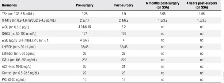
Examination was unremarkable, although she did have a small goiter and tachycardia. In order to really irm up the results, thyroid hormones were repeated in another laboratory showing once more a high TSH with high fT4 and fT3 (Table 1). The sex hormone-binding globulin (SHBG), the TSH alpha subunit (α-SU) (repeated twice) and the α-SU/TSH molar ratio
were also elevated (Table 1). The existence of possible interferences between TSH and fT4 was excluded after analyzing these results by equilibrium of dialysis. As the patient was still symptomatic despite treatment with pro-panolol, she was started on low-dose carbimazole (5 mg, twice a day) to avoid higher TSH stimulation. Further studies were organized, including magnetic resonance imaging (MRI), a thyroid releasing hormone stimulation (TRH) test, a thyroid ultrasound and a thyroid scintigra-phy. The MRI showed a pituitary microadenoma expan-ding into the pituitary fossa with no extension to either the suprasellar cistern or the optic chiasm (Figure 1). The response to the TRH test was lat (Table 2). The ultra-sound conirmed the presence of a 2.5 x 2.1 x 1.9 cm solid nodule in the right lobe. The thyroid scintigraphy also showed a right cold nodule and a ine-needle aspi-ration punction was performed to rule out malignancy.
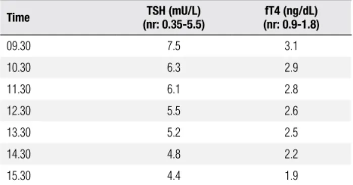
To further delineate whether the pituitary mass was a thyrotropinoma or an incidentaloma, an octreotide sup-pression test was performed. As it showed an appropriate suppression after 6 hours (Table 3), transsphenoidal re-section of the tumor was planned. To achieve euthyroi-dism before the surgical resection, a therapy with lanre-otide (20 mg per month) was started for three months. Other preoperative investigations included an anterior pituitary screening, showing intact axes (Table 1).
Table 1. Time-course of biochemical determinations
Hormones Pre-surgery Post-surgery 6 months post-surgery (on SSA) 4 years post-surgery (on SSA)
TSH (nr: 0.35-5.5 mU/L) 9.28 7.9 3.95 1.66
fT4/fT3 (nr: 0.9-1.8 ng/dL/2.3-4.3 pg/mL) 2.3/7.7 2.1/6.2 1.3/3.2 1.5/3.6
αSU (nr: 0.5-3 μg/L) 4.01/6.45 3.2 nd nd
SHBG (nr: 30-100 nmol/L) 127 109 nd nd
αSU (μg/l)/TSH (mU/L) x10 (nr: < 1) 4.3/6.9 4 nd nd
LH/FSH (nr: > 30 mU/mL) 35/45 33/46 nd nd
Estradiol (nr: < 30 pg/mL) 33 32 nd nd
IGF-1 (nr: 105-353 ng/mL) 232 229 nd nd
ACTH (nr: 10-80 ng/L) 36 31 nd nd
Cortisol (nr: 6.5-22.5 ng/dL) 22 23 nd nd
PRL (3-30 ng/mL) 16 19 nd nd
Cop
yright
© ABE&M t
odos os dir
eit
os r
eser
vados
.
(20 mg per month). Further treatment options such as radiotherapy were considered. Nowadays, 4 years later, she remains on medical treatment and radiotherapy has been ruled out, as the tumor volume has reduced and she is successfully asymptomatic with TSH levels in the normal range.
DISCUSSION
Thyrotropinomas are rare pituitary adenomas and an even more rare cause of hyperthyroidism. The bioche-mical features are quite characteristic, as the levels of circulating free thyroid hormones (fT3, fT4) are ele-vated in the presence of normal or high serum TSH concentrations. The irst case of TSH-oma was docu-mented in 1960 by measuring serum TSH levels with a bioassay (1) and since then, approximately 300 cases have been described. With the introduction of ultra-sensitive immunometric TSH assays, nowadays it is possible to diagnose TSH-omas earlier, even before the stage of macroadenoma.
Failure to recognize a TSH-oma may result in dra-matic consequences such as improper surgery or abla-tion that may cause the pituitary tumor volume to fur-ther expand and become an invasive macroadenoma (2). This could probably be due to similar mechanisms that lead to aggressive transformation of pituitary cells after adrenalectomy in Nelson’s syndrome (3).
Molecular mechanisms leading to the formation of thyrotropinomas are currently unknown. Somatic mu-tations of thyroid hormone receptors, increased expres-sion of basic ibroblast growth factor and loss of hetero-zygosity, and particular polymorphisms of somatostatin receptor type 5 are thought to be involved in tumor pathogenesis (4-6).
In patients with TSH-oma, signs and symptoms of hyperthyroidism are frequently associated with those related to tumor expansion such as headache and visual ield defects (7). Signs of thyrotoxicosis may vary from severe to absent, although clinical features are usually milder than expected for the levels of thyroid hormo-nes. This is probably due to the longstanding duration of the disease. As 30% of thyrotropinomas co-secrete other hormones additional symptoms of hormone hypersecretion are often associated. On physical exa-mination, the presence of goiter is the rule (more than 90% of patients with TSH-oma present with goiter), even in patients with previous thyroidectomy/ablation, as thyroid residue can regrow due to TSH
hyperstimu-Table 2. Response to the TRH test (after 200 μg of TRH) TSH (mU/L)
(nr: 0.35-5.5) (nr: 0.9-1.8)fT4 (ng/dL)
0 min 10.6 3.7
20 min 10.4 3.2
60 min 10.7 3.5
nr (normal baseline range).
Table 3. Octreotide Suppression Test (after 100 μg subcutaneous of Octreotide)
Time (nr: 0.35-5.5)TSH (mU/L) (nr: 0.9-1.8)fT4 (ng/dL)
09.30 7.5 3.1
10.30 6.3 2.9
11.30 6.1 2.8
12.30 5.5 2.6
13.30 5.2 2.5
14.30 4.8 2.2
15.30 4.4 1.9
nr (normal baseline range).
Figure 1. MRI at diagnosis, showing a pituitary microadenoma.
Cop
yright
© ABE&M t
odos os dir
eit
os r
eser
vados
.
lation (8). This was also found in our patient despite her thyroid having been ablated in the past.
When suspecting a TSH-oma, and given its low pre-valence, inadequate measurement of TSH and thyroid hormones should always be taken into consideration, and laboratory measurements should be repeated, as it was in our case. This could, for example, occur in me-dical therapy with amiodarone, in the presence of anti-T4 autoantibodies, in familial dysalbuminemic hyper-thyroxinemia and in association with increased levels of hormone-binding globulins (thyroxine binding globu-lin, albumin or transthyretin) (9). Once this possibili-ty is excluded, it is important to make an appropriate differential diagnosis between a TSH-secreting pitui-tary adenoma and the syndrome of thyroid hormone resistance (RTH) (10), as in both syndromes, patients present with high peripheral thyroid hormone levels, inappropriately normal or elevated levels of TSH and symptoms of thyrotoxicosis.
TSH, FSH and LH share a common α-subunit (α
-SU) that is co-secreted with the pituitary hormones. The measurement of this subunit can be very useful to diagnose thyrotropinomas. Therefore, high levels of
α-SU or an α-SU (µg/L)/TSH (mU/L) x 10 > 1 (11)
are indicative of thyrotropinoma in more than 90% of the cases (although a normal result does not exclude this diagnosis). In our patient, both α-SU levels and
the α-SU/TSH molar ratio were concordant with the
existence of a thyrotropinoma. Moreover, other ma-rkers of thyroid hormone action, such as the sex hor-mone-binding globulin (SHBG), can help us with the diagnosis, (specially differentiating thyrotropinomas from thyroid hormone resistance), as they are known to be elevated in 80% of TSH-omas.
Regarding dynamic tests, both stimulatory and inhibitory tests have been proposed for the diagnosis of thyrotropinomas. Classically, the T3 suppression test (80-100 µg of T3 per day for 8-10 days) has been used to assess the presence of a thyrotropinoma. This test is specially sensitive and speciic when studying a patient with previous thyroid ablation or differentiating TSH-omas from secondary pituitary hyperplasia (8). Althou-gh a complete inhibition of TSH secretion after this test has never been recorded in patients with a thyrotropi-noma, it is contraindicated in elderly patients or those with coronary heart disease. Therefore, the TRH test has also been widely used, although it is less sensitive in patients with prior thyroid ablation. In the vast ma-jority of patients with thyrotropinoma, TSH and α-SU
levels do not increase (or increase less than 2-fold) after TRH administration, in our case TSH did not increase at all. Moreover, in patients with hyperthyroidism, dis-crepancies between TSH and α-SU responses to TRH
are pathognomonic of TSH-omas co-secreting other pituitary hormones.
To complete the diagnosis, and in suspecting a TSH-oma, a MRI is very useful, and it is nowadays the preferred tool for visualization of a thyrotropino-ma. However, pituitary tumors on MRIs have to be interpreted carefully, as pituitary incidentalomas can be found in up to 10% of the population (12). In most cases (80%) (13), as previously stated, a macroadenoma is found, although due to the ultrasensitive TSH assays microadenomas are now increasingly reported (10%-20%) (14).
In our patient, given all the results (elevated α-SU
concentrations, high α-SU/TSH molar ratio, TSH
un-responsiveness to TRH stimulation, microadenoma in the MRI) and the absence of biochemical data in other family members, the diagnosis of thyrotropinoma was favored, and the possibility of a RTH was ruled out.
Cop
yright
© ABE&M t
odos os dir
eit
os r
eser
vados
.
become the drug of choice. This is because, nowadays, it is known that most thyrotropinomas (90%) show sensiti-vity to native somatostatin and its analogs. Indeed, they can be useful helping either in the differential diagnosis of problematic cases of central hyperthyroidisms (when administered for at least 2 months prior to surgery) or as long-term treatment (added to surgery or radiotherapy) (19). This way, they can revert thyroid hormone levels in about three quarters of the patients and make the tumor shrink in one third of the patients.
Regarding medical treatment, it is important to keep thyroid hormone levels at upper limits of normal to prevent an additional stimulus of the pituitary tumor to expand (20).
In conclusion, TSH-omas are rare adenomas. Diag-nosis may often be delayed because the symptoms are at-tributed to more common causes of thyrotoxicosis. This delay and misdiagnosis, coupled with the usually aggres-sive nature of these tumors, allows them to become large and invasive. The primary goal in treatment must be sur-gical adenomectomy, and when it is not successful radio-therapy or medical treatment should be added. Our case is specially interesting, given the low prevalence of this pathology, its diagnosis at the stage of microadenoma (despite prior thyroid ablation and the delay in the diag-nosis) and its successful long term medical treatment with SSA, with no need of further radiotherapy.
Disclosure: no potential conlict of interest relevant to this article was reported.
REFERENCES
1. Jailer JW, Holub DA. Remission of Grave’s disease following ra-diotherapy of a pituitary neoplasm. Am J Med. 1960;28:497-500. 2. Beck-Peccoz P, Brucker-Davis E, Persani L, Smallridge RC,
Wein-traub BD. Thyrotropin-secreting pituitary tumors. Endocr Rev. 1996;17:610-38.
3. Sanno N, Teramoto A, Osamura RY. Thyrotropin-secreting pituita-ry adenomas. Clinical and biological heterogeneity and current treatment. J Neurooncol. 2001;54:179-86.
4. Ando S, Sarlis NJ, Oldield EH, Yen PM. Somatic mutation of TR-beta can cause a defect in negative regulation of TSH in a TSH-se-creting pituitary tumor. J Clin Endocrinol Metab. 2001;86:5572-6.
5. Filopanti M, Ballarè E, Lania AG, Bondioni S, Verga U, Locatelli M, et al. Loss of heterozygosity at the SS receptor type 5 locus in human GH-and TSH-secreting pituitary adenomas. J Endocrinol Invest. 2004;27:937-42.
6. Ezzat S, Horvath E, Kovacs K, Smyth HS, Singer W, Asa SL. Basic ibroblast growth factor expression by two prolactin and thyrotro-pin-producing pituitary adenomas. Endocr Pathol. 1995;6:125-34. 7. Brucker-Davis F, Oldield EH, Skarulis MC, Doppman JL,
Wein-traub BD. Thyrotropin-secreting pituitary tumors: diagnostic cri-teria, thyroid hormone sensitivity, and treatment outcome in 25 patients followed at the National Institutes of Health. J Clin Endo-crinol Metab. 1998;84:476-86.
8. Socin HV, Chanson P, Delemer B, Tabarin A, Rohmer V, Mockel J, et al. The changing spectrum of TSH-secreting pituitary adeno-mas: diagnosis and management in 43 patients. Eur J Endocrinol. 2003;148:433-42.
9. Khandwala H, Chatterjee VKK. Inappropriate secretion of thyroid-stimulating hormone. Can Med Assoc J. 2006;175:351-3. 10. Refetoff S, Weiss RE, Usala SJ. The syndrome of resistance to
thyroid hormone. Endocr Rev. 1993;14:348-99.
11. Kourides IA, Ridway EC, Weintraub BD, Bigos ST, Gershengorn Mc, Maloof F. Thyrotropin-induced hyperthyroidism: use of alpha and beta subunit levels to identify patients with pituitary tumors. J Clin Endocrinol Metab. 1977;45:534-43.
12. Hall WA, Luciano MG, Doppman JL, Patronas NJ, Oldield EH. Pituitary magnetic resonance imaging in normal human volunte-ers: occult adenomas in the general population. Ann Intern Med. 1994;120:817-20.
13. Abucham J, Vieira TC. Glycoprotein-secreting pituitary adeno-mas: pathogenesis, diagnosis and treatment. Arq Bras Endocri-nol Metab. 2005;49(5):657-3.
14. Gesundheit N, Petrick PA, Nissim M, Dahlberg PA, Doppman JL, Emerson CH, et al. Thyrotropin-secreting pituitary adenomas: cli-nical and biochemical heterogeneity: case reports and follow-up of nine patients. Ann Intern Med. 1989;111:827-35.
15. McCutcheon IE, Weintraub BD, Oldield EH. Surgical treatment of thyrotropin-secreting pituitary adenomas. J Neurosurg. 1990;73:674-83.
16. Duarte FH, Jallad RS, Salgado LR, Bronstein MD. TSH-secreting pituitary tumors: two case reports and literature review. Arq Bras Endocrinol Metab. 2009;53(9):1157-66.
17. Bertherat J, Brue T, Enjalbert A, Gunz G, Rasolonjanahary R, War-net A, et al. Somatostatin receptors on thyrotropin-secreting pituitary adenomas: comparison with the inhibitory effects of octreotide upon in vivo and in vitro hormonal secretions. J Clin Endocrinol Metab. 1992;75:540-6.
18. Beck-Peccoz P, Persani L. Medical management of thyrotropin-secreting pituitary adenomas. Pituitary. 2002;5:83-8.
19. Mannavola D, Persani L, Vannucchi G, Zanardelli M, Fugazzola L, Verga U, et al. Different responses to chronic somatostatin ana-logues in patients with central hyperthyroidism. Clin Endocrinol. 2005;62:176-81.