Prosthodontics
Júlio César Brigolini de Faria(a) Laís Regiane Silva-Concílio(a) Ana Christina Claro Neves(a) Milton Edson Miranda(b) Marcelo Lucchesi Teixeira(b)
(a)Department of Prosthodontics, University of Taubaté, Taubaté, SP, Brazil.
(b)Department of Prosthodontics, São Leopoldo Mandic, Campinas, SP, Brazil.
Corresponding author: Laís Regiane Silva-Concílio
Rua Expedicionário Ernesto Pereira, 110 Taubaté - SP - Brazil
CEP: 12020-270
E-mail: regiane1@yahoo.com
Received for publication on Jun 10, 2010 Accepted for publication on Dec 21, 2010
Evaluation of the accuracy of different
transfer impression techniques for
multiple implants
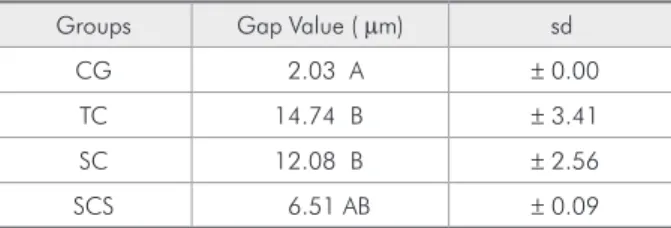
Abstract: The aim of this study was to evaluate the accuracy of three implant transfer impression techniques. Four groups (n = 5) were deined, according to the technique: TC – tapered copings without splint; SC – square copings without splint; SCS – square copings splinted with dental loss and acrylic resin, and CG (control group) – master model with four external hexagonal implants and a superstructure. Individual trays and polyether were used for the impression. All casts were checked for their it into the master superstructure; for this, all four screws were placed in the implants. Digital photos were taken and images were analyzed using UTHSCSA ImageTool software. Statistical analyses were per-formed using one-way analysis of variance and Student’s t test (p < 0.05). The means and standard deviation were (µm): CG = 2.03 ± 0.00, TC = 14.74 ± 3.41, SC = 12.08 ± 2.56, and SCS = 6.51 ± 0.09. The control group was found to be statistically different from the TC and SC groups. Within the limitations of this study, all groups presented clinically ac-ceptable standard gap values, and the SCS group showed no statistical difference in relation to the CG (control group), demonstrating more ac-curacy and idelity to transfer implants.
Descriptors: Dental implants; Dental Prosthesis; Dental impression technique.
Introduction
An important factor for success in implant-supported prosthesis is the passive adaptation of the prosthetic superstructure.1-8 Failure to
pro-duce a passive it can generate considerable stress,such as prosthetic screw loosening or fracture of screw or implant, and even bone loss around the implant, interfering with the osseointegration process.4 Various methods
have been suggested for evaluating implant framework it: alternate in-ger pressure, direct vision and tactile sensation, radiographs, one-screw test, and screw resistance test. Alternate inger pressure and one-screw test are especially important to verify misalignment in vertical (x) axis.9
Reproduction of the intra-oral relationship of implants through im-pression procedures is the irst step in achieving an accurate and pas-sive it prosthesis.10 This transference is still complicated by the number,
angulation, depth and position of the implants.10-12 As in conventional
di-rect or open tray technique and didi-rect-splinted tech-nique.13-16
There may be clinical situations in which the use of the closed tray technique is indicated, such as when there is limited inter-arch space, dificult access to posterior implants and angulated im-plants.15,16 The open tray technique allows the
cop-ing to remain in the impression. This reduces the ef-fect of implant angulations, decreases deformation of the impression material upon recovery from the mouth, and eliminates the concern for replacing the coping back into its respective space in the impres-sion. However, there are several parts that must be controlled when fastening is being performed, some rotational movement of the impression coping is re-quired when securing the implant analog, and blind attachment of the implant analog to the impression coping are some disadvantages of this technique.15-25
Branemark et al. emphasized the importance of us-ing impression copus-ings that are splinted with den-tal loss scaffolding covered with autopolymerizing acrylic resin for transfer impression.1 Nowadays,
this same technique has been employed by others with minor modiications and has proven to be a se-cure impression procedure.19,26
The accuracy of casts requires an appropriate selection of impression materials and trays. Stud-ies have shown that elastomeric impression materi-als, especially silicone and polyether, are the most recommended for transfer impression procedures, due to their increased linear stability, lower residual shrinkage during storage, greater rigidity and lack of rotation resistance of the coping inside the im-pression leading to a more accurate cast.27,28 When
compared to hydrocolloids, elastomeric materials are preferred because of their dimensional stability and resistance.27-30 Polysulides are more dificult
to deal with, having an unpleasant smell and slow polymerization reaction. Condensation silicones are dimensionally less stable than polysulides, and polyethers are regarded as the materials of choice. In this study, to avoid interference in the impressions due to material variations, all of them were made with the same material.17,20
The aim of this study was to evaluate the accu-racy obtained with three different implant transfer
impression techniques.
Materials and Methods
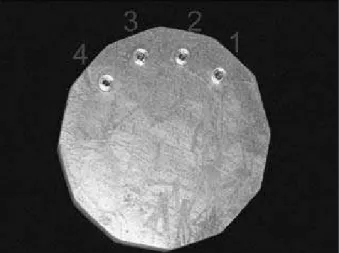
A surgical bar of the Neopronto System (Neo-dent, Curitiba, PR, Brazil), used as a guide for im-plant placement, was used to control the parallel-ism and distance between implants. Four external hexagonal 3.75 mm x 13 mm Titamax implants (Neodent, Curitiba, PR, Brazil) were placed using perforations made with surgical drills (Neodent, Curitiba, PR, Brazil) in an uncolored autopolymer-izing acrylic resin model (Reliance Dental Mfg. Co., Worth, IL, USA). Implants were numbered from 1 to 4 in a clockwise manner. This model was named the Master model (Figure 1).
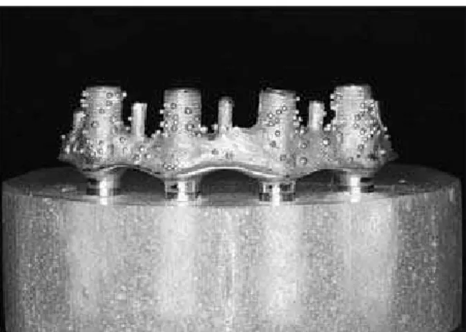
A protocol bar was waxed using metallic Tilite UCLA cylinders (Neodent, Curitiba, PR, Brazil) on top of a master model. This waxed bar was sec-tioned and splinted again to reduce residual stress, and the master model/superstructure complex was included in the Microine 1700 quartz-containing investment mould (Talladium, Curitiba, PR, Brazil) and cast with Tilite alloy (Talladium, Curitiba, PR, Brazil), using the lost-wax casting technique. Fin-ishing and polFin-ishing were performed using 50-µm aluminum oxide airborne-particle abrasion (Assler, São Paulo, SP, Brazil), aluminum oxide stone (JON, São Paulo, SP, Brazil) and ultrasonically cleansed (Thornton Ltda., Vinhedo, SP, Brazil) in distilled water for 10 min. This cast was employed as the
control group (Figure 2).
An impression of the control group was utilized to prepare ive individual trays for each impression technique using chemically-activated acrylic resin (Reliance Dental Mfg. Co., Worth, IL, USA). For the group with square copings, the trays had to be perforated to allow access to the coping screws. Four groups were deined, according to the transfer impression technique:
• TC Group – tapered copings without splint (Neodent, Curitiba, PR, Brazil);
• SC Group – square copings without splint (Neo-dent, Curitiba, PR,Brazil);
• SCS Group – square copings splinted with dental loss (Gillette, São Paulo, SP, Brazil) and acrylic resin Duralay (Reliance Dental Mfg. Co., Worth, IL, USA) and
• the Control Group (as described above).
In every case, the Impregum polyether material (3M Espe, Seefeld, Starnberg, Germany) was used for the impression; this was mixed and injected us-ing an impression syrus-inge (JON, São Paulo, SP, Bra-zil), according to the instructions provided by the manufacturer. All procedures were performed by the same operator. For each technique, ive differ-ent impressions were prepared. Models were poured with type IV die stone (Polidental, Cotia, SP, Brazil), according to the manufacturer’s recommendations.
The vertical misit between the master super-structure and the implants was checked for all
speci-mens, and the region of misit was determined to be the region between the lower external margin of the superstructure and the top external margin of the implant. The reading was done in the counter-clock-wise direction and was performed on both the ves-tibular and lingual faces. To maintain the samples over the superstructure, all screws were applied us-ing a manual torque of 30Ncm (ITL Dental, Irvine, CA, USA).
Using a metallic base, the casts and a Canon EOS 30D n camera with an MP-E 65 mm f2.8 1-5X Macro Lens (Canon, New York, NY, USA) were placed in a standard position, and photos of the vestibular and lingual faces focusing on the central point of each implant were taken with 5 X zoom. Three photos were taken for each sample. The imag-es were analyzed using UTHSCSA ImageTool soft-ware (Evans Technology Inc., Roswell, GA, USA). A digital caliper (Mitutoyo, Suzano, SP, Brazil) with an opening of 0.001 µm was used as a measure of reference. Gap values (µm) were measured and com-pared to reference measures and to the misit of the superstructure in the images of the master super-structure over the casts.
The Shapiro-Wilk test was performed to conirm that the marginal gap data were normally distrib-uted (α = 0.05). Mean values and standard devia-tion (sd) were calculated, and statistical inferences among the groups were made using one-way analy-sis of variance and Student’s t test (α = 0.05) (Bioes-tat 5.0, Maringá, PR, Brazil).
Results
Signiicant differences in gap values were found among the groups (Table 1). Means, standard de-viations (sd) and intergroup analysis are shown in Table 2. The result of the Student t test showed that the CG was statistically different from the TC and SC groups.
Figure 2 - Metallic protocol bar (superstructure) positioned on the master model: control group.
Table 1 - One-way ANOVA test.
Degree of freedom
Sum of squares
Mean
square F value P value
Between
Discussion
Care with implant transfer to obtain working casts and, consequently, passive adaptation of the prosthetic superstructure to the implant is vital to maintain the integrity of the prosthesis-implant-bone-periodontal tissues complex.1,3,8-10,18
Accord-ing to Eames et al.,28 to obtain suitable and reliable
superstructures, it is important to maintain idelity with the impression procedures. This is achieved with the following steps: impression, cast acquisi-tion, waxing, embedding and casting. Even with all the reported techniques for transfer methods, accu-racy is not always achieved.7,10
The choice of the type of impression varies ac-cording to the complexity of the work, impression technique chosen, tray model and implant systems and/or prosthetic components used.15,29 Currently,
the main impression techniques used are: closed tray with tapered copings and open tray with square copings, which may be used together or not.17,20,24
The transfers used in this study were tapered and square, united or not, according to the available lit-erature.11,15-17,19,20-22,26
According to some authors there is no perfect it between the abutment and the implant.9,10 However,
large gaps can cause problems because they favor bacterial colonization increasing inlammation of the tissues surrounding the implants;9 marginal its
of up to 100 µm are clinically acceptable,9 and these
problems may be closely linked to transfer
impres-sion techniques.
In our study, the TC and SC groups were sta-tistically different from the CG, where the CG was found to be more precise. The SCS group showed no statistical difference when compared with the CG. This technique resulted in values that were close to those of the CG and, as such, was more accurate. Such results are in accordance with the available lit-erature.10,16,19,21 All groups, including the statistically
different, TC and SC, showed acceptable results in the clinical evaluation of implant framework it.9
Greater accuracy in the SCS group is related to the fact that transfers are removed together with the model and do not need to be repositioned inside the impression, as in the TC technique. This advantage minimizes errors.19,20 Hence, in the SCS technique,
a rigid connection between the transfers is accom-plished with a chemically-activated acrylic resin, avoiding movement and rotation of the transfers in-side the model.10,16,21,26 These observations were not
found in other studies, where the three techniques showed similar results.11,15,22,24,25
Based on our results, the SCS technique is more accurate than the other techniques evaluated. How-ever, important variables such as saliva, dificulty in the adaptation of the impression and/or prosthesis components, limitation in mouth opening and an-gulation of the implants were not present in this ex-perimental study. More studies are thus necessary to validate our results and to test other variables.
Conclusions
Within the limitations of this study, the follow-ing conclusions were drawn:
1. All groups showed clinically acceptable standard gap values;
2. the SCS group showed no statistical difference in relation to the CG (control group), showing more accuracy and idelity to transfer implants.
Table 2 - Gap values (means ± sd; µm) observed among groups.
Groups Gap Value ( µm) sd
CG 2.03 A ± 0.00
TC 14.74 B ± 3.41
SC 12.08 B ± 2.56
SCS 6.51 AB ± 0.09
References
1. Branemark P-I, Zarb GA, Albrektsson T. Tissue-integrated prostheses. 1st ed. Chicago: Quintessence; 1985. 352 p. 2. Nissan J, Gross M, Shifman A, Assif D. Stress levels for
well-fitting implant superstructures as a function of tightening force levels, tightening sequence, and different operators. J Prosthet Dent. 2001 Jul;86(1):20-3.
3. Skalak R. Biomechanical considerations in osseointegrated prostheses. J Prosthet Dent. 1983 Jun;49(6):843-8.
4. Rangert B, Jemt T, Jorneus L. Forces and moments on Branemark implants. Int J Oral Maxillofac Implants. 1989 Fall;4(3):241-7.
5. Jemt T, Book K. Prosthesis misfit and marginal bone loss in edentulous implant patients. Int J Oral Maxillofac Implants. 1996 Sep-Oct;11(5):620-5.
6. Millington ND, Leung T. Inaccurate fit of implant super-structures. Part 1: Stresses generated on the superstructure relative to the size of fit discrepancy. Int J Prosthodont .1995 Nov-Dec;8(6):511-6.
7. Waskewicz GA, Ostrowski JS, Parks VJ. Photoelastic analy-sis of stress distribution transmitted from a fixed prostheanaly-sis attached to osseointegrated implants. Int J Oral Maxillofac Implants. 1994 Jul-Aug;9(4):405-11.
8. Sahin S, Cehreli MC. The significance of passive framework fit in implant prosthodontics: current status. Implant Dent. 2001;10(2):85-92.
9. Kan JY, Rungcharassaeng K, Bohsali K, Goodacre CJ, Lang BR. Clinical methods for evaluating implant framework fit. J Prosthet Dent. 1999 Jan;81(1):7-13.
10. Lee H, So JS, Hochstedler JL, Ercoli C. The accuracy of im-plant impressions: a systematic review. J Prosthet Dent. 2008 Oct;100:285-91.
11. Carr AB. Comparison of impression techniques for a two-implant 15-degree divergent model. Int J Oral Maxillofac Implants. 1992 Winter;7(4):468-75.
12. Conrad HJ, Pesun IJ, Delong R, Hodges JS. Accuracy of two impression techniques with angulated implants. J Prosthet Dent. 2007 Jun;97(6):349-56.
13. Burns J, Palmer R, Howe L, Wilson R. Accuracy of open tray implant impressions: an in vitro comparison of stock versus
custom trays. J Prosthet Dent. 2003 Mar;89(3):250-5. 14. Cox JR, Brandt RL, Hughes HJ. A clinical pilot study of
the dimensional accuracy of double-arch and complete-arch impressions. J Prosthet Dent. 2002 May;87(5):510-5. 15. Herbst D, Nel JC, Driessen CH, Becker PJ. Evaluation of
impression accuracy for osseointegrated implant supported superstructures. J Prosthet Dent. 2000 May;83(5):555-61. 16. Vigolo P, Majzoub Z, Cordioli G. Evaluation of the accuracy
of three techniques used for multiple implant abutment impres-sions. J Prosthet Dent. 2003 Feb;89(2):186-92.
17. Humphries RM, Yaman P, Bloem TJ. The accuracy of implant master casts constructed from transfer impressions. Int J Oral Maxillofac Implants. 1990 Winter;5(4):331-6.
18. Henry PJ, Tan AE, Uzawa S. Fit discrimination of implant-supported fixed partial dentures fabricated from implant level impressions made at stage I surgery. J Prosthet Dent. 1997 Mar;77(3):265-70.
19. Assif D, Marshak B, Schmidt A. Accuracy of implant impres-sion techniques. Int J Oral Maxillofac Implants. 1996 Mar-Apr;11(2):216-22.
20. Carr AB. Comparison of impression techniques for a five-implant mandibular model. Int J Oral Maxillofac Implants. 1991 Winter;6(4):448-55.
21. Naconecy MM, Teixeira ER, Shinkai RS, Frasca LC, Cervieri A. Evaluation of the accuracy of 3 transfer techniques for implant-supported prostheses with multiple abutments. Int J Oral Maxillofac Implants. 2004 Mar-Apr;19(2):192-8. 22. Phillips KM, Nicholls JI, Ma T, Rubenstein J. The Accuracy
of Three Implant Impression Techniques: A Three-dimen-sional Analysis. Int J Oral Maxillofac Implants. 1994 Sep-Oct;9(5):533-40.
23. Chee W, Jivraj S. Impression techniques for implant dentistry. Br Dent J. 2006 Oct 7; 201(7): 429-432. Review.
24. Spector MR, Donovan TE, Nicholls JI. An evaluation of im-pression techniques for osseointegrated implants. J Prosthet Dent. 1990 Apr;63(4):444-7.
25. Del’Acqua MA, Arioli-Filho JN, Compagnoni MA, Mollo Jr F de A. Accuracy of impression and pouring techniques for an implant-supported prosthesis. Int J Oral Maxillofac Implants. 2008 Mar-Apr; 23(2):226-36.
26. Assif D, Nissan J, Varsano I, Singer A. Accuracy of implant impression splinted techniques: effect of splinting material. Int J Oral Maxillofac Implants 1999 Nov-Dec;14(6):885-8. 27. Wee AG. Comparison of impression materials for direct
multi-implant impressions. J Prosthet Dent. 2000 Mar;83(3):323-31. 28. Eames WB, Wallace SW, Suway NB, Rogers LB. Accuracy
and dimensional stability of elastomeric impression materials. J Prosthet Dent. 1979 Aug;42(2):159-62.
29. Tjan AH, Whang SB, Tjan AH, Sarkissian R. Clinically ori-ented evaluation of the accuracy of commonly used impression materials. J Prosthet Dent. 1986 Jul;56(1):4-8.