339
Paget’s disease with sacral involvement
Radiol Bras. 2010 Set/Out;43(5):339–342 Case Report • Relato de Caso
Paget’s disease with sacral involvement: a case report*
Doença de Paget com acometimento sacral: relato de casoFernanda Nogueira Holanda Ferreira Braga1, Marcio Vale Braga2, Francisco Andrade Neto3
The authors report a case of a 71-year-old male patient diagnosed with Paget’s disease of sacrum. Imaging study was performed with radiography, scintigraphy, computed tomography and magnetic resonance imaging, and the diagnosis was confirmed by biopsy. The patient progressed with a good response to monthly treatment with ibandronate 150 mg, presenting a significant reduction in biochemical markers of disease.
Keywords: Osteitis deformans; Paget’s disease of bone; Sacrum; Metabolic bone diseases; Bone resorption.
Os autores relatam o caso de um paciente do sexo masculino, 71 anos de idade, com diagnóstico de doença de Paget óssea sacral. Foi realizado estudo com radiografia, cintilografia, tomografia computadorizada e ressonância magnética, e o diagnóstico foi confirmado por análise histopatológica. O paciente evoluiu com boa resposta ao uso de ibandronato 150 mg, mensalmente, com redução significativa dos marcadores bio-químicos da doença.
Unitermos: Osteíte deformante; Doença de Paget óssea; Sacro; Doenças ósseas metabólicas; Reabsorção óssea. Abstract
Resumo
* Study developed at Universidade Estadual do Ceará (UECE), Fortaleza, CE, Brazil.
1. Master, Professor Assistant of the Discipline of Rheuma-tology, Course of Medicine, Universidade Estadual do Ceará (UECE), Fortaleza, CE, Brazil.
2. MD, Specialist in Radiology, Professor of the Discipline of Radiology, Course of Medicine, Universidade de Fortaleza (Uni-for), Fortaleza, CE, Brazil.
3. Graduate Student of Medicine, Universidade Estadual do Ceará (UECE), Fortaleza, CE, Brazil.
Mailing address: Dra. Fernanda Nogueira Holanda Ferreira Braga. Coordenação de Medicina, Centro de Ciências da Saúde, Universidade Estadual do Ceará (UECE). Avenida Paranjana, 1700, Campus do Itaperi. Fortaleza, CE, Brazil, 60740-000. E-mail: [email protected]
Received May 10, 2010. Accepted after revision July 23, 2010.
366.5 U/L (reference value: 129 U/L); ALP bone fraction, 287.8 U/L (80%); 24-hour calciuria, 410 mg (reference value: up to 220 mg).
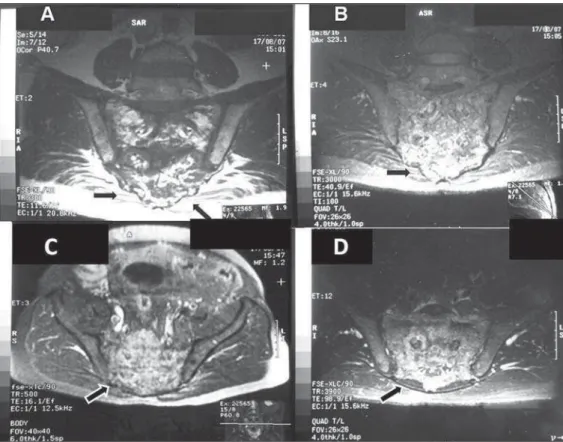
The hip radiograph is shown on Figure 1, bone scintigraphy on Figure 2, multislice computed tomography (CT) of the sacrum on Figure 3, and Figure 4 shows pelvic magnetic resonance imaging (MRI), with their respective findings.
Sacral biopsy was requested, consider-ing the clinical-radiological suspicion of bone Paget’s disease in an elderly, male patient with ill-defined pain, and with the
Braga FNHF, Braga MV, Andrade Neto F. Paget’s disease with sacral involvement: a case report. Radiol Bras. 2010;43(5): 339–342.
lumbar pain. The patient denied other symptoms, and the clinical examination did not demonstrate any alterations.
The physical examination of the joint revealed a positive Patrick’s test on the left coxofemoral joint, with pain at palpation at a point near the trochanteric bursa. Lasègue’s maneuver was negative. Ab-sence of pain at flexion, extension and pal-pation of the lumbar spine. Normal re-flexes. Preserved muscular strength and sensitivity.
Serum tests demonstrated the follow-ing results: alkaline phosphatase (ALP),
0100-3984 © Colégio Brasileiro de Radiologia e Diagnóstico por Imagem INTRODUCTION
Paget’s disease is a common focal, pro-gressive osteometabolic disorder of bone remodeling characterized by both an in-crease in osteoclastic resorption and sec-ondary bone formation, resulting in a dis-organized and fragile lamellar bone mo-saic susceptible to deformities and frac-tures(1–3). The authors report the case of a
patient with sacral involvement by Paget’s disease and describe the respective radio-logical findings.
CASE REPORT
A male 71-year-old patient complaining of acute moderate pain in the left coxofemo-ral region, with ill-defined irradiation to the left thigh, in association with mechanical
340
Braga FNHF et al.
Radiol Bras. 2010 Set/Out;43(5):339–342
Figure 2. Whole-body Tc-99m scintigraphy demonstrates heterogeneous radiopharmaceutical distribu-tion in the skeleton, with increased tracer uptake in the sacrum (arrows), right acetabulum (anterior pelvis) and bilaterally in the sacroiliac region (posterior pelvis).
the disease progression. Bone scintigraphy was similar to the pre-treatment scinti-graphic study. MRI demonstrated a small decrease in contrast uptake in the sacral region. Contrast uptake was not observed in the posterior acetabular column as well as in the adjacent soft tissues.
DISCUSSION
Paget’s disease is a chronic osteometa-bolic disorder characterized by abnormali-ties in all the phases of bone remodeling, resulting from osteoclastic and osteoblas-tic hyperactivity(1–4). The axial skeleton is most frequently affected, the pelvis being involved in about 70–90% of cases, and the spine in up to 53% of cases(3,4). Vertebral spine involvement occurs at one or more levels, with the lumbar level being de-scribed in up 58% of cases, thoracic level, in 45%, and cervical level, in 44%(3,4).
However, in many cases the disease is asymptomatic and is incidentally diag-nosed by radiological findings in studies performed for other reasons(3). Paget’s
dis-ease of bone is currently described as the
Figure 3. A,B: 3D multislice CT image. C,D: Respectively, axial and sagittal sections of the sacrum. To-mographic sections demonstrating significant enhancement of gross, medullary bone trabeculate in the sacrum, with a predominantly scle-rotic pattern (arrows), loss of the normal trabecular architecture, and with no evidence of significant de-struction of bone cortical or adjacent extraosseous soft tissues. On C, image demonstrating the right iliac involvement (asterisk) adjacent to the sacrum, with the previously mentioned characteristics. No evi-dence of fracture trace is observed.
objective of ruling out the presence of as-sociated neoplasia(3–5) as well as
histo-pathologically confirming the presence of the disease. The histopathological analysis demonstrated irregular, thickened bone tra-beculae forming a meshwork of intercom-municating spaces with remodeling changes, showing ossification lines with different orientations, mosaic pattern, with medullary, peritrabecular fibrosis and os-teoclastic cells suggestive of Paget’s
dis-ease. Absence of malignancy is observed. The treatment selected was oral iban-dronate 150 mg monthly doses. After six months of treatment, the patient presented reduction in total ALP levels to 132 U/L (a 63.98% reduction), and in the ALP bone fraction to 51 U/L (an 82.27% reduction), resulting in clinical management of the disease(1).
341
Paget’s disease with sacral involvement
Radiol Bras. 2010 Set/Out;43(5):339–342
second most prevalent osteometabolic dis-ease worldwide, after osteoporosis(1–4).
The most frequent symptom is bone or joint pain, with a prevalence in about 50% of cases(1,3), primarily explained by
second-ary degenerative joint disease, periosteal or cartilaginous disease, microfractures and bone deformities. Other less described causes for pain are pathological fracture, nervous compression, intervertebral in-volvement, spondylolysis, spondylolisthe-sis and sarcomatous degeneration(1–4).
Periosteal apposition with endosteal ab-sorption constitute the most frequent mechanism leading the affected bone to expand, which can be demonstrated by ra-diological imaging. In this setting, osteo-clasts are increased in number and size. There is an osteoblastic dysfunction, with hyperactive, although morphologically normal, osteoblasts, leading to a chaotic development of a new periosteal and en-dosteal bone, with loss of the normal lamel-lar pattern(1–3).
At the initial lytic phase, radiography of the involved bones demonstrates well-de-fined radiolucent areas without sclerosis. With the disease progression, at the mixed
phase, gross trabeculae and cortical thick-ening are observed. At the final, blastic phase, areas of sclerosis and increased bone volume are observed(3–5). In this phase, the
main differential diagnosis is with metasta-sis, particularly in breast and prostate(5). CT
is better to demonstrate the loss of normal bone trabeculae, also with focal areas of lysis, sclerosis and cortical bone expansion as reported in the present case(3,4). In the
present case, plain hip radiography did not show to be sufficiently elucidative for the diagnosis, leading the assisting physician to supplement the investigation with the other imaging resources reported and de-scribed in the literature(1–8).
Tc-99m scintigraphy is highly sensitive, but poorly specific for detecting bone re-modeling, demonstrating focal intensity with radiopharmaceutical uptake in the af-fected bones, suggesting the presence of disease even before the symptoms on-set(1,4–6).
MRI is indicated to investigate differen-tial diagnoses such as malignancy, and to evaluate medullary infiltration in the ver-tebral presentation of the disease(3,5,7,8).
Hyperintense signal corresponding to an
increase in fat within the medullary space of the involved bone may be identified at all MRI sequences, characterizing an atro-phic bone marrow, with a possible narrow-ing of the canal caused by the cortical thickening. Heterogeneous signal intensity is another pattern that can be found on and T2-weighted sequences. On the T1-weighted sequence, the bone marrow may present decreased signal intensity, with foci of normality, which rules out the presence of malignant degeneration because of the absence of expansile lesions. On the other hand, T2–weighted sequence demonstrates heterogeneously hyperintense signal in the bone marrow, corresponding to its fi-brovascular component. Finally a third pre-sentation is identified by the low signal intensity of the bone marrow at all the se-quences, representing sclerosis and charac-terizing the inactive blastic phase. The medullary enhancement can be seen with the utilization of gadolinium-based con-trast agent(4,5,7,8).
Bone biopsy is useful to confirm the di-agnosis in cases of dubious lesion presen-tation, as well as in patients with high risk for developing associated neoplasias, either
342
Braga FNHF et al.
Radiol Bras. 2010 Set/Out;43(5):339–342
as a primary bone tumor or as a metastasis in the vertebral spine(3,4). Neoplastic
trans-formation of the lesions, although rare, are considered as a complication of Paget’s disease, so bone biopsy becomes essential in this differentiation(3,4,8).
Currently, bisphosphonates constitute the medication of choice for the specific treatment in an attempt to re-establish the normal bone remodeling, reducing the bone turnover(1–3,5). Ibandronate may be
ad-ministered either in oral monthly doses or by intravenous bolus injection, with proven safety and effectiveness against pla-cebo(1,2).
The clinical follow-up is made with evaluation of ALP levels, the most sensi-tive marker for osteoblastic activity that must be performed every 3–6 months(1,2). Follow-up with CT and MRI can prove the pharmacological efficacy, as well as evalu-ating the possible complications of the dis-ease itself, such as malignant degeneration or pathological fractures(1,4,5,8).
The ill-defined, left-sided coxofemoral
pain initially reported by the patient trig-gered a comprehensive diagnostic investi-gation. Such complaint, whose site did not coincide with the radiological findings, was later attributed to left-sided trochant-eric bursitis that was managed with clini-cal symptomatic treatment and physio-therapy. Once the treatment for the bone disease was established, the patient did not complain of pain anymore.
Finally, the authors emphasize that par-ticular attention should be paid to male and elderly patients with pain in the lumbar spine and in the coxofemoral joint, because of the potential risk for development of neoplasias in this group of patients(4,5,8). The radiologist must be attentive to the possible presentations and complications of the disease, even in uncommon sites, trying whenever possible correlating them with the patients’ clinical symptoms.
Acknowledgment
To Dr. Marta Maria Chagas Medeiros, Associated Professor of Rheumatology at
Universidade Federal do Ceará, for the re-vision and suggestions on the present study.
REFERENCES
1. Griz L, Caldas G, Bandeira C, et al. Paget’s dis-ease of bone. Arq Bras Endocrinol Metab. 2006; 50:814–22.
2. Cundy T, Bolland M. Paget disease of bone. Trends Endocrinol Metab. 2008;19:246–53.
3. Dell’Atti C, Cassar-Pullicino VN, Lalam RK, et al. The spine in Paget’s disease. Skeletal Radiol. 2007;36:609–26.
4. Smith SE, Murphey MD, Motamedi K, et al. From the archives of the AFIP. Radiologic spectrum of Paget disease of bone and its complications with pathologic correlation. Radiographics. 2002;22: 1191–216.
5. José FF, Pernambuco ACA, Amaral DT. Doença de Paget do osso. Einstein. 2008;6(Supl 1):S79– S88.
6. Love C, Din AS, Tomas MB, et al. Radionuclide bone imaging: an illustrative review. Radio-graphics. 2003;23:341–58.
7. Roberts MC, Kressel HY, Fallon MD, et al. Paget disease: MR imaging findings. Radiology. 1989; 173:341–5.