BREAST ULTRASOUND: EVALUATION OF ECHOGRAPHIC CRITERIA
FOR DIFFERENTIATION OF BREAST LESIONS*
Maria Julia Gregorio Calas1
, Hilton Augusto Koch2
,Maria Virginia Peixoto Dutra3
OBJECTIVE: Breast cancer is one of the most important causes of death in women. The association of dif-ferent diagnostic methods has been successfully employed as a means to enhance early diagnosis. In this scenario, the interventional and diagnostic breast ultrasound has played a significant role. This study has two main objectives: to identify echographic criteria related to benignancy and malignancy and to analyze echographic characteristics, evaluating their role as malignancy predictors. MATERIALS AND METHODS: Echographic morphological criteria adopted in the images description were: shape, limits, contour, echogenicity, echotexture, echotransmission, lesion orientation, and secondary signs. Validation was sought on 450 echographic images compared with follow-up and histopathological results. RESULTS: Main benignancy criteria were: well defined shape, regular contour, precise limits, lesions isoechoic to fatty tissue, homogeneous echotexture, and horizontal orientation. The criteria more typically related to malignancy were: ill-defined shape, irregular contour, partially precise limits, and hypoechogenicity. Contour irregularity has presented the greatest sensitivity (92.7%) as well as the highest negative predictive value (98.2%), while vertical orientation of the lesion has presented the greatest specificity (99.3%), and ill-defined shape, the highest positive predictive value (91.0%). CONCLUSION: The standardization of the method for characterization and description of breast ultrasound images has resulted in reports uniformization and optimization, allowing more appropriate therapeutic decisions.
Keywords: Ultrasound; Breast lesions; Diagnosis.
Ultra-sonografia mamária: avaliação dos critérios ecográficos na diferenciação das lesões mamárias. OBJETIVO: O câncer de mama é uma das causas mais importantes de mortalidade feminina. Na busca do diagnóstico cada vez mais precoce, a associação de métodos diagnósticos tem sido utilizada com êxito, tendo grande participação a ultra-sonografia mamária diagnóstica e intervencionista. Este trabalho tem como objetivos: identificar os critérios ecográficos mais relacionados com benignidade e malignidade e avaliar as propriedades das características ecográficas, verificando o seu poder de predição de malignidade. MATE-RIAIS E MÉTODOS: Os critérios morfológicos ecográficos utilizados nas descrições das imagens foram: forma, limites, contorno, ecogenicidade, ecotextura, ecotransmissão, orientação e sinais secundários. A validação foi buscada em 450 imagens ecográficas, comparadas aos resultados de seguimento ou de histopatologia de peça cirúrgica. RESULTADOS: Os principais critérios de benignidade foram: forma definida, contorno regular, limites precisos, lesões isoecóicas ao tecido adiposo, ecotextura homogênea e orientação horizontal. Os critérios mais característicos de malignidade foram: forma indefinida, contorno irregular, limites parcialmente precisos e lesões hipoecóicas. O contorno irregular apresentou a maior sensibilidade (92,7%) e o maior valor preditivo negativo (98,2%) para malignidade, a orientação vertical apresentou a maior especificidade (99,3%), e a forma indefinida, o maior valor preditivo positivo (91,0%). CONCLUSÃO: O método padronizado para a caracterização e descrição das imagens ultra-sonográficas mamárias apresentado resultou em uniformidade e otimização dos laudos, viabilizando as condutas mais adequadas.
Unitermos: Ultra-sonografia; Lesões mamárias; Diagnóstico. Abstract
Resumo
* Study developed, as a part of a Master’s Degree Disserta-tion, in the Sector of Radiology at Faculdade de Medicina da Uni-versidade Federal do Rio de Janeiro, Rio de Janeiro, RJ, Brazil. 1. Master’s Degree in Medicine by Faculdade de Medicina da Universidade Federal do Rio de Janeiro.
2. Titular Professor of Radiology at Faculdade de Medicina da Universidade Federal do Rio de Janeiro.
3. Post-Graduation Professor at Woman and Child Health – Instituto Fernandes Figueira, Fundação Oswaldo Cruz, Centro de Estudos e Pesquisas da Mulher.
nique recognized for its proved contribu-tion to the early deteccontribu-tion and decrease in breast cancer mortality. However, the accu-racy of this method is highly dependent on the breast parenchyma composition and tissular characteristics(1–4).
The association of diagnostic methods has been successfully employed in the
search of a more and more early diagnosis of this pathological entity(1–4). Breast ultra-sound, both for diagnostic and interven-tional purposes, plays a significant role as a method supplementary to the clinical mammography, and has become a well es-tablished and invaluable method for diag-nosis of breast diseases(1–4).
The first reference to breast ultrasound in the literature appears in 1951, with a study by Wild and Neal, describing in vivo sonographic features of two breast tumors, one malignant and another benign(5). With
Mailing Address: Dra.Maria Julia Gregorio Calas. Rua Siqueira Campos, 238, ap. 810, Copacabana. Rio de Janeiro, RJ, 22031-070 – Brazil. E-mail: [email protected]
Received January 11, 2006. Accepted after revision March 31, 2006.
INTRODUCTION
tech-the introduction of tech-the gray scale in tech-the seventies by Kossof, Jellins et al.and, along the last decades, with the utilization of dynamic study, high-frequency linear trans-ducers (7.5 MHz to 13 MHz) and electronic focus, the breast ultrasound has been estab-lished as a method for diagnostic evalua-tion in the mammary propedeutics(5).
This method is well tolerated and ac-cepted by patients for not requiring ioniz-ing radiation nor compression, besides be-ing fast and easy to perform(1–4). Addition-ally, today ultrasound is the only real-time imaging method widely available, repre-senting an excellent modality for guidance of interventional invasive procedures, al-lowing the choice of the shortest route be-tween the skin and the area of interest, with higher swiftness and minimum discomfort for the patient(6).
The echographic interpretation is based on the knowledge of the normal mammary structure, its variants and multiple aspects of breast diseases. Abnormal images have been evaluated and defined according to morphological characteristics. The capac-ity for evaluating morphological criteria of echographic images and differentiating between benign and malignant alterations has been subject for several studies, but there are controversies in the literature re-garding the predictive capacity of each echographic characteristic(7–11).
Many authors consider that the combi-nation of echographic criteria results in higher sensitivity and specificity of the findings when compared with the evalua-tion of an isolate characteristic(7,8). For any isolate echographic characteristic to have practical applicability in the differentiation between malignant and benign lesions, this characteristic must be a finding with a high rate of interobserver agreement(7,8). Consid-ering that ultrasound is an operator-depen-dent method, the professional qualification, besides the operator experience, is clearly mandatory, allowing not only the images visualization, but also the utilization of morphological characteristics for differen-tiation between malignant and benign le-sions(7–11). A quantitative approach is sug-gested by other studies, aiming at analyz-ing tumors margins and texture on the ba-sis of several mathematical techniques. These techniques have been employed as
powerful ancillary diagnostic tools (com-puter aided diagnostics – CAD-type sys-tems) for assessing different types of medi-cal images, considering that quantification does not depend on the operator experi-ence(12).
The standardization of terms employed for describing breast lesions is important since it allows a more objective analysis of images, creating an uniform nomenclature capable of indicating the degree of malig-nancy suspicion according to the morphol-ogy of the echographic findings and, con-sequently, allowing a more accurate orien-tation for the conduct to be adopted(13,14).
The present study aims at identifying those echographic criteria which are most closely related to lesions benignancy and malignancy, besides evaluating echo-graphic characteristics to determine their predictive value for malignancy.
MATERIALS AND METHODS
Our casuistic included 637 cases of pa-tients who had been referred by request of their doctors for undergoing ultrasound-guided fine needle aspiration (206 cases), and ultrasound-guided core-biopsy (431 cases), in the period between January and December of 2003.
All examinations were performed with a GE Logic MD 400 equipment with a multi frequency transducer, with orthogo-nal photographic recording of each image, besides appropriate material for each pro-cedure. The photographic records review, as well as the evaluation for the presence of echographic criteria on images, was per-formed by one of the authors of the present study who has ten-year experience in breast imaginology and invasive procedures.
Of the 637 cases, 187 were excluded because the material was considered as unsatisfactory for the purposes of the present study. The remaining 450 cases are distributed as follows: 9 cases of simple cysts, 42 of complicated cysts, 2 of com-plex cysts and 397 cases of solid breast lesions.
This study was developed in a private service of diagnostic imaging to which the patients were referred by request of their doctors. It was up to the patients’ doctors to make a decision about their treatment on
the basis of information originated by the examinations (results of the invasive pro-cedures), independently from the present study.
For the 450 selected cases, the standard test consisted of the histopathological re-sults in the surgical cases (provided by the patients’ doctors), and of the echographic follow-up of the lesions in later examina-tions performed during a six-to twenty-four-month period, in the non-surgical cases. Information on the follow-up was obtained in the service data-base or was provided by the patients´ doctors.
an-B A
Figure 7. Echotexture: homogeneous (A) and het-erogeneous (B).
Figure 6. Echogenicity: anechoic (A), hypoechoic (B), isoechoic (C), and hyperechoic (D).
D
A B C
Figure 4. Regular (A) and partially regular margins (B), also denominated macrolobular.
A B
Figure 2. Ill-defined shape.
gular and microlobular). Images with par-tially precise and imprecise limits were grouped as lesions with imprecise limits. As regards echo transmission, the charac-teristics were grouped as present echo transmission in the presence of acoustic shadow or posterior enhancement, and ab-sent echo transmission, in the absence of retrotumoral acoustic phenomenon. The software Epiinfo 6.04b CDC-USA, World Health Organization (Oct.1997) was em-ployed for such evaluation.
The project was submitted to the Com-mittee on Ethics in Research of Hospital Universitário Clementino Fraga Filho at Universidade Federal do Rio de Janeiro, and was approved with no restriction.
RESULTS
On ultrasound, the size of the 450 le-sions ranged between 4 mm and 55 mm. In seven cases, the lesions could not be mea-sured because of their large volumes.
The results of the procedures (fine-needle aspiration and core biopsy) were compared by means of the standard test (surgery and follow-up), resulting in 82 (18.2%) cases of cancer, and 368 (81.8%) cases of benign lesions.
The most frequent benign lesions were fibroadenomas (43.1%), followed by non-proliferative lesions (35.4%). Amongst the malignant findings, the infiltrating ductal carcinoma was prevalent, corresponding to 72.7% of cases (Figures 10A and 10B).
Figure 1. Defined shape: round (A), oval-shaped (B).
A B
Figure 3. Limits: precise (A), partially precise (B) and imprecise (C).
A B C
Figure 5. Margins: irregular, with microlobular (A), spiculated (B), angular (C), and indistinct aspect (D).
D
A B C
A B
Figure 9. Orientation: horizontal (A) and vertical (B).
Figure 8. Echo transmission: absent (A), acoustic enhancement (B), bilateral acoustic shadowing (C) and central acoustic shadowing (D).
C D
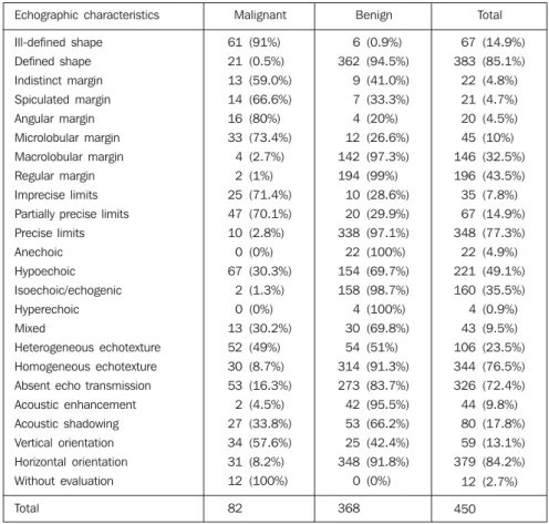
Table 1 List of echographic characteristics of breast lesions and standard test.
Echographic characteristics
Ill-defined shape
Defined shape
Indistinct margin
Spiculated margin
Angular margin
Microlobular margin
Macrolobular margin
Regular margin
Imprecise limits
Partially precise limits
Precise limits
Anechoic
Hypoechoic
Isoechoic/echogenic
Hyperechoic
Mixed
Heterogeneous echotexture
Homogeneous echotexture
Absent echo transmission
Acoustic enhancement
Acoustic shadowing
Vertical orientation
Horizontal orientation
Without evaluation
Total
Malignant
61 (91%)
21 (0.5%)
13 (59.0%)
14 (66.6%)
16 (80%)
33 (73.4%)
4 (2.7%)
2 (1%)
25 (71.4%)
47 (70.1%)
10 (2.8%)
0 (0%)
67 (30.3%)
2 (1.3%)
0 (0%)
13 (30.2%)
52 (49%)
30 (8.7%)
53 (16.3%)
2 (4.5%)
27 (33.8%)
34 (57.6%)
31 (8.2%)
12 (100%)
82
Benign
6 (0.9%)
362 (94.5%)
9 (41.0%)
7 (33.3%)
4 (20%)
12 (26.6%)
142 (97.3%)
194 (99%)
10 (28.6%)
20 (29.9%)
338 (97.1%)
22 (100%)
154 (69.7%)
158 (98.7%)
4 (100%)
30 (69.8%)
54 (51%)
314 (91.3%)
273 (83.7%)
42 (95.5%)
53 (66.2%)
25 (42.4%)
348 (91.8%)
0 (0%)
368
Total
67 (14.9%)
383 (85.1%)
22 (4.8%)
21 (4.7%)
20 (4.5%)
45 (10%)
146 (32.5%)
196 (43.5%)
35 (7.8%)
67 (14.9%)
348 (77.3%)
22 (4.9%)
221 (49.1%)
160 (35.5%)
4 (0.9%)
43 (9.5%)
106 (23.5%)
344 (76.5%)
326 (72.4%)
44 (9.8%)
80 (17.8%)
59 (13.1%)
379 (84.2%)
12 (2.7%)
450
Figure 10.A: Hypoechoic, lobulated nodule, precise limits, homogeneous echotexture, horizontal orientation and bilateral acoustic shadowing (fibroadenoma).
B: Hypoechoic nodule, microlobular and indistinct margins, partially precise limits, heterogeneous echotexture, vertical orientation (infiltrating ductal carcinoma).
B A
In the studied casuistic, the patients’ ages ranged between 16 and 88 years (mean age 52 years). Amongst patients who pre-sented with carcinomas, the ages ranged between 29 and 88 years (mean age 60 years). On the other hand, the ages of pa-tients with benign lesions ranged between 16 and 81 years (mean age 45 years).
A list of the breast lesions echographic characteristics is on Table 1, where the column “Benign” includes lesions in fol-low-up or those histopathologically diag-nosed as benign, and the column “Malig-nant” includes those lesions considered as malignant at surgery.
Amongst the lesions presenting a de-fined shape, 94.5% (362/383) were benign, and amongst those with ill-defined shape, 91.0% (61/67) were malignant.
Amongst lesions with regular margins, 98.2% (336/342) were benign. Amongst lesions with irregular margins, 70.4% (76/ 108) were malignant, the microlobular type being the most frequently found — 30.5% of cases (33/108).
Amongst lesions with precise limits, 97.1% (338/348) were benign, and amongst those with imprecise limits, 70.6% (72/ 102) were malignant.
Amongst anechoic and hyperechoic images, 100% of lesions were benign. As regards hypoechoic images, 69.7% were benign, and 30.3% were malignant. Amongst isoechoic images, 98.7% were benign, and 1.3% malignant. Of those im-ages presenting mixed echogenicity, 69.8% were benign, and 30.2% malignant.
Amongst the images with homogeneous echotexture, 91.3% (314/344) were benign,
acoustic shadow, in 66.2% of benign le-sions.
Amongst images with horizontal orien-tation, 91.8% (348/379) were benign. Ver-tical orientation was present in 57.6% (34/ 59) of the malignant lesions. In 12 cases, images orientation could not be analyzed because lesion were on a deep plane and presented a marked acoustic shadow, com-plicating a correct evaluation of the lesion and 49.0% (52/106) of those with
hetero-geneous echotexture were malignant, and 51% (54/106 were benign.
size, and, consequently, a precise evalua-tion of the lesion orientaevalua-tion.
Secondary signs were found just in 14 (17%) cases of malignancy, and were ab-sent in 100% of benign cases.
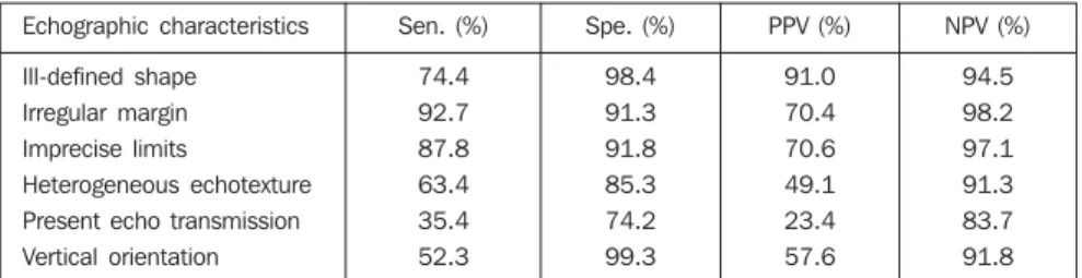
The evaluation of characteristics more closely related to lesions malignancy was considered for analyzing the diagnostic properties, as per Table 2. The most sensi-tive characteristic was the irregular mar-gins. Two characteristics presented a very high specificity: vertical orientation (99.3%) and ill-defined shape (98.4%). The nega-tive predicnega-tive values were high, in contrast to the positive predictive values in the pres-ence of echo-transmission (23.4%), and heterogeneous echotexture (49.1%).
DISCUSSION
The quality of a procedure is highly dependent on the operator’s knowledge of the equipment, the appropriate technique, the results interpretation, the patient’s his-tory, besides the malignant, benign and functional alterations of the breast(2,4,15).
Notwithstanding all the studies re-viewed have utilized high-resolution equip-ment and trained professionals, the diver-gences observed in the literature are due to different methodologies applied, either uti-lizing different morphological characteris-tics or different criteria for distinguishing malignant from benign lesions(7–9,16–19). This diversity reflects the fact that not always all the morphological characteristics of an im-age with all their variables can be utilized(19). Some features are unique to ultrasound, such as orientation and echogenicity, and some are fundamental to interpreting im-ages, such us shape and margins(20).
Amongst the studies selected for the purpose of comparative analysis, all of them utilized the description of the contour or margin, followed by echo transmission,
Table 2 Echographic characteristics properties suggestive malignancy.
Echographic characteristics
Ill-defined shape
Irregular margin Imprecise limits
Heterogeneous echotexture Present echo transmission
Vertical orientation
Sen. (%)
74.4
92.7 87.8
63.4 35.4
52.3
Spe. (%)
98.4
91.3 91.8
85.3 74.2
99.3
PPV (%)
91.0
70.4 70.6
49.1 23.4
57.6
NPV (%)
94.5
98.2 97.1
91.3 83.7
91.8
Sen., sensitivity; Spe., specificity; PPV, positive predictive value; NPV, negative predictive value.
echogenicity, echotexture and orientation. The features less utilized by the authors were: limits, compressibility, branching and size of the lesion(7–17,19,21–24).
Some authors(10,15,19,20,24,25) describe not only morphological signs of the lesions but also the alterations in surrounding tissues, the parenchyma architectural distortion being the most frequently described sec-ondary sign.
An universal standardization of terms for description of echographic features is still pending, however, with the publication of the Illustrated Breast Imaging and Re-porting Data System (BI-RADS®): Ultra-sound by the American College of Radiol-ogy(26), most probably this standardization and consequent uniformization will be ac-cepted.
Although studies involving interpreta-tion and classificainterpreta-tion of sonographic im-ages recommend an analysis of the concor-dance between observers, and limitation of the generalization based on studies by only one observer, Skaane and Engedal(17), Zonderland et al.(19), Buchberger et al.(27), and Chen et al.(28) have considered that studies interpreted by only one observer might be consistent in the application of criteria for a lesion definition.
The study developed by Chen et al.(28) suggests that sonographic characteristics considered in the lesions differentiation might vary in tumors, depending on their size. However, they consider that the most significant feature in lesions differentia-tion, independently from their size, is the contour or margins, according to the results reported by Skaane and Engedal(17).
Paulinelli et al.(9) evaluated the influ-ence of the age and size of the tumor on the interpretation of the sonographic character-istics of solid breast nodules. These authors have concluded that the presence of irregu-lar margins, internal heterogeneous echoes
and posterior shadowing in benign tumors is directly proportional to the patient’s age. The presence of internal heterogeneous echoes, anterior halo and thickening of Cooper ligaments in malignant tumors is directly proportional to the size of the tu-mor.
Additional studies are necessary to reach a consensus on which echographic characteristics would be more significant, less subjective, and more reproducible, and, besides, it is necessary to analyze other factors which could affect the sonographic images interpretation and the risk of malig-nancy(9,11,14,20).
For most of the authors(16,17,19,21,22,25, 27–29), regular or macrolobular margins
rep-resented the main criterion for defining a lesion as benign. The shape described as defined, oval, rounded or ellipsoid was the second most important feature, in agree-ment with the results of the present study, where the regular and macrolobular mar-gins were identified in 98.2% of the benign cases, and the defined shape, in 94.5%.
The malignancy characteristics most frequently described in the literature(9,10, 16–19,21,22,25,27–29) were irregular margins,
presence of posterior acoustic shadowing, vertical orientation and irregular shape.
In the present study, the more character-istic signs of malignancy were: ill-defined shape, irregular margins, partially precise limits and hypoechoic lesions. Secondary signs were present in only 14 of 82 cases of malignancy, and absent in the 368 be-nign cases. The literature suggests a crite-rion for benignity in the absence of such signs(11,15,25,29), and a criterion for malig-nancy in their presence.(15,17,29).
In the 12 cases where orientation could not be identified due the size and localiza-tion of the lesion, the term “indeterminate” orientation might be employed, according to Zonderland et al.(19). This pattern sug-gests a criterion for malignancy, since the 12 cases of indeterminate orientation had their malignancy confirmed on the surgical piece.
In the present study, the criteria non-sig-nificant for echographic differentiation were: echotexture and echo transmission. Amongst 106 images with heterogeneous echotexture, 52 cases (49%) were malig-nant and 54 cases (51%) were benign. As regards posterior acoustic findings, acous-tic shadowing was present in 66.2% of the benign lesions and in 33.8% of the malig-nant lesions, in contrast to other authors observations(7,10,11,17,19,21–24,27–29).
A contradiction is observed in the litera-ture. While the features most utilized by the authors for echographically describing a lesion were echo transmission, echogeni-city and echotexture, such features were not significant for differentiating a malignant from a benign lesion.
The definition of the echotexture of a lesion as homogeneous or heterogeneous depends on the diversity of the tissues in such lesion. For example, a lesion might be heterogeneous for presenting areas of ne-crosis (carcinomas) or due to fibroadenomas (benign) hyalinization(28).So, the utiliza-tion of this single characteristic might lead to misinterpretation. The absence of a prog-nostic value of this variable was described by Stavros et al.(11) and Skaane and Enge-dal(17). Also, it is important to note that in the new standardization of the American College of Radiology(26), this feature was not taken into consideration.
Echogenicity might be defined as a shade of the gray scale demonstrating a lesion and representing one of the main problems in the description of such
le-sion(21,24). In the absence of standardization, the echogenicity becomes a source of sub-jectivity, since the gray scale parameters utilized for this type of characterization are still to be known.
In the study developed by Stavros et al.(11), the lesions echogenicity was com-pared with the normal adjacent adipous tis-sue, i.e., with a structure of echogenicity approximating the gray scale spectrum, that was uniform and present in all of the pa-tients. For Stavros et al.(11), the hyper-echogenicity of a lesion represented the feature with higher negative predictive value (100%). In a different way, Soon et al.(24) have compared the lesions echogeni-city with the adjacent glandular tissue in 393 cases of carcinoma, and have found two cases (0.5%) of carcinomas as hyper-echoic lesions. For Zonderland et al.(19),the gain adjustment in the equipment, as well as the breast thickness, might affect the echogenicity description.
In the study developed by Chen et al.(28), echogenicity was the main feature in the differentiation between infiltrating carcino-mas and carcinocarcino-mas in situ, the latest pre-senting more isoechoic when compared with infiltrating carcinomas.
The echo transmission is a function of the equipment, the transducer, the compres-sion exerted during the procedure, the size, localization and even the type of the le-sion(21).
The echo transmission has emerged in the beginnings of the breast ultrasonogra-phy as an essential, almost pathognomonic
sign of malignancy, indispensable for diag-nosis(5). As a matter of fact, the experience has proved that this is a sign found in 20% to 60% of cases of breast cancer, especially in those where the tumor is larger than 2 cm(21). This is the reason, mainly in cases of non-palpable tumors (with < 1 cm), for which the absence of echo transmission does not allow neither the exclusion of malignancy, nor assurance of benignity. An article published by Weinstein et al.(30), presents a group of benign lesions which might present posterior acoustic shadow-ing, like fibroadenomas, radiate scar, dia-betic mastopathy, steatonecrosis, post-sur-gical scar, focal fibrosis, and sclerosing adenosis.
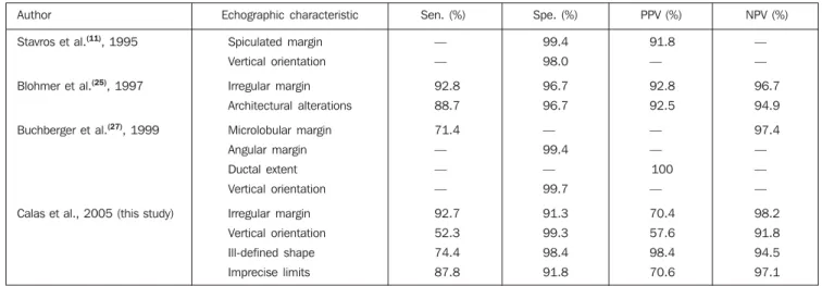
As regards diagnostic properties found by several authors and in the present study (Table 3), the irregular margins (microlob-ular, spiculated or angular aspect) and ori-entation were the echographic characteris-tics with highest rates of sensitivity, speci-ficity and predictive values.
In summary, one may conclude that the main features for differentiating malignant from benign lesions are margins and shape of the lesion, with the first one presenting the highest sensitivity, and the second, the highest specificity.
Therefore, the quantitative study of the margins of a lesion might be a powerful tool for helping the observer to differenti-ate malignant tumors from the benign ones. Alvarenga et al.(31), utilizing a method based on mathematical morphology for images segmentation, have found 95.7%
Table 3 Properties of malignancy echographic characteristics, by author.
Author
Stavros et al.(11), 1995
Blohmer et al.(25), 1997
Buchberger et al.(27), 1999
Calas et al., 2005 (this study)
Echographic characteristic
Spiculated margin
Vertical orientation
Irregular margin
Architectural alterations
Microlobular margin
Angular margin
Ductal extent
Vertical orientation
Irregular margin
Vertical orientation
Ill-defined shape
Imprecise limits
Sen. (%)
—
—
92.8
88.7
71.4
—
—
—
92.7
52.3
74.4
87.8
Spe. (%)
99.4
98.0
96.7
96.7
—
99.4
—
99.7
91.3
99.3
98.4
91.8
PPV (%)
91.8
—
92.8
92.5
—
—
100
—
70.4
57.6
98.4
70.6
NPV (%)
—
—
96.7
94.9
97.4
—
—
—
98.2
91.8
94.5
97.1
sensitivity and 96.7% specificity in the dif-ferentiation of tumors by means of margins analysis.
The next step in this study will be to develop an interobserver study (with quali-tative criteria), comparing it with the results obtained with quantitative methods applied to margins and echogenicity of lesions. Based on this comparison, we will be able to evaluate the contribution that the quan-tification may add to the final diagnosis of such lesions.
REFERENCES
1. Paulinelli RR, Moreira MAR, Freitas Júnior R. A importância do diagnóstico precoce do câncer de mama. Femina 2004;32:233–237.
2. Fine RE, Staren ED. Updates in breast ultrasound. Surg Clin North Am 2004;84:1001–1034. 3. Paulinelli RR, Moreira MAR, Freitas Júnior R.
Ultra-sonografia no diagnóstico do câncer de mama: realidade atual e possibilidades para o futuro. Rev Bras Mastol 2003;13:168–174. 4. Fonseca ALA. Ultra-sonografia da mama. In:
Pas-qualette HA, Koch HA, Soares-Pereira PM, Kemp C, editores. Mamografia atual. 1ª ed. Rio de Janeiro: Revinter, 1998:205–215.
5. Dempsey PJ. The history of breast ultrasound. J Ultrasound Med 2004;23:887–894.
6. Lucena CEM. Procedimentos intervencionistas mamários guiados por ultra-som. Femina 2002; 30:537–541.
7. Arger PH, Sehgal CM, Conant EF, Zuckerman J, Rowling SE, Patton JA. Interreader variability and predictive value of US descriptions of solid breast masses: pilot study. Acad Radiol 2001;8: 335–342.
8. Baker JA, Kornguth PJ, Soo MS, Walsh R, Men-goni P. Sonography of solid breast lesions: ob-server variability of lesion description and assess-ment. AJR Am J Roentgenol 1999;172:1621– 1625.
9. Paulinelli RR, Freitas-Júnior R, Moreira MAR, et al. Risk of malignancy in solid breast nodules ac-cording to their sonographic features. J Ultra-sound Med 2005;24:635–641.
10. Paulinelli RR, Vidal CSR, Ruiz AN, Moraes VA, Bernardes Júnior JRM, Freitas Júnior R. Estudo prospectivo das características sonográficas no diagnóstico de nódulos sólidos da mama. Rev Bras Ginecol Obstet 2002;24:195–199. 11. Stavros AT, Thickman D, Rapp CL, Dennis MA,
Parker SH, Sisney GA. Solid breast nodules: use of sonography to distinguish between benign and malignant lesions. Radiology 1995;196:123–134. 12. Azevedo CM, Alvarenga AV, Pereira WCA, Infan-tosi AFC. Análise computacional de imagens por ultra-som de lesões da mama em pacientes mas-tectomizadas e em pacientes com lesões sistêmi-cas. Rev Imagem 2004;26:279–286.
13. Calas MJG, Castro R, Manoel VR, Pasqualette HA, Soares-Pereira PM. Proposta de normatiza-ção dos laudos de ultra-sonografia mamária. Fe-mina 2002;30:103–110.
14. Pasqualette HAP, Soares-Pereira PM, Calas MJG, et al. Revisão e validação de uma proposta de classificação de laudos de ultra-sonografia mamá-ria. Rev Bras Mastol 2003;13:159–167. 15. Kossoff MB. Ultrasound of the breast. World J
Surg 2000;24:143–157.
16. Rahbar G, Sie AC, Hansen GC, et al. Benign ver-sus malignant solid breast masses: US differen-tiation. Radiology 1999;213:889–894. 17. Skaane P, Engedal K. Analysis of sonographic
features in the differentiation of fibroadenoma and invasive ductal carcinoma. AJR Am J Roent-genol 1998;170:109–114.
18. Watson L. The role of ultrasound in breast imag-ing. Radiol Technol 2000;71:441–459. 19. Zonderland HM, Hermans J, Coerkamp EG.
Ul-trasound variables and their prognostic value in a population of 1103 patients with 272 breast cancers. Eur Radiol 2000;10:1562–1568. 20. Mendelson EB, Berg WA, Merritt CR. Toward a
standardized breast ultrasound lexicon, BI-RADS: ultrasound. Semin Roentgenol 2001;36: 217–225.
21. Michelin J, Levy L. Ultra-sonografia da mama – diagnóstica e intervencionista. 1ª ed. Rio de Ja-neiro: Medsi, 2001.
22. Murad M, Bari V. Ultrasound differentiation of benign versus malignant solid breast masses. J Coll Physicians Surg Pak 2004;14:166–169. 23. Taylor KJ, Merritt C, Piccoli C, et al. Ultrasound
as a complement to mammography and breast ex-amination to characterize breast masses. Ultra-sound Med Biol 2002;28:19–26.
24. Soon PSH, Vallentine J, Palmer A, Magarey CJ, Schwartz P, Morris DL. Echogenicity of breast cancer: is it of prognostic value? Breast 2004;13: 194–199.
25. Blohmer JU, Schmalisch G, Kürten A, Chaoui R, Lichtenegger W. Relevance of sonographic crite-ria for differential diagnosis of mammary tu-mours. Eur J Ultrasound 1997;6:35–41. 26. American College of Radiology. Illustrated Breast
Imaging Reporting and Data System Atlas (BI-RADS®): Ultrasound. 4th ed. Reston: American
College of Radiology, 2003.
27. Buchberger W, Dekoekkoek-Doll P, Springer P, Obrist P, Dünser M. Incidental findings on sono-graphy of the breast: clinical significance and diagnostic workup. AJR Am J Roentgenol 1999; 173:921–927.
28. Chen SC, Cheung YC, Su CH, Chen MF, Hwang TL, Hsueh S. Analysis of sonographic features for the differentiation of benign and malignant breast tumors of different sizes. Ultrasound Obstet Gyne-col 2004;23:188–193.
29. Chao TC, Lo YF, Chen SC, Chen MF. Prospec-tive sonographic study of 3093 breast tumors. J Ultrasound Med 1999;18:363–370.
30. Weinstein SP, Conant EF, Mies C, Acs G, Lee S, Sehgal C. Posterior acoustic shadowing in benign breast lesions: sonographic-pathologic correla-tion. J Ultrasound Med 2004;23:73–83. 31. Alvarenga AV, Infantosi AFC, Azevedo CM,