UNIVERSIDADE DE LISBOA
FACULDADE DE CIÊNCIAS
DEPARTAMENTO DE BIOLOGIA ANIMAL
The role of Down syndrome cell adhesion molecule 1 for the
bacterial microbiota of Tribolium castaneum
Luís Manuel Macedo da Silva
Mestrado em Biologia Evolutiva e do Desenvolvimento
Dissertação orientada por:
Professor Dr. Joachim Kurtz | University of Münster, Germany
Professor Dr. Élio Sucena | University of Lisbon, Portugal
Acknowledgement
I would first like to thank Professor Dr. Joachim Kurtz for the opportunity to do the thesis in his group and for all the support he provided along the way.
I would especially like to thank Dr. Sophie Armitage for her tireless guidance and open door whenever I had questions or problems.
I am also grateful to the Tribolium group, namely Barbara, Kevin, Diana, Nora and Rasha for the help in the laboratory, as well as all the fruitful discussions and troubleshooting throughout the project.
I would also like to thank Professor Élio Sucena and Sólveig Thorsteinsdóttir for all the challenges and help they provided throughout the Master.
Finally, I must also thank my friends and family for all the support, long conversations and mostly for the patience they had.
Abstract
Hosts establish different types of interactions with the elements of their microbiota. These interactions can be broadly classified as positive, neutral or negative and shaped the evolution of the immune system. Negative interactions may lead to host-parasite coevolution. While the host may only survive and reproduce by exhibiting more efficient immune responses, the parasite requires novel ways of dodging the host's immune system. On the other hand, positive interactions can evolve to the point where the lack of specific microorganisms may interfere with tissue development, immunity or resistance to pathogens. Neutral interactions are not only difficult to define but are also thought to be dynamic and could evolve to either positive or negative interactions. Consequently, the host needs to be able to differentiate between harmful and beneficial microbes. This identification is thought to be performed through pattern recognition receptors that recognise microbe-associated molecular patterns and other environmental cues, enabling an appropriate response from the host.
Dscam1 is a gene expressed in neurons and essential to the correct development of neuronal
circuits. Through alternative splicing, it is able to originate around 18.000 isoforms in Drosophila
melanogaster and 15.000 in Tribolium castaneum. More recently, its expression was observed in
immune cells and tissues of some invertebrates. Recent studies have shown differential isoforms expression after infection by different classes of bacteria. Hence, it was suggested that Dscam1 may work as a pattern recognition receptor, opsonin and/or bacteria regulator. Concerning bacteria regulation, there are studies in Anopheles gambiae that show a proliferation of gut bacteria after a
Dscam1 knock-down. In the same model, overexpression of bacteria-induced Dscam1 isoforms
resulted in a reduction of bacteria in the haemolymph. The above-mentioned results suggest that Dscam1 is involved in negative regulation of bacteria. Nonetheless, there is also evidence suggesting a decrease in the intracellular parasites and no effect on extracellular bacteria after Dscam1 knock-down in Laodelphax striatellus.
Hence, I aimed to clarify the role of Dscam1 in the dynamics of Tribolium castaneum bacterial microbiota. This was achieved through Dscam1 knock-down and bacterial load analysis through RTqPCR for the bacterial 16S rRNA at three time-points of the beetle development: 15-days old larvae, 23-days old pupae and 30-days old adults.
Our results show that a Dscam1 knock-down does not affect the total amount of bacteria in the beetle in any of the three developmental time-points analysed. This is in agreement with previous studies. Nevertheless, a correlation between the relative expression of the bacterial 16S rRNA and
Dscam1 was observed. The correlation had a negative tendency: a higher expression of Dscam1 was
correlated with a lower expression of 16S rRNA and therefore less bacteria in the system. Such a tendency was also observed in Dong and colleagues’ studies on adult mosquitoes. A knock-down may not be sufficient to disturb a robust system capable of bacterial regulation in this model. Further analyses of the composition of bacterial microbiota are required to clarify the role of Dscam1 in bacterial regulation in T. castaneum. It is for example possible that changes occur in the proportion of different bacterial taxa.
Resumo
Organismos multicelulares estabelecem diversos tipos de interações com microrganismos. Estas interações podem ser positivas, neutras ou negativas para o hospedeiro. As diferentes interações moldaram a evolução do sistema imunitário. Interações negativas levam a uma co-evolução parasita-hospedeiro. Os hospedeiros necessitam de apresentar mecanismos e respostas imunes mais eficientes para sobreviverem e reproduzirem-se, enquanto que os parasitas requerem novas formas de escapar ou resistir ao sistema imune do hospedeiro e infeta-lo. Contudo, grande parte dos organismos estabelecem interações neutras ou benéficas para o hospedeiro. Num cenário de mutualismo, a interação pode ser necessária ao desenvolvimento de estruturas ou crucial para que o hospedeiro apresente uma imunidade eficiente. No caso das interações neutras, pensa-se que sejam dinâmicas e facilmente possam evoluir para uma interação positiva/negativa. Desta forma, os diversos tipos de interação levaram à evolução do sistema imunitário e à necessidade de uma forma de diferenciação entre microrganismos benéficos e prejudiciais por parte do hospedeiro.
Uma possível explicação para a diferenciação entre diferentes tipos de interação foi sugerida recentemente por Lazzaro and Rolff. Esta hipótese sugere que o sistema imunitário do hospedeiro é capaz de ajustar o tipo de resposta conforme o rácio de sinais a que é sujeito. Enquanto que uma ferida levaria à expressão de sinais de danos, um simbionte apenas apresentaria os seus padrões moleculares associados a micróbios a receptores nas células do hospedeiro. Contudo, um parasita apresentaria não só os padrões moleculares associados a micróbios, como também causaria danos no tecido infetado, levando à expressão de sinais de danos pelas células. Assim, o estabelecimento de interações positivas e respostas imunes apropriadas dependem do reconhecimento por parte do hospedeiro e possivelmente de um balanço entre diferentes sinais. A diferenciação entre diferentes microrganismos deverá depender de receptores que permitam o reconhecimento e levem à produção uma resposta adequada. Dscam1 tem sido sugerido como possível receptor de reconhecimento dada a grande quantidade de isoformas que é capaz de produzir por alternative splicing.
Down syndrome cell adhesion molecule 1 (Dscam1) é um gene expresso em neurónios e em
alguns invertebrados em células e tecidos do sistema imunitário. Por alternative splicing é capaz de originar cerca de 18.000 isoformas em Drosophila melanogaster e 15.000 em Tribolium castaneum. Esta proteína é essencial para o desenvolvimento de circuitos neuronais A grande variabilidade de isoformas permite a identificação e consequente repulsão de dendrites-irmãs. Isto assegura que as dendrites cubram a maior área possível e assim estabeleçam um correto circuito neuronal. A análise da estrutura proteica de Dscam1 permitiu identificar dois epítopos (I e II). O epítopo I está envolvido em interações homofílicas no sistema nervoso e é bastante conservado, coerente com a sua função no desenvolvimento de circuitos neuronais e elevada pressão seletiva para um grupo limitado de isoformas. Por outro lado, o epítopo II apresenta grande variabilidade na sua sequência. Dada esta variabilidade, foi proposto que este epítopo poderá estar envolvido no reconhecimento de microrganismos. Recentemente, vários estudos têm proposto não só uma função no reconhecimento de microrganismos, mas também como possível opsonina e/ou regulador de bactérias no hospedeiro. Sabe-se que diferentes bactérias induzem a expressão de diferentes isoformas de Dscam1. Contudo, ainda permanece um mistério como é que é produzida esta expressão diferencial e qual a sua função na imunidade de invertebrados. No mosquito Anopheles gambiae, um knock-down de Dscam1 levou a uma redução no index de fagocitose, bem como uma proliferação das bactérias na hemolinfa. Seis anos mais tarde, ao sobre-expressarem uma isoforma de Dscam1 induzida na presença de bactérias, observaram uma redução nas bactérias no mosquito. Porém, há resultados contraditórios. Em
Laodelphax striatellus após o knock-down de Dscam1, observa-se uma redução na infeção por
Deste modo, o objetivo desta tese foi clarificar o papel de Dscam1 na microbiota bacteriana de T. castaneum. Para tal, procedeu-se ao knock-down de Dscam1 e posteriores análises à quantidade total de microbiota bacteriana em três pontos de desenvolvimento: estádio de larva (15 dias pós-oviposição); estádio de pupa (23 dias pós-oviposição) e estádio de adulto (30 dias pós-oviposição). Através de RTqPCR foi possível quantificar quer o knock-down de Dscam1, quer a quantidade de bactérias em cada tratamento utilizando como proxy o gene bacteriano 16S rRNA.
Os resultados deste projeto mostram que em Tribolium castaneum, um knock-down de
Dscam1 parece não afetar a quantidade total de bactérias. Isto foi observado para os três pontos de
desenvolvimento analisados. Estes resultados sugerem que neste modelo, Dscam1 poderá não regular a flora bacteriana como observado em mosquito. No entanto, foi observada uma forte correlação entre a expressão relativa do 16S rRNA bacteriano e a expressão relativa do Dscam1 para dois dos três pontos de desenvolvimento. A correlação é de caráter negativo: uma maior expressão de Dscam1 está correlacionada com uma menor expressão de 16S rRNA e por consequente, menor quantidade de bactérias no sistema. Esta tendência foi observada por Dong e colegas em mosquitos adultos. Assim, é possível que Dscam1 faça parte de um sistema robusto que reconhece/regula bactérias e não é afetado por baixos níveis de Dscam1. É ainda possível que caso Dscam1 tenha uma função no reconhecimento/regulação de bactérias, haja outros receptores com função redundante que possam compensar a falta deste em particular.
De forma a responder a estas questões, um estudo mais exaustivo onde se analisasse também a composição bacteriana após knock-down de Dscam1 seria necessário. É possível que não se observe alterações na quantidade total de bactérias, mas sim diferentes proporções de cada classe após redução de Dscam1. Seria expectável que classes reguladas por Dscam1 teriam as suas proporções aumentadas face a outras classes. Dada a evidência de forte competição bacteriana dentro da microbiota, é possível que não se observe diferenças significativas no número total de bactérias, mas sim diferenças na sua identidade.
Table of Contents
Acknowledgement ... II Abstract ... III Resumo ... IV Table of Contents ... VI List of figures ... VIII List of tables ... IX List of abbreviations ... X
1. Introduction ... 1
1.1 - Evolution of immunity ... 1
1.2 - Insect immunity... 2
1.3 Dscam1: Down syndrome cell adhesion molecule 1 ... 4
1.3.1 Dscam1 in immunity ... 5
1.4 Aim of the thesis... 7
2. Materials and Methods ... 8
2.1 Model organism: Tribolium castaneum ... 8
2.2 Time-point experiment hypothesis and preliminary tests... 8
2.2.1 Is it possible to detect expression for all genes in RTqPCR using Power Microbiome™ RNA isolation kit? ... 9
2.2.2 Does microbiota-enriched food increase the amount of bacteria detectable via RTqPCR? ... 9
2.2.3 Does individuals’ surface sterilization affect levels of bacterial 16S rRNA detected via RTqPCR? ... 10
2.2.4 Do both dsRNA constructs result in a successful Dscam1 knock-down on 11-days old larvae? ... 10
2.3 Effect of Dscam1 knock-down on Tribolium castaneum microbiota dynamics ... 11
2.4 Data analysis and software ... 12
2.4.1 RTqPCR data ... 12
2.4.2 Statistical analysis ... 12
3. Results and Discussion ... 13
3.1 Preliminary tests ... 13
3.1.1 Detection of genes expression through RTqPCR ... 13 3.1.2 Does microbiota-enriched food increase the amount of bacteria detectable via RTqPCR? . 13
3.1.3 Does individuals’ surface sterilization affect levels of bacterial 16S rRNA detected via
RTqPCR? ... 14
3.1.4 Do both dsRNA constructs result in a successful Dscam1 knock-down after dsRNA injection on 11-days old larvae? ... 16
3.2 Effect of Dscam1 knock-down on Tribolium castaneum microbiota dynamics ... 17
4. Concluding remarks ... 22 5. References ... 23 6. Appendices ... 30 6.1 Detailed protocols... 30 6.1.1 RNA extraction ... 30 6.1.2 Reverse transcription ... 31 6.1.3 Quantitative PCR ... 32 6.1.4 dsRNA production ... 32
6.1.5 Ethanol precipitation of RNA oligonucleotides ... 34
6.2 List of equipment... 34
6.3 List of kits ... 35
6.4 List of Primers ... 36
6.5 Supplementary data ... 37
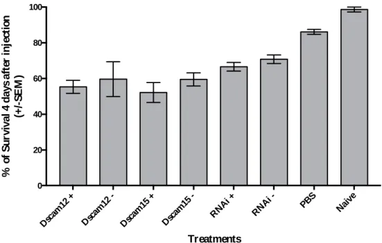
6.5.1 - Larvae survival four days after injections (Preliminary experiment 3.1.4) ... 37
6.5.2 - Survival for each treatment/developmental time-points (Experiment 3.2) ... 37
6.5.3 - Dscam1 knock-down statistics at each developmental time-point ... 38
6.6 Abnormal RTqPCR Rp49 amplification curve ... 39
6.5.1 Can it be a problem with the primers binding during the reverse transcription or the qPCR? ... 39
6.5.2 May the new SYBR green require different protocol conditions? ... 41
List of figures
Figure Page
1.1 Host differentiation between pathogens and symbionts through elicitors ratio. 2
1.2 Schematic representation of genomic and mRNA Dscam1 sequences from T. castaneum and known ocurrences of Dscam1 in arthropods.
5
3.1 Mean 2-∆Ct values from RTqPCR for bacterial 16S rRNA expression across microbiota-enriched flour
and regular flour treatments in pupal and adult stage.
15
3.2 Relative expression of the bacterial 16S rRNA and Dscam1 across a surface sterilization treatment and two control treatments.
15
3.3 Expression of Dscam1 relative to the RNAi control after injection of Dscam1 dsRNA. 17
3.4 Relative expression of the bacterial 16S rRNA across developmental time-points and treatments 19
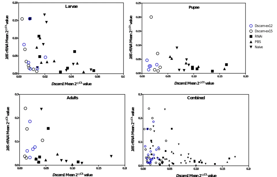
3.5 Relative expression of the bacterial 16S rRNA against relative expression of Dscam1. 21
6.1 Larvae survival four days after injection. 37
6.2 Survival for each treatment at different beetle developmental time-points. 37
6.3 Abnormal RTqPCR amplification curve for Rp49. 40
6.4 Schematic illustration of the different steps during the troubleshooting on the problem with Rp49 amplification curve
40
6.5 Mean rp49 expression of three samples after reverse transcription using random hexamers and oligo(dT) primers.
41
6.6 1.5% TBE agarose gel for 400ng of RNA samples. 43
6.7 Electrophoregram output from 2100 Bioanalyzer for the RNA samples 1,2,3 and a older one, R3. 43
6.8 Electrophoregram output from 2100 Bioanalyzer for the RNA samples extracted with and without the phenol:chloroform:isoamy alcohol step during RNA isolation.
45
6.9 1.5% TBE agarose gel and 2100 Bioanalyzer output for RNA samples extracted with and without the phenol-chloroform based step during RNA isolation.
46
6.10 Rp49 expression for each of the three samples after reverse transcription with different RNA
quantities.
48
6.11 Rp49 expression for each of the three samples after reverse transcription of 250 and 500ng total RNA
and different cDNA dilutions.
List of tables
Table Page
3.1 RTqPCR Ct values at which Rp49, Rpl13a, Dscam1 and bacterial 16S rRNA expression were detected across the three beetle developmental stages.
13
6.1 List of machines and equipment used. 34
6.2 List of kits used. 35
6.3 Primer sequences used in the study. 36
6.4 P-value and knock-down efficiency for each of treatment on each time-point. 38
6.5 Comparison between the different qPCR protocols 41
6.6 RNA concentration of samples extracted with and without the phenol:chloroform:isoamy alcohol step during RNA isolation.
List of abbreviations
AMP Antimicrobial peptide
asnA Asparginase synthetase
DSCAM Down syndrome cell adhesion molecule DSCAM1 Down syndrome cell adhesion molecule form 1
dsRNA Double strand RNA
IMD Immune deficiency pathway
LPS Lipopolysaccharide
MAMP Microbe associated molecular pattern
min Minutes
PCR Polymerase chain reaction
PRR Pathogen recognition receptor
qPCR Quantitative polymerase chain reaction RFLP Restriction fragment length polymorphism
RNAi RNA interference
ROS Reactive oxygen species
Rp49 Ribosomal protein 49 Rpl13a Ribosomal protein L13a
RT Reverse transcription
RTqPCR Real-time quantitative polymerase chain reaction
s Seconds
TGIP Trans-generational immune priming
1. Introduction
1. Introduction
1.1 - Evolution of immunity
Since early in eukaryotic evolution, multicellular organisms have been exposed to a high diversity of microorganisms. The term microbiota encompasses all the mutualistic, commensal and pathogenic microorganisms that live inside or on a host (Lederberg & McCray 2001). These microbes can include protozoa, bacteria, viruses or fungi. Classically, host-microbe interactions are seen as mostly agonistic and a decisive evolutionary driving-force (Woolhouse et al. 2002; Obbard et al. 2009). Pathogens evolved to lucratively parasitize the host, negatively affecting their fitness (Hornef et al. 2002). The host-parasite antagonism may lead to an evolutionary arms race, known as Red Queen hypothesis (Van Valen 1973). Hosts may only survive and reproduce by exhibiting more efficient immune mechanisms, while parasites require novel ways of dodging the hosts' immunity to successfully infect them. It is fair to assume host-parasite coevolution presents a strong selective pressure in the natural environment and has led to the selection of a broad range of responses and in part to the evolution of the immune system (Dybdahl & Lively 1998; Kaufman 2010; Obbard et al. 2009). Nonetheless, most microbes are actually neutral or beneficial to the host (Lee & Mazmanian 2010). Almost every environmentally exposed host surface is bursting with commensal and beneficial microbes but most reside in their gut (Turnbaugh et al. 2007; Sommer & Bäckhed 2013). In
Drosophila, there is evidence for the contribution of commensal bacteria for mate preference (Sharon
et al. 2010). In mice, commensal microbiota found in the ocular surface seem to elicit an interleukin-17 response, central to the tissue immune homeostasis (Leger et al. 20interleukin-17). Nonetheless, in some cases it may be difficult to draw the line between a neutral and positive/negative interaction and its dynamics may allow a neutral interaction to evolve to either a mutualistic or pathogenic scenario (Hooper & Gordon 2001). Regarding mutualistic interactions, they may evolve to the point where the lack/absence of the microbe can lead to the incorrect development of tissues, immunological disequilibrium or even higher host susceptibility to pathogens (Sommer & Bäckhed 2013; Mazmanian et al. 2005; Teixeira et al. 2008; Weiss et al. 2013; Dong et al. 2009). As an example, the bacteria
Vibrio fischeri releases tracheal cytotoxins that act in synergy with lipopolysaccharides (LPS) as a
morphogen, inducing the morphogenesis of the light organ in the squid Euprymna scolopes (Koropatnick et al. 2004). In mice, the gut bacteria Bacteroides fragilis releases a polysaccharide essential to the correct maturation of T cells, as well as the lymphoid organogenesis (Mazmanian et al. 2005). Wolbachia is a genus of wide-spread endosymbionts among arthropods that can have both parasitic and mutualistic interactions. Its presence has been linked to enhanced resistance towards a variety of RNA viruses in Drosophila (Teixeira et al. 2008). Hence it is fair to assume, that symbionts are equally important to the shaping of the immune system (Bosch 2014).
Taking the insects as an example, the gut colonization can be achieved by very distinct paths: host diet, vertical transmission (parental-offspring), cropophagy and in social insects also by horizontal (social) transmission (Engel & Moran 2013). Moreover, the insect guts can be particularly unstable over the lifetime of an organism as a result of the remodelling upon metamorphosis and moulting. In Tribolium castaneum there is evidence for changes in the bacterial phyla proportions across development (Futo 2016). During the remodelling events, the gut microbiota is nearly or completely eliminated (Engel & Moran 2013) and gained later in life (Hammer et al. 2014).
Although, the bacterial microbiome is environmentally acquired, it does not simply reflect the microbial intake of the host (Chandler et al. 2011). Hence, the host exerts significant control over its composition. A study in Hydra has shown that species-specific microbial communities are shaped
1. Introduction
through the expression of species-specific antimicrobial peptides (AMPs) patterns (Fraune & Bosch 2007; Franzenburg et al. 2013). In Drosophila, the immune pathways responsible for this regulation can also be modulated, namely through the repression of AMPs transcription by caudal (Ryu et al. 2008). Host microbial regulation may lead to population and species-specific bacterial phylogenetic clusters. The latter was defined as core microbiota (Turnbaugh et al. 2007) and was found in some populations and species (Dishaw et al. 2014; Brucker & Bordenstein 2012; Ochman et al. 2010; Thongaram et al. 2005). However, the concept is still controversial since there are also conflicting results (Chandler et al. 2011; Corby-Harris et al. 2007; Wong et al. 2013) and the study of natural microbial communities in the laboratory is problematic. Furthermore, the microbiota can be relatively flexible and shaped by external factors, such as host diet, host pathobiology or the environment itself (Turnbaugh et al. 2007; Yun et al. 2014).
Thus, it seems that the host immune system evolved to not only destroy prejudicial microbes, but also to maintain and regulate strong and stable relationships with beneficial ones (Bosch 2014). Both driving-forces are critical to the maintenance of the host's homeostasis but how it evolved to identify each one of the two types of microbes still remains unclear. A possible explanation for the differentiation between harmful and beneficial microbes was recently suggested by Lazzaro and Rolff (2011). According to this hypothesis, a pathogen would release microbe-associated molecular patterns (MAMPs) but also induce the infected tissue to release "danger signals" (Fig. 1.1). On the other hand, a symbiont would release MAMPs but not cause any damage and therefore no "danger signals" would be released from the tissue cells. A natural wound would only induce the production of "danger signals". This way, the balance between different elicitors could indicate what type of response should take place (Lazzaro & Rolff 2011).
Figure 1.1 - Host differentiation between pathogens and symbionts through elicitor ratios. A wound only induces the
production of "danger signals" by the host tissue, while symbionts only display MAMPs without causing damage to the host. Pathogens invasion exhibits both MAMPs and induces "danger signals" production, leading to the activation of an immune response by the host. The balance between the different elicitors may indicate what kind of response should take place. Figure adapted from Lazzaro & Rolff (2011).
1.2 - Insect immunity
Insects have evolved different lines of defences that interact in synergy to protect the organism: i) behavioural mechanisms, such as pathogens avoidance; ii) physical barriers, for instance the chitin exoskeleton; and lastly, iii) a wide range of immunological responses (Siva-Jothy et al. 2005).
Once a parasite has breached the insect outer barriers, such as the cuticle, the immune system can be activated. Host immune system activation is dependent on pathogen recognition. Recognition is
1. Introduction
based on MAMPs not found in the host, allowing a distinction between host cells and different classes of microorganisms (Lazzaro & Rolff 2011; Medzhitov & Janeway 1997). MAMPs may include lipopolysaccharides and peptidoglycans. After pattern recognition through host pattern recognition receptors (PRRs), an appropriate response can be initiated (Schmid-Hempel 2005).
Invertebrates lack the adaptive branch of the immune system, as well as all their elements. Consequently they rely on a wide range of innate immune responses (Schmid-Hempel 2005). These immune responses can be categorized into cellular or humoral. Cellular immunity is based on haemocytes, the phagocytic cells of invertebrates. After pathogen recognition, phagocytosis takes place. Depending on the pathogen features, such as size, different strategies may be applied. In the presence of small pathogens, multiple haemocytes can attack the intruder, forming nodules, called nodulation. In the presence of larger pathogens, the haemocytes will form a capsule around it, called encapsulation. Both nodules and capsules go through a process of melanisation, where the pathogen is enclosed with melanin and destroyed with reactive oxygen species (ROS) (Lavine & Strand 2002; Marmaras & Lampropoulou 2009). ROS are one of the main insect humoral responses. They are a by-product during the activation of the melanisation cascade in the haemocytes (Cerenius & Söderhäll 2004). Another crucial mechanism of humoral immunity is the production of AMPs. These are found throughout the tree of life (Zasloff 2002), as a fundamental innate defence mechanism. These small molecules interfere with the metabolism of various bacterial pathogens in processes such as nucleic acid synthesis or enzyme activity (Mylonakis et al. 2016). In insects, AMPs are produced in the fat body at each infection and secreted into the haemolymph. Their activity remains for several days after the infection, protecting the organism from a re-infection by the pathogen (Makarova et al. 2016). In
Drosophila, different classes of bacteria are thought to activate different pathways through PRRs
binding (Lemaitre & Hoffmann 2007). Toll, Immune deficiency (IMD) and JAK/STAT pathways are known to differentially regulate AMP synthesis (Hillyer 2016). This results in the expression of an AMP pattern more effective against the class of pathogens currently infecting the host. However, not all insects express AMPs so distinctly. T. castaneum seems to have a more promiscuous AMP activation, through the usage of both Toll and IMD pathways (Yokoi et al. 2012).
Since invertebrates lack the adaptive machinery, it was thought that invertebrates could only rely on the generalized immune responses mentioned above (Schmid-Hempel 2005). However, a study (Kurtz & Franz 2003) has shown that in fact invertebrates also possess a form of immune memory, later called immune priming (Little & Kraaijeveld 2004). In this study, copepods Macrocyclops
albidus were infected with the tapeworm Schistocephalus solidus. Two treatment groups took place.
Both groups were first primed with the parasite and 2 days later one of the group was infected by a sibling S. solidus (genetically similar to the priming parasite), while the other group was infected by an unrelated S. solidus (genetically non-similar). There was a reduction in the reinfection success and intensity for the first group, compared to the latter. Hence, the immune system of the copepods was able to respond more efficiently after being presented for a second time with a sibling parasite (Kurtz & Franz 2003), which indicates that a form of specific memory exists. Immune priming has been defined as an enhanced protection resulting from past experience with a pathogen (Kurtz 2005). Evidence has been found suggesting this phenomenon may happen not only in invertebrates but also in vertebrates and plants (Paust & Von Andrian 2011; Spoel & Dong 2012). Invertebrate immune priming has now been demonstrated in a wide range of species (Contreras‐Garduño et al. 2016; Milutinović et al. 2016). This phenomenon can occur not only within a generation but also across generations leading to stronger immune reactions or improved resistance of the offspring, which is then called trans-generational immune priming (TGIP) (Zanchi et al. 2011; Eggert et al. 2014; Roth et al. 2010; Trauer-Kizilelma & Hilker 2015). In T. castaneum it was shown that oral priming previous to infection with Bacillus thurigiensis leads to a shift in the gene expression (Greenwood et al. 2017),
1. Introduction
primed individual. The gene expression is also dependent on the infection route taken by the pathogen and the host population (Behrens et al. 2014).
It has been proposed that microbiota may also have a role in oral immune priming. Rodrigues
et al. (2010) observed that primed Anopheles gambiae mosquitoes subjected to antibiotics treatment
had an increase in Plasmodium parasites, as well as lower phagocytic activity, compared to the treatment without antibiotics (Rodrigues et al. 2010). In addition, Futo and colleagues (2015) observed that T. castaneum larvae with lowered microbial loads (after sterilization) showed decreased survival upon a secondary challenge with Bacillus thurigiensis compared to non-sterilized larvae. Furthermore, if the larvae were allowed to re-colonise themselves with gut microbiota after sterilisation and before priming-challenge, the survival after challenge was the same as the group where the microbiota were not manipulated (Futo et al. 2015). Both results suggest that host-associated microbiota is crucial to immune priming and may boost the immune response. Also, a study in bumblebees (Koch & Schmid‐ Hempel 2012) showed that different gut microbiota composition may lead to specific resistance against different pathogen strains, reinforcing the importance of microbiota in relation to immunity.
1.3 Dscam1: Down syndrome cell adhesion molecule 1
DSCAM was originally found in humans as a new class of neural cell adhesion molecules expressed within the nervous system (Yamakawa et al. 1998). Given its location on chromosome 21 and specific expression, it is thought to contribute to the neuroanatomic abnormalities found in Down syndrome (Yamakawa et al. 1998; Delabar et al. 1992).
A Drosophila melanogaster homolog of human DSCAM was later isolated and named Dscam (Schmucker et al. 2000). Both transmembrane proteins are members of the Immunoglobulin superfamily, one of the largest class of surface receptor proteins found in animals (Schmucker & Chen 2009). There are four Dscam paralogs in Drosophila (form 1-4). However, only Dscam1 possess a high diversity of isoforms originated through alternative splicing of the high variably cluster of exons (exons 4, 6, 9 and 17) (Schmucker et al. 2000; Hattori et al. 2008). Dscam1 structure consists of ten immunoglobulin-like domains, six type III fibronectin domains, a single transmembrane segment and a cytoplasmic domain. In D. melanogaster it is predicted to encode 18,612 distinct axon guidance receptors through alternative splicing of 95 exons (Schmucker et al. 2000). Although, the domain structure of the vertebrate DSCAM and Dscam1 is quite similar, the latter presents a bigger sequence diversity in their extracellular region (Schmucker et al. 2000). The alternative exons that encode for hypervariability seem to be present in insects and crustaceans but not in vertebrates (Brites et al. 2013). Up to now, this hypervariable form has been found in numerous pancrustaceans, including T.
castaneum (Ng et al. 2014; Watson et al. 2005). However, the number of alternatively spliced exons
varies throughout the pancrustaceans (Armitage & Brites 2016). A phylogenetic analysis of Dscam hypothesised that this hypervariable form may have appeared in the common ancestor of Pancrustacea (Armitage et al. 2012; Armitage & Brites 2016) (Figure 1.2C).
Dscam1 is expressed throughout sensory and central nervous system neurons (Fuerst et al. 2009; Schmucker & Chen 2009). Its first discovered role was to mediate the sister-dendrites repulsion in neuronal wiring (Schmucker et al. 2000; Lawrence Zipursky & Grueber 2013). During neuronal wiring, specific cells express Netrin-B as a guidance cue perceived by the netrin-receptor Frazzled in the neurons' membrane (Hiramoto et al. 2000). At the same time, unique patterns of Dscam1 isoforms are also expressed in the cell membranes of neurons. This patterning allows the neuron to discriminate between its own neurites and neurites from another neuron (Lawrence Zipursky & Grueber 2013). Dscam1 in synergy with the Netrin/Frazzled system assures the spreading of the dendrites throughout
1. Introduction
the neuronal field, maximizing territory coverage (Lawrence Zipursky & Grueber 2013; Hiramoto et al. 2000; Matthews & Grueber 2011). In absence of Dscam1, the dendrites tend to "hypertarget" the source of Netrin and not establish a correct neural circuit (Matthews & Grueber 2011). Homophilic binding of the extracellular region has been shown to result in the repulsion of neurons, whereas heterophilic binding in the nervous system leads to tiling of two neurons (Hughes et al. 2007). In
Drosophila, when the ectodomain diversity was eliminated, defects during neural wiring arose (Hattori
et al. 2007). Meijers and colleagues analysed the protein structure of Dscam at Drosophila interspecies level and noticed that the epitope I, involved in homophilic interactions in the nervous system, presents a highly conserved amino acid sequence. This observation is consistent with a high selective pressure to a limited set of isoforms for neural wiring. On the other hand, the epitope II is predicted to be involved in heterophilic bindings and possesses a higher sequence diversity, consistent with a secondary function as PRR (Meijers et al. 2007).
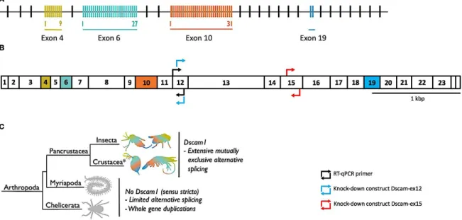
Figure 1.2 - Schematic representation of genomic and mRNA Dscam1 sequences from T. castaneum and known occurrences of Dscam1 in arthropods. (A) illustrates T. castaneum Dscam1 gene (accession number: TC012539), while (B)
represents its mRNA with the corresponding exon numbers. Alternatively spliced regions are coloured. Black arrows indicate the position of the RTqPCR primers for expression analysis. Blue (Dscam-ex12) and red (Dscam-ex15) arrows indicate the position of knock-down constructs for RNAi mediated depletion of Dscam1 in T. castaneum. (C) Until now, Dscam1 has only been found in pancrustaceans. Myriapods and chelicerates have diversified the Dscam gene family through other routes. *Crustacea is considered a paraphyletic group containing the hexapods; phylogeny follows (Legg et al. 2013). Scale indicates 1kbp. Figure adapted from Armitage et al. (2017).
1.3.1 Dscam1 in immunity
More recently, Dscam1 mRNA was found in haemocytes and fat bodies, immune cells of pancrustaceans (Neves et al. 2004; Watson et al. 2005), as well as the secreted tail-less Dscam1 protein in the haemolymph (Watson et al. 2005). Through microarrays it was estimated that these immune cells can produce up to ~ 18,000 receptor isoforms of Dscam1 in Drosophila. The splice variants found in haemocytes and fat bodies differ from the ones found in the brain (Watson et al. 2005). In T. castaneum, 69 variable exons (Figure 1.2A) can give rise to 15,066 different Dscam1 isoforms (Armitage & Brites 2016) . It was proposed that due to their hypervariability, they could have a role as PRR and therefore in immunity (Boehm 2007; Watson et al. 2005).
1. Introduction
In the past decade, several studies have been showing a relation between Dscam1 and immunity. For instance, flies with Dscam1 knock-down haemocytes are unable to efficiently phagocytose bacteria compared to the control flies (Watson et al. 2005). In crayfish, Dscam1 was found to be induced in haemocytes upon contact to MAMPs like lipopolysaccharides and b-1, 3-glucans (Li et al. 2015). It was reported that different Dscam1 isoforms bind to a different degree to
Escherichia coli, suggesting a possible role as specific opsonin (Watson et al. 2005). Furthermore,
infection of A. gambiae individuals with different parasites induced the expression of different Dscam1 splice isoforms (Dong et al. 2006). Recently Fu and colleagues have shown (Fu et al. 2014) in primed Litopenaeus vannamei that a knock-down of Dscam1 leads to a decrease in the phagocytosis of the white spot syndrome virus (WSSV) upon secondary infection. Consequently, the groups treated with Dscam1-siRNA injections showed a lower survival compared to the control-siRNA groups. Although, this last study concerns virus infection, it is the first study showing a direct effect of Dscam1 in immune priming (Armitage et al. 2015). In contrast, a study with the shrimp Artemia
fransciscana did not observe any significant difference in Dscam1 expression during TGIP, either
within the generation challenged with Vibrio campbellii or its offspring (Norouzitallab et al. 2016). Studies (Brites et al. 2011; Chávez‐Galarza et al. 2013) have shown that Dscam1 may be under balancing selection. It is known that genes involved in pathogen recognition, such as the major histocompatibility complex, tend to show long-term balancing selection (Takahata et al. 1992; Ferrer-Admetlla et al. 2008).
Dscam1 intracellular diversity also originates from exon inclusion/exclusion and type III polyadenylation (Chou et al. 2011; Yu et al. 2009). These may result in different protein products being produced (Lutz & Moreira 2011). Therefore, the same locus may be able to generate both tail-less and membrane-bound forms (Chou et al. 2011). Different combinations of Dscam1 transmembrane and cytoplasmic domains variants may explain how the receptor is able to be involved in different systems and in response to specific stimulus activate distinct signal transduction pathways inside each one of them (Yu et al. 2009).
In shrimp, upon infection with WSSV there is an increase in both tail-less and transmembrane Dscam1 (Chiang et al. 2013). Moreover, when facing a natural pathogen as the bacterium Vibrio
harveyi, Dscam1 isoforms show a more specific and stronger pattern response. In the latter, there was a particular up-regulation of alternative tail-less variants that contain an immune-related endocytosis/phagocytosis motif (Hung et al. 2013). Parallel to this, there is an up-regulation of the splicing activator B52 and down-regulation of the splicing repressor Hrp36. This expression changes may lead to the increased production of membrane-bound and tail-less Dscam found in shrimp after WSSV infection (Chiang et al. 2013). In addition, IMD and Toll pathway are thought to mediate species-specific defences in A. gambiae infected with Plasmodium and bacteria. The latter is possible through the transcriptional regulation of the splicing factors Caper and IRSF1 that leads to the production of pathogen-specific splice variants of Dscam1 (Dong et al. 2012).
Regarding the secreted form of Dscam1, it was first hypothesized to be produced through proteolytic cleavage of the membrane-bound Dscam1 (Watson et al. 2005). In shrimp, several alternative stop codons were found inside the Dscam1 gene (Ng et al. 2014) reinforcing the hypothesis that tail-less Dscam may be produced directly through type III polyadenylation (Chou et al. 2009). Its hypothesized role may be also similar to adaptive immunity elements. Soluble Dscam1 binds to
Escherichia coli (Watson et al. 2005) and was found in the D. melanogaster phagosome proteome
(Stuart et al. 2007). Additionally, its epitope configuration hypothetically allows a Dscam1 isoform to form a homodimer that through the hypervariable epitope II may be capable of recognition and opsonisation of pathogens (Meijers et al. 2007; Stuart & Ezekowitz 2008). Studies have already shown specificity on the isoform production upon pathogen infection (Dong et al. 2006; Hung et al. 2013; Chiang et al. 2013).
1. Introduction
Over the last years, some studies have been positioning Dscam1 as an important element in bacteria regulation. In 2006, Dong and colleagues (Dong et al. 2006) showed in A. gambiae that after
Dscam1 knock-down, there was a reduction in the phagocytosis index and the bacteria in the
haemolymph proliferated. Through sequence analyses of the bacterial 16S rRNA, they were able to relate them with three Gram-negative bacteria species. In addition, an increased number of
Plasmodium oocysts on the midgut were found. Six years later, the same group overexpressed
different Dscam1 forms naturally induced by Plasmodium and bacteria. Curiously, they observed that the bacteria-induced form (exon 4.14) led to a reduction in the amount of midgut bacteria in comparison to the control and Plasmodium-induced forms of Dscam1 (Dong et al. 2012). It is known that Dscam1 is expressed in haemocytes (Watson et al. 2005), cells capable of phagocytosis. Phagocytosis should be dependent on MAMPs recognition by PRRs. If in fact Dscam1 is responsible for a large PRR repertoire, then a knock-down may preclude the identification and therefore the regulation of the microbiota.
In contrast, Zhang (Zhang et al. 2016) has shown opposite results for the influence of Dscam1 on extracellular bacteria. In the latter, Laodelphax striatellus with a knock-down of Dscam1 showed a decrease in the infection of both rice strip virus and Wolbachia compared to the control. However, no change was observed in the number of extracellular bacteria (Zhang et al. 2016). It is possible that Dscam1 may have an intracellular role and its knock-down may have disrupted the intracellular environment, making it less suitable for intracellular parasites.
In short, invertebrates' complex microbial communities appear to be interconnected with the host's immunity and occasionally even operate as an extension of it (Sommer & Bäckhed 2013). A rich microbiota seems to be decisive not only for basal immunity but also for specific mechanisms, such as immune priming (Futo et al. 2015). Dscam1 may play a crucial role in pancrustacean immunity as a PRR, opsonin and regulator of bacterial microbiota, contributing to the host homeostasis (Boehm 2007; Watson et al. 2005; Dong et al. 2006).
1.4 Aim of the thesis
The overall aim of this thesis is to understand the role of Dscam1 in the whole-body bacterial microbiota of T. castaneum. It has been suggested that Dscam1 may have a role in pancrustacean immune recognition (Boehm 2007) and might be essential to the regulation of bacterial microbiota (Dong et al. 2006). However, there is contradictory evidence for the latter function (Dong et al. 2006; Dong et al. 2012; Zhang et al. 2016). Therefore, I aim to clarify if Dscam1 regulates bacterial microbiota in T. castaneum by knocking down Dscam1 and quantify the total amount of bacteria throughout T. castaneum development.
2. Materials and Methods
2. Materials and Methods
2.1 Model organism: Tribolium castaneum
Insects are the most diverse group of animals on Earth and represent more than half of the living species known to Mankind. Insects strong interactions with microbes allow the dissection of immune reactions that are easily overlooked in vertebrate systems (Chambers & Schneider 2012) and may influence the immune outcome of infections and diseases in higher vertebrates as the human (Mukherjee et al. 2015).
Coleoptera is the most evolutionary successful order of animals, covering the majority of the habitats (Hunt et al. 2007). Tribolium castaneum (Herbst 1797), a member of Coleoptera, is a severe agricultural pest that has shown resistance to all classes of insecticides used against it (Richards et al. 2008). It is an important model organism in ecology and evolution and has allowed the development of a vast range of concepts, such as the competition theory (Park 1962). This beetle presents advantages such ease of culture, short generation time, high fecundity and the availability of several genetic tools, such as genetic markers (Demuth et al. 2007). As in Caenorhabditis elegans, RNA interference (RNAi) is systemic in Tribolium, allowing knock-down of specific gene products in any developmental point (Tomoyasu & Denell 2004). Moreover, its genome was recently sequenced facilitating genetic studies with the organism (Richards et al. 2008). Additionally, T. castaneum possess a hypervariable Dscam1 capable of producing 15.066 different isoforms (Watson et al. 2005). This organism also exhibits a dynamic microbiota across development. Actinobacteria, Bacteroidetes, Firmicutes and Proteobacteria are the predominant phyla found in this organism and their proportion changes across developmental stages (Futo 2016).
T. castaneum Cro 1 population established from 165 beetle pairs wild collected in Croatia in
2010 (Milutinović et al. 2013), was used in the following experiments. As a food source and substrate, organic wheat flour with 5% brewer’s yeast was used, which prior to use was frozen at -20°C and then heated up to room temperature (~20°C). The population was kept under controlled conditions: 30ºC, 70% humidity and on a 12-hour light-dark cycle (Bucher 2009).
2.2 Time-point experiment hypothesis and preliminary tests
Studies have shown that contradictory results for Dscam1 role as bacteria regulator in the gut and haemolymph (Dong et al. 2006; Dong et al. 2012; Zhang et al. 2016). Therefore, the aim of the main experiment was to clarify Dscam1 role in T. castaneum microbiota across its development, through Dscam1 knock-down. The bacterial 16S rRNA was used as a proxy for the total amount of whole-body bacteria in the beetle. Three developmental time-points were analyzed: 15-days old larvae, 23-days-old pupae and 30-days old adults. Prior to the start of this experiment, several questions had to be answered to assure the methodology was optimized. The several points are described below.
2. Materials and Methods
2.2.1 Is it possible to detect expression for all genes in RTqPCR using Power
Microbiome™ RNA isolation kit?
A previous study from Futo et al. (2015) used the Power Microbiome RNA isolation kit (MO BIO Laboratories, Inc.) for RNA extraction, since it performed better in the bacterial 16S rRNA extraction from the beetle. However, earlier tests shown Dscam1 may present low expression. Therefore, it was important to test if we could get a reliable signal from all genes in study using this particular RNA extraction kit. In this study were used as reference gene for the amount of beetle RNA, the ribosomal protein 49 (Rp49) and the ribosomal protein L13a (Rpl13a). As target genes were used the bacterial 16S rRNA and Dscam1. Rp49 and Rpl13a are used as reference genes for the amount of beetle RNA in the experiment.
For this, we analysed the following three beetle developmental stages: ~15-days old individuals (larval stage), ~25 days-old individuals (pupal stage) and ~35 days-old individuals (adult stage). Each developmental stage was composed by three replicates of 10 pooled individuals. The animals were separated through the use of 280μm and 710μm sieve and their age was estimated by separation through the sieves and size of the individuals. Directly after the separation, they were frozen in liquid nitrogen and stored at -80ºC. The RNA was then extracted with the Power Microbiome™ RNA isolation kit (MO BIO Laboratories, Inc.) under sterile conditions. RNA concentration and absorbance ratios were measured with Qubit® and Implen Nanophotometer®. A reverse transcription of 500ng of RNA was performed with Superscript III kit (Invitrogen™) to 20μl of final volume of cDNA. Afterwards, a RTqPCR for all four genes at the different developmental stages was performed on a 96-well plate using the following protocol: pre-incubation at 95ºC for 3 minutes followed by 40 cycles of 10 seconds at 95ºC, 20s at 58ºC and 2s at 72ºC. The fluorescence acquisition was performed in each cycle at 72ºC. In order to confirm the identity of the PCR products, a melting curve was derived using the temperature range between 95 and58ºC, as in Futo et al. (2015). A control with water instead of cDNA was used as a control for the sterility of the RTqPCR solutions. Detailed protocols and list of primers used can be found in the section 6.1 and 6.4 of the appendices, respectively. Primers position in the mRNA are illustrated in Figure 1.2B.
2.2.2 Does microbiota-enriched food increase the amount of bacteria detectable via
RTqPCR?
We hypothesized that if beetles were raised on flour enriched with microorganisms that they would develop an increased bacterial load, which might be beneficial in the case that Dscam1 knock-down reduces the bacterial microbiota. To test this hypothesis, two different media were prepared: i) microbiota-enriched-media, where 100 adults and 100 larvae were placed in 100g of flour with 5% yeast for 3 days before the oviposition, adapted from Futo et al. (2015); and ii) regular-media, only flour with 5% yeast without any exposure to larvae or adults. A ~2000 individuals’ subpopulation of approximately 1-month old adults were then allowed to oviposit in the two flours types for 24 hours. On the eleventh day after oviposition larvae were transferred to a 96-well plate to simulate the main experiment conditions. Three different stages were analysed: larvae (15-days old), pupae (23-days old) and adults (30-days old). For each combinatorial treatment (developmental stage x media), 3 biological replicates of 10 pooled individuals were considered. RNA extraction, reverse transcription and RTqPCR for 16S rRNA and rp49 was performed as the previous experiment. The larval samples did not contain 500ng of RNA, so these samples reverse transcription was performed with only 100ng of RNA.
2. Materials and Methods
2.2.3 Does individuals’ surface sterilization affect levels of bacterial 16S rRNA detected
via RTqPCR?
Previous analysis of the bacterial 16S rRNA expression were performed without any type of surface sterilization of the individual before RNA extraction. Therefore, it was unknown to us if we were mostly detecting external or internal bacterial 16S rRNA expression. Considering we are particularly interested in the gut and haemolymph microbiota because it is the bacteria that presumably would be affected by Dscam1 knock-down, it was important to test if the surface microbiota was large enough to camouflage any Dscam1 knock-down effect on internal microbiota. To access this question, we performed larvae sterilization in order to see if there is any reduction in the expression levels of
16S rRNA and Dscam1. If bacteria on the external insect cuticle represents a significant proportion of
the total bacteria, we predict that we would see a weaker 16S rRNA signal after surface sterilization compared to the controls.
In this study, one sterilization treatment and two control treatments took place. The sterilization treatment was submitted to the following methodology, adapted from Futo et al. (2015). Larvae (~15-days old) were separated from the flour with a 710μm sieve and subjected to 20 minutes on ice for anaesthetic purposes. After this, three replicates of 10 pooled larvae were positioned between two nets and submerged for 20 seconds in each of the following solutions: Ethanol 70%, MilliQ-water, sodium hypoclorite (NaClO) at 2% and two final washes in MilliQ-water. This process was performed separately for each sample group of 10 pooled larvae and solutions were changed between each one. Directly afterwards, they were frozen in liquid nitrogen and stored at -80°C until the next day, when the RNA extraction took place for all the treatments. Two controls treatments took place: Control and Ice. The Control treatment used the same methodology as the rest of the above-described experiments, i.e., the larvae were directly frozen, while larvae in the Ice treatment were placed for 20 minutes on ice before being frozen in liquid nitrogen. The latter was used to see if there was any effect from the anaesthesia on ice on gene expression. RNA extraction, reverse transcription and RTqPCR for bacterial 16S rRNA, Dscam1 and rp49 was performed as above.
2.2.4 Do both dsRNA constructs result in a successful Dscam1 knock-down on 11-days
old larvae?
In order to proceed to the main experiment, it was important to assure we could get an effective
Dscam1 knock-down in 11-days old larvae. For this, two different dsRNA constructs were used to
knock-down Dscam1. Exon 12 and 15 regions were selected for RNAi mediated knock-down because both are non-alternatively spliced regions within the T. castaneum Dscam1 gene (Peuß et al. 2016) (Figure 1.2B). A region of the Escherichia coli gene aspargine synthetase (Nakamura et al. 1981) (asnA) that is not found in T. castaneum, was used as a control for the dsRNA machinery. This control was named RNAi treatment. Injections with phosphate-buffered saline (PBS) were performed in one of the treatments as a control for the injection itself. A last group of animals that were not subjected to any type of injection was added as control in the experiment and was named naive treatment. The dsRNA injections were performed on 11-days old larvae. Two different dsRNA concentrations were tested: ~2600 ng/μl and ~1700 ng/μl.
Thus, eight treatments took place:
1) Dscam-ex12+, knock-down for Dscam1 exon 12 at the concentration of ~2600ng/μl; 2) Dscam-ex12-, knock-down for Dscam1 exon 12 at the concentration of ~1700ng/μl; 3) Dscam-ex15+, knock-down for Dscam1 exon 15 at the concentration of ~2600ng/μl;
2. Materials and Methods
4) Dscam-ex15-, knock-down for Dscam1 exon 15 at the concentration of ~1700ng/μl; 5) RNAi+, asnA at the concentration of ~2600ng/μl;
6) RNAi-, asnA at the concentration of ~1700ng/μl; 7) PBS;
8) Naive.
For each treatment, whole-body samples from 3 biological replicates of 10 pooled individuals were considered. Injections took place between the 1st and 2nd integument with a glass capillary and without the usage of carbon dioxide or any other chemical for anesthetic purposes. Directly afterwards, they were individualized in a 96-well plate with flour and 5% yeast, posteriorly taped and ~8 holes per well were made to allow for air exchange. Each 96-well plate had an equal number of individuals from each treatment randomly distributed through the plate. The order of the injections was also performed randomly. Injections were performed in 3 blocks in consecutive days.
The plates were then incubated at 30ºC, 70% humidity and on a 12-hour light-dark cycle for four days. After the four days, survival for each group was quantified and the larvae were frozen in liquid nitrogen and stored at -80ºC. The RNA extraction, reverse transcription and RTqPCR for all four genes (rp49, rpl13a, Dscam1 and bacterial 16S rRNA) was performed as in the previous experiments. A 1.5% TBE agarose gels were performed to assess dsRNA and RNA quality prior to injections and prior to reverse transcription, respectively.
2.3 Effect of Dscam1 knock-down on Tribolium castaneum microbiota dynamics
As described before, the intention of this experiment is to clarify the role of Dscam1 in the microbiota dynamics of T. castaneum, through the knock-down of Dscam1. Hence, three developmental stages were analysed: Larval (15-days old); pupal (23-days old); and adult stage (30-days old). Five injection treatments took place:
1) Dscam-ex12, knock-down for Dscam1 exon 12; 2) Dscam-ex15, knock-down for Dscam1 exon 15;
3) RNAi, control for dsRNA machinery activation using asnA gene; 4) PBS, control for the injection itself;
5) Naive, individuals not subjected to any kind of injection.
The concentration of dsRNA used was ~1700ng/μl. For each combinatorial treatment (developmental stage x injection treatment) 5 replicates of 10 pooled individuals were considered. Injections were performed in 5 blocks in consecutive days. Both injections and individualization in the 96-well plate were performed as in the preliminary test on the Dscam1 knock-down.
The plates were then incubated at 30ºC, 70% humidity and on a 12-hour light-dark cycle for four days. A block of injections was randomly removed from the incubator 4, 12 and 19 days after the injections for larval, pupal and adult stage analysis. For pupal and adult samples, each replicate had 5 females and 5 males. RNA extraction, reverse transcription and RTqPCR for bacterial 16S rRNA,
Dscam1, rp49 and rpl13a was performed as above. Each RTqPCR plate had a common sample
obtained from a unique RT, working as a control for variation between plates. A 1.5% TBE agarose gels were performed to assess dsRNA and RNA quality prior to injections and prior to reverse transcription, respectively.
2. Materials and Methods
2.4 Data analysis and software
2.4.1 RTqPCR data
The data obtained from the RTqPCR assays was analysed through the Ct comparative method (Schmittgen & Livak 2008). In this project, the Ct value was defined as the crossing point qPCR cycle. Absolute gene expression was presented as 2-Ct, whereas relative gene expression was given as below:
𝑅𝑒𝑙𝑎𝑡𝑖𝑣𝑒 𝑒𝑥𝑝𝑟𝑒𝑠𝑠𝑖𝑜𝑛 = 2−∆𝐶𝑡 = 2−(𝐶𝑡 𝑔𝑒𝑛𝑒 𝑜𝑓 𝑖𝑛𝑡𝑒𝑟𝑒𝑠𝑡 − 𝐶𝑡 𝑟𝑒𝑓𝑒𝑟𝑒𝑛𝑐𝑒 𝑔𝑒𝑛𝑒)
A geometric mean of the different replicates 2-C or 2-ΔCt was calculated for each treatment, as well as the correspondent standard error. Hence, a higher 2-Ct corresponds to a higher expression from the gene in analysis, while a higher 2-ΔCt correlates with a higher expression of the gene of the interest comparatively to the reference gene(s).
The fold change in expression due to the Dscam1 knock-downs was calculated as below: 𝐹𝑜𝑙𝑑 𝑐ℎ𝑎𝑛𝑔𝑒 = 2−∆∆𝐶𝑡
= 2−[(𝐶𝑡 𝑔𝑒𝑛𝑒 𝑜𝑓 𝑖𝑛𝑡𝑒𝑟𝑒𝑠𝑡 − 𝐶𝑡 𝑟𝑒𝑓𝑒𝑟𝑒𝑛𝑐𝑒 𝑔𝑒𝑛𝑒)𝑇𝑟𝑒𝑎𝑡𝑒𝑑−(𝐶𝑡 𝑔𝑒𝑛𝑒 𝑜𝑓 𝑖𝑛𝑡𝑒𝑟𝑒𝑠𝑡 − 𝐶𝑡 𝑟𝑒𝑓𝑒𝑟𝑒𝑛𝑐𝑒 𝑔𝑒𝑛𝑒)𝐶𝑜𝑛𝑡𝑟𝑜𝑙
The knock-down treated groups were compared to the RNAi control group. Since, the fold change due to the knock-downs represents a reduction in the gene expression, the inverse of the fold change was calculated in the sections below.
2.4.2 Statistical analysis
The expression of the reference genes rp49 and rpl13a was used to normalise the expression of the target genes Dscam1 and bacterial 16S rRNA. The several statistical tests/models were performed on the 2-Ct /2-ΔCt expression values using the software RStudio (Version 1.0.143) (Team 2015).
Additionally, the data obtained from RTqPCR for comparisons between treated and untreated samples was analysed with REST2009 Software (Qiagen GmbH). This software calculations are based on the model by Pfaffl (Pfaffl et al. 2002). The program determines whether there is a significant difference in the expression of target genes comparatively to the expression of reference genes between treated and untreated samples. The program takes in account the different reaction efficiencies of both reference and target genes.
3. Results and Discussion
3. Results and Discussion
3.1 Preliminary tests
3.1.1 Detection of gene expression through RTqPCR
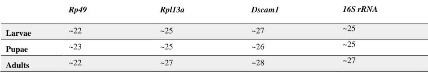
The aim of this preliminary test was to confirm that we could get a reliable signal from all the four genes used in the main experiment when using RNA extracted with the Power Microbiome™ RNA isolation kit. The Ct values for each of the four genes, is described in the Table 3.1. In general, the reference genes Rp49 and Rpl13a were detectable at earlier cycle numbers, which is expected from genes coding for widely-expressed ribosomal proteins (Cardoso et al. 2014; Scharlaken et al. 2008). On the other hand, Dscam1 presents a later cycle number signal, possibly due to its limited and specific expression in certain beetle tissues (Watson et al. 2005; Neves et al. 2004). Lastly, the bacterial 16S rRNA presents a rather early average signal for a bacterial gene. The RNA isolation kit used is optimized to extract bacterial RNA (Futo et al. 2015) and that could explain its early cycle number when compared to beetle genes. Since we obtained reliable signals from all the genes, we continued the experiments using this RNA isolation kit.
Table 3.1 – RTqPCR Ct values at which Rp49, Rpl13a, Dscam1 and bacterial 16S rRNA expression were detected across the three beetle developmental stages. Each sample is composed of 10 pooled individuals.
3.1.2 Does microbiota-enriched food increase the amount of bacteria detectable via
RTqPCR?
Dscam1 may have a positive effect (Zhang et al. 2016) on symbionts. In this case, a
knock-down of Dscam1 would presumably lead to their reduction. A strong reduction in the whole-body bacteria might not be observed in the RTqPCR. Therefore, it was important to test if it would be possible to increase the beetle bacterial load detectable in the RTqPCR in case there is a strong reduction in the bacteria after Dscam1 knock-down in the main experiment. For this, individuals grown in regular flour were compared to individuals grown in a microbiota-enriched flour and their bacterial 16S rRNA expression was analysed for differences in three developmental stages: larval, pupal and adult stage.
In this experiment, we did not observe any significant difference in Rp49 expression across treatments (Kruskal-Wallis: p-value = 0.2752). Since the qPCR signal for larval samples only appeared after the cycle 35, they were removed from the analysis. This result may be due to the lack of RNA in these samples for cDNA reverse-transcription and consequently RTqPCR. Concerning the adult and pupal samples, no significant difference was observed in the bacterial 16S rRNA relative expression between individuals grown on regular flour and grown on microbiota-enriched flour
Rp49 Rpl13a Dscam1 16S rRNA
Larvae ~22 ~25 ~27 ~25
Pupae ~23 ~25 ~26 ~25
3. Results and Discussion
(REST, pupae: p-value=0.643; adults: p-value=0.226) (Figure 3.1). These results suggest there is no effect of the microbiota-enriched media on the bacterial load of the individuals in the pupal and adult stage. It is possible that the individuals in the regular flour already possess a large bacterial load and an increase is not possible even if there are more microorganisms available in the media.
Nonetheless, we cannot discard other hypotheses. It is possible that the microbiota-enriched media does not possess more microorganisms than the regular flour. Prior to the oviposition in these media, larvae and adults were left living on the flour. These individuals may have consumed a significant part of the flour resources and their microorganisms. Therefore, in the end there might be no difference between the two types of flour. To test this hypothesis, we would have to quantify the bacteria in both flours. An optimum experimental scenario would be to add directly the microorganisms to the flour and then compare the individuals that lived on either one of them. However, this was not possible to perform and we do not know if that would affect the results in the main experiment.
Hence, taking the results obtained from the pupal and adult stage, the hypothesis that a microbiota-enriched flour could increase the bacterial load observed in a RTqPCR was refuted. Therefore, we proceeded to the main experiment using regular flour.
3.1.3 Does surface sterilization affect levels of bacterial 16S rRNA detected via
RTqPCR?
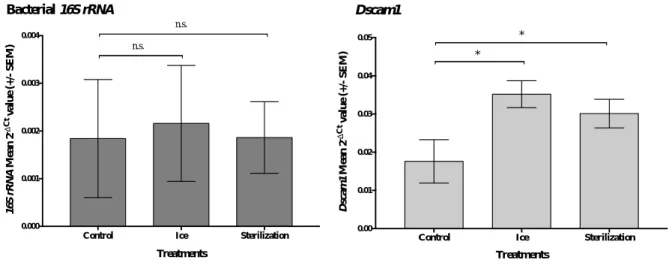
The aim of this experiment was to test if the bacteria in the cuticle that is not affected by the knock-down of Dscam1 would camouflage any effect caused by the knock-down on the internal microbiota. For this purpose, a Sterilization treatment and two controls took place. While the individuals of the Sterilization treatment were anesthetized on ice and posteriorly sterilized, the individuals of the Ice treatment were only subjected to the anaesthesia on ice. The Control treatment was not treated at all.
Regarding this experiment, there is no significant difference in the reference gene rp49 across treatments (Kruskall- Wallis: p-value = 0.1479). For each biological replicate, Dscam1 and 16S rRNA relative expression to the reference gene rp49 was calculated and plotted (Figure 3.2). Both Ice and Sterilization treatment were compared to the Control treatment through the use of REST software.
The relative expression of the bacterial 16S rRNA in Ice and Sterilization treatment did not differ from the Control treatment (REST, Ice: p-value = 0.508; Sterilization: p-value = 0.666). At least two hypotheses can explain this result. The first is that the surface sterilization methodology used may not have worked. In this scenario, 16S rRNA expression would naturally not differ between treatments. Although we cannot discard the possibility that this type of sterilization may not work on T.
castaneum larval cuticle, given the extensive literature on successful surface sterilization of arthropods
through similar methods (Dong et al. 2006; Montagna et al. 2015; Chandler et al. 2011) we are more inclined to the second and following hypothesis. It is possible that the methodology used has been able to remove the majority or total amount of bacteria in the beetle cuticle but this quantity is too low compared to the bacteria found inside of the beetle. Moreover, the RTqPCR using 16S rRNA may not provide enough discriminatory power to observe small-scale changes in the total amount of bacteria. Also, the bacterial load on and inside the beetle is not standardized and it may present a wide variation across individuals what by itself can overshadow fine differences between treatments.
In the case of Dscam1, differences were observed across treatments. According to REST statistics, there is a significant difference between both treatments with anaesthesia on ice and the control non-treated on ice: Ice (REST, p-value = 0,005) and Sterilization (REST, p-value = 0,017).