CASE REPORT
From the Disciplines of General Surgery and Urology, Surgery Department, Hospital das Clínicas, Faculty of Medicine, University of São Paulo.
Received for publication on February 25, 2002.
BILATERAL GIANT RENAL ANGIOMYOLIPOMA
ASSOCIATED WITH HEPATIC LIPOMA IN A PATIENT
WITH TUBEROUS SCLEROSIS
Edison Daniel Schneider-Monteiro, Antonio Marmo Lucon, André Avarese de Figueiredo, Aldo Junqueira Rodrigues Junior and Sami Arap
SCHNEIDER-MONTEIRO ED et al. - Bilateral giant renal angiomyolipoma associated with hepatic lipoma in a patient with tuberous sclerosis. Rev. Hosp. Clín. Fac. Med. S. Paulo 58(2):103-108, 2003.
OBJECTIVE: To report a case of bilateral giant renal angiomyolipoma associated with tuberous sclerosis, with successful treatment, and to review the literature concerning angiomyolipoma treatment.
CASE REPORT: Patient with tuberous sclerosis and angiomyolipoma diagnosed by ultrasonography during her pregnancy. At that time, the angiomyolipoma on the right side was 9 cm in diameter. Conservative management was selected during her pregnancy. The patient returned 7 years later, with a 24.7 x 19.2 x 10.7 cm tumor on the right side and another of 13 x 11.5 x 6.5 cm on the left side, in addition to multiple small angiomyolipomas. A nephron-sparing surgery with tumoral enucleation was performed on the right side, and after 3 months, the tumor on the left side was removed. Renal function in the post-operative period was preserved, and contrast medium progression was uniform and adequate in both kidneys.
CONCLUSION: We conclude that an angiomyolipoma larger than 4 cm should be removed surgically, since they have a greater growth rate and pose a risk of hemorrhage. Resection of smaller tumors is safe and has decreased morbidity. Tumoral enucleation is an effective treatment method that preserves kidney function.
DESCRIPTORS: Angiomyolipoma. Tuberous sclerosis. Hepatic lipoma. Nephron-sparing surgery. Therapeutic.
INTRODUCTION
Renal angiomyolipoma is a benign tumor comprised of 3 different types of tissue: fatty, smooth muscle, and vascular. It is estimated to occur in 0.3% of the population and comprises 3% of the solid renal masses1,2. It
af-fects 2 distinctive populations: the bearers and the non-bearers of tuber-ous sclerosis. Tubertuber-ous sclerosis is a dominant autosomal congenital dis-ease, having an estimated frequency in the western society of 1:10.000. It re-sults from alterations in the 9q34 or 16p13.3 chromosome and is normally characterized by a classic triad of
men-tal retardation, epilepsy, and
seba-ceous adenomas3-5. Angiomyolipoma
associated with tuberous sclerosis oc-curs with greatest frequency between the second and third decade while in its isolated form. It primarily affects women between the fourth and seventh
decade of life6. The most common
signs and symptoms are abdominal pain, palpable abdominal mass,
hematuria and other consequences of intra-tumoral hemorrhage7. The latter
occurs in approximate 25% of the pa-tients; in 10% of them, it can lead to hypovolemic shock when in the acute phase8,9. The symptoms and
complica-tions of angiomyolipoma are related to its size and rapidity of growth. Lesions greater than 4 cm in size indicate a greater risk of complication, such as hemorrhage. According to the litera-ture, management depends on its size, which indicates the best treatment to be given10.
of them being larger than that in the present case. We have found only 1 case of similar association of renal angiomyolipoma, tuberous sclerosis,
and hepatic lipoma11. We undertook
this bibliographic research using the PUBMED database and the following keywords: renal angiomyolipoma and tuberous sclerosis, giant renal angio-myolipoma, renal angioangio-myolipoma, tuberous sclerosis and lipoma, angiomyolipoma, and hepatic lipoma. We present 1 case of giant (> 12 cm) bilateral renal angiomyolipoma asso-ciated with hepatic lipoma that was di-agnosed in a pregnant woman with tu-berous sclerosis.
CASE REPORT
A female patient, 34 years old, who had bearded tuberous sclerosis and had experienced convulsive crises since she was 8 years old, presented during pregnancy (20 weeks) reporting left lumbar pain. On physical examination, she presented no abnormalities, but the abdominal sonography showed a renal mass of 9 cm diameter on the right. Magnetic resonance imaging confirmed the presence of a tumor of the dimensions given above in her right kidney, suggesting a diagnosis of renal angiomyolipoma. It was decided to leave her under observation during pregnancy and to resolve the case af-ter delivery. The patient did not return for follow up after the birth, but came back, asymptomatic, to the clinic 7 years later, reporting that 3 months previously, a right abdominal mass of large proportions had been discovered during a physical exam. She also said she had lost 8 kg during the 3 months. She denied any family history of tu-berous sclerosis. We observed around the nose multiple sebaceous adenomas and warts under the toe nails. Hy-pochromic patches (café-au lait) were noted on her trunk, and a mass was
present on her right flank. On physi-cal examination, there was a hepato-megaly, and the tumoral mass was pal-pable in the right flank and iliac re-gion, extending to the mesogastrium and going beyond the midline; it had a smooth surface of solid consistence, of low mobility, and clear limits.
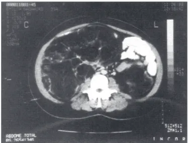
Abdominal sonography showed a solid mass of 24.7 x 19.2 x 10.7 cm in
size and of 2638 cm3 in volume,
though it was impossible to identify its origin. Computed tomography showed topical kidneys with non-spe-cific hypoattenuated cortical nodular images of less than 1 cm, possibly cor-responding to cysts or angiomyo-lipoma as well as hypo-attenuant solid nodules with very high fat content. Some of the tumors of considerable size with evident blood vessels in the interiors were pushing the intestine forward. The largest at the lower pole of the right kidney measured 20 x 15 x 16 cm extending as far as the pelvic region (Fig. 1). The presence of he-patic lipoma was also demonstrated; there were 3 rounded hypoattenuated images in the right lobe, less than 1 cm in size, which could represent small cysts or lipomas. Further, there was an-other hypoattenuated, cleared,
delim-ited nodule of fat density in segment III.
Kidney function was normal, and the urine analyses showed discrete proteinuria (0.34 g/24h). Radionuclide renography with DMSA showed bilat-eral depressed renal tubular function, primarily in the right kidney, signs suggestive of sequels of pyelonephri-tis of the left kidney and bilateral dila-tation of the collecting system.
The patient underwent medial lon-gitudinal laparotomy and enucleation of the tumors of the right kidney, pre-serving the renal tissue as much as pos-sible. The largest tumor in the inferior pole and small tumoral masses were withdrawn, weighing altogether 2020 g and measuring 23.0 x 21.0 x 11.5 cm. There was no intention of com-pletely withdrawing all small intra-renal tumors, because this might dam-age the renal function. A further he-patic lipoma in segment III of 1.5 cm was removed. The pathology exami-nation revealed angiomyolipoma with predominance of fat component and hepatic lipoma. The renal tumor re-acted positively to HMB-45, an impor-tant angiomyolipoma marker, but the hepatic lipoma didn’t. After 3 months, the patient underwent another
tion for the enucleation of the tumors on the left kidney, the technique be-ing repeated and the renal tissue pre-served. The various irregular tumor fragments weighed a total of 475 g and the largest of the fragments measured 13.0 x 9.5 x 5.5 cm.
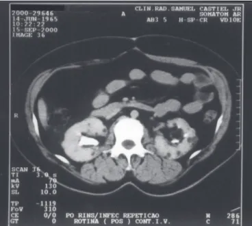
After surgery, renal function main-tained normal. Computed tomography revealed good renal function and mul-tiple small solid nodules on both kid-neys. After 2 years, computed tomog-raphy was repeated and revealed pre-served renal function and nodules of the same size of the last exam (Fig. 2).
DISCUSSION
Tuberous sclerosis is a dominant autosomal congenital disease that is characterized clinically by the classi-cal triad of mental retardation, epi-lepsy, and sebaceous adenoma12.
Char-acteristically, tuberous sclerosis occurs in the brain, where whitish nodules are found in the distorted cortical cytoar-chitecture, with deformed, bizarre neu-rons that are frequently enlarged.
Re-nal angiomyolipomas, cardiac rhab-domyomas, and pancreatic cysts may be associated with the tuberous scle-rosis13. From 40% to 80% of the
bear-ers of tuberous sclerosis present with renal angiomyolipoma with multiple bilateral asymptomatic tumors, which may be associated with cysts and more rarely renal carcinoma6, 12. In those
pa-tients with angiomyolipoma without tuberous sclerosis, the tumor is gener-ally single, larger, and
assympto-matic6,8,14. The angiomyolipoma may
affect the perirenal fat and local lym-phatic vessels, and the presence of tumors in extra renal sites is considered multicentric and not metastatic. An-giomyolipoma may occur in the lungs, liver, Fallopian tubes, vagina, spermatic cord, penis, or nasal cavi-ties14. The association of
angiomyo-lipoma and hepatic angiomyo-lipoma is rarely observed in patients with tuberous sclerosis11.
Although angiomyolipoma is comprised of 3 distinct groups of tis-sue, pleomorphism and aneuploid mi-totic figures may occur in some cases. Electronic microscopy not
infre-quently reveals muscular cells with li-pid content as if simulating a cell in-termediate between smooth muscular and fat cell. These findings corrobo-rate the hypothesis that the different types of tissue originate from one stem
cell 15. Because of the presence of
multinucleated giant cells, it may be difficult to distinguish the tumor from liposarcoma, malignant fibrous histio-cytoma, leiomyosarcoma, or sarco-matoid carcinoma. This difficulty em-phasizes the importance of HMB-45 immunoreaction, because a mono-clonal antibody reacts specifically with premelanosomes in the smooth muscle cells. No other benign or ma-lignant renal tumors show staining with HMB-45. This confirmation is important for the correct definition of the nature of the tumor and the most appropriate procedures for the pa-tient16, 17.
The natural history of angiomyo-lipoma is not fully known; however, some authors have presented some im-portant observations. Steiner and col-leagues studied 35 patients with a to-tal of 48 lesions, 34 of which were fol-lowed clinically for 4 years. Of these, 64% presented no change during fol-low up, and it was found that tumors initially larger than 4 cm were more prone to growth. No metachromic le-sions or renal carcinoma were observed in this group of patients. It was further noticed that growth of tumors did not seem to be due to intra-tumoral bleed-ing, but rather arose as a result of the growth of its tissue. The growth may be slow or rapid, there being no spe-cific fact known to account for this rate of growth 18. Ewalt and colleagues
studied the growth of renal lesions in children suffering from tuberous scle-rosis and observed that the majority of the lesions were angiomyolipoma and had the tendency to grow; they did not observe any case of regression among these tumors, in contrast with what oc-curs with renal cysts. The largest
growth observed in the 60 patients was 4 cm in 1 year (0 to 4cm and in one other case from 5 to 9 cm). The young-est patient with angiomyolipoma was 2 years old. It was further observed that the growth of various lesions in the same kidney proceeded at differ-ent rates, the number of lesions and their rate of growth being progressive and dependent of age. It was sug-gested that the loss of the function of the tumor suppressive gene owing to a second mutation may explain why some tumors grow while others main-tain stable. The hormonal influences of the steroid receptors in the muscle cells may explain the differences in tumor behavior seen during different periods of life, with greater tumor growth in post-puberal period and during pregnancy 12. Other studies add
further that angiomyolipoma in pa-tients with tuberous sclerosis grow over time, that tumors greater than 4 cm generally occur post-puberty19, and
that angiomyolipoma has greater like-lihood of growing in women than in men because according to hormonal theory, there are progesterone receptors in the muscle cells of the tumor 20. In
the literature, there was one report of 6 cases of association of pregnancy and angiomyolipoma with acute bleeding, including 2 cases of acute
rupture calling for nephrectomy21.
What may be regarded as certain in the natural history of the disease is the re-lationship between size and risk of complications, with tumors having di-ameters greater than 4 cm having the greatest risk of rupture.
As for clinical manifestations, the main symptom is abdominal pain, more specifically lumbar pain. Other possible symptoms and signs are pal-pable mass, hemorrhage, hematuria, nausea and vomiting, systemic arterial hypertension, anemia, fever, shock, re-nal failure and urinary infection18. In
those patients with lesions smaller than 4 cm the diagnosis is in the
ma-jority of cases fortuitous during a physical exam. The symptoms are more frequent in patients with larger tumors22; about 25% of patients with
angiomyolipoma present with intense abdominal pain or with shock due to intra-tumoral hemorrhage and undergo
emergency nephrectomy8.
At the present time, diagnosis is made based on abdominal sonography and computed tomography, by which the presence of fat cells in the compo-sition of the tumor is discovered. This finding is confirmed in the computed tomography by means of the measure-ment of image density that should be less than or equal to – 50 Hounsfield. Doubt concerning diagnosis persists in those cases in which there is little fat content or in the presence of previous bleeding, which may conceal the pres-ence of fat cells in the tumor 6,10,23.
The treatment has been fully dis-cussed in the literature. The decision regarding treatment methodology is made primarily based on of the size of the angiomyolipoma, the symptoms, the rate of growth, complications, and the degree of diagnostic certainty re-garding radiological results. Prior to 1976, 93% of all angiomyolipoma that were unrelated to tuberous sclero-sis were treated by total nephrectomy. More recently, on the basis of sonography and computed tomogra-phy, which have created the possibil-ity of diagnostic certainty, the objec-tive in the majority of the cases and as far as possible is conservative treat-ment. Selective arterial embolization has shown itself effective in the treat-ment of acute hemorrhage with or with-out later surgery or as initial treatment
of the angiomyolipoma8,10. Although
arterial embolization is minimally in-vasive, it does not preserve renal func-tion; it only has a temporary effect; it requires close clinical observation be-cause of associated complications; and as a rule, it is inefficient when used alone. Regarding surgical treatment,
tumorectomy, partial nephrectomy, or total nephrectomy may be carried out. The surgical treatment that preserves the largest amount of renal tissue is tumor enucleation, which has been un-dertaken with excellent results, even for giant angiomyolipoma (larger than 20 cm), being practically applicable to patients with tuberous sclerosis who present multiple and bilateral le-sions10,24,25. Total nephrectomy should
be used very rarely; it is only justified in cases of uncontrollable bleeding, when there is risk to the patient’s life, in central tumors, in the presence of extensive necrosis or when there is in-flammation of the renal tissue, or when there is a diagnosis of renal carcinoma in the same kidney. This last scenario is debatable in the light of recent pro-posals for surgery for renal carcinoma with preservation of renal tissue 5,23,26.
More recently, cryotherapy has been suggested as a therapeutic option and may be associated with laparoscopy27.
maximal preservation of renal tissue (enucleation or partial nephrectomy) is achieved10.
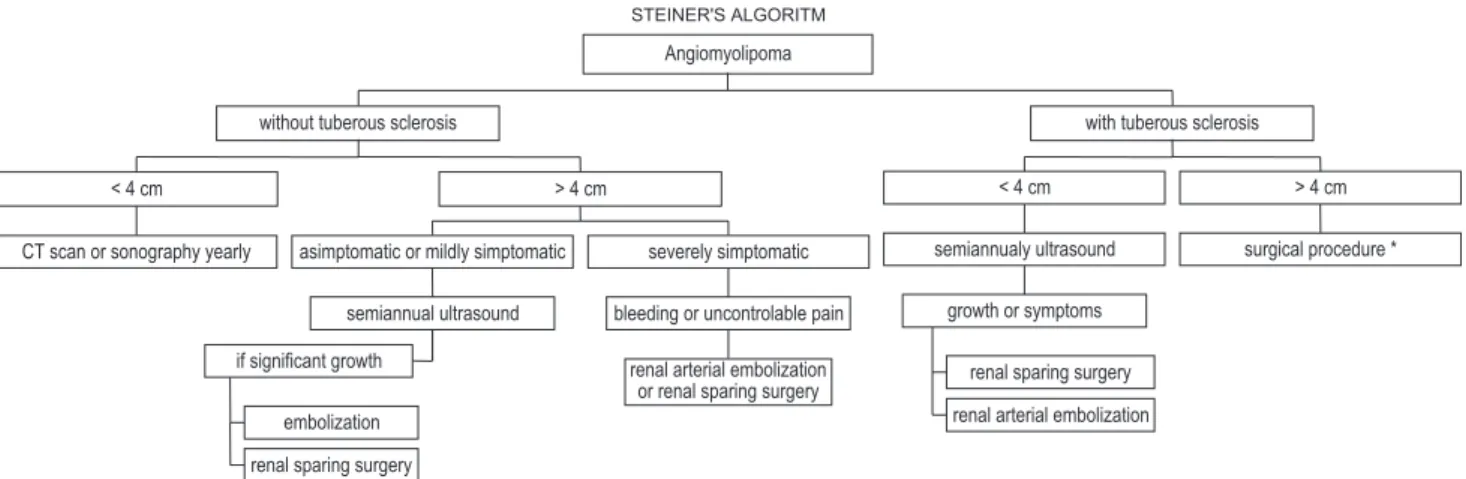
Steiner and colleagues have pro-posed changes in Oesterling´s algo-rithm, thus differentiating the treat-ment for patients with tuberous scle-rosis (Fig. 3)18. This differentiation
seems to be preferable to prevent the tumor from attaining larger dimen-sions, especially with cases associated with tuberous sclerosis in which
con-servative surgery is more difficult and causes greater losses of blood due to the size of the tumor.
In conclusion, the basis of manage-ment of angiomyolipoma is the at-tempted preservation of renal tissue, which can be effectively achieved with nephron-sparing surgical procedures such as tumor enucleation. Under some conditions, however, it is necessary to do selective angioembolization, par-tial nephrectomy, or even total
ne-phrectomy. Especially in patients with tuberous sclerosis with large bilateral and multiple tumors, the aim of treat-ment is the preservation of the great-est possible degree of efficient renal function. Therefore, tumoral enuclea-tion is one of the best choices in these cases and is perfectly feasible. Smaller tumors can be excised and submitted to experimental studies using laparoscopy or cryotherapy.
Figure 3. Steiner’s algaritm18.
*Consider immediate prophylatic renal arterial embolization or renal sparing surgery.
RESUMO
SCHNEIDER-MONTEIRO ED e col. – Angiomiolipomas renais gigantes bilateralmente associados a lipoma hepático em pacientes com escle-rose tuberosa. Rev. Hosp. Clín. Fac. Med. S. Paulo 58(2):103-108, 2003.
OBJETIVO: Relatar um caso de angiomiolipoma gigante, bilateral, as-sociado a esclerose tuberosa, tratado com sucesso e revisar a literatura concernente ao tratamento do angio-miolipoma.
RELATO DO CASO: Paciente portadora de esclerose tuberosa, com
diagnóstico de angiomiolipoma reali-zado por ultra-sonografia durante ges-tação. O tumor apresentava 9cm de diâmetro, à direita. Optou-se por con-duta conservadora durante a gestação, e a paciente retornou somente 7 anos após, com tumor de 24,7 x 19,2 x 10,7 cm à direita e outro à esquerda de 13 x 11,5 x 6,5 cm, além de múltiplos angiomiolipomas pequenos. Realizada inicialmente ressecção tumoral à direi-ta, por enucleação, com preservação do parênquima renal, e 3 meses após à esquerda. A função renal pós-operató-ria se manteve inalterada, e ambos os rins apresentaram uniformidade e
pro-gressão do contraste adequados.
CONCLUSÃO: Concluímos que os angiomiolipomas maiores que 4cm de-vem ser tratados cirurgicamente por-que têm maior risco de crescimento e hemorragias. As ressecções de tumores menores são mais seguras e têm menor morbidade. A enucleação dos tumores é forma eficaz de ressecção dos mes-mos, com preservação de parênquima renal.
DESCRITORES:
REFERENCES
1 . HADJU SI, FOOTE Jr. F W - Angiomyolipoma of the kidney: report of 27 cases and review of the literature. J Urol 1969; 102:396-401.
2 . MAZEMAN E, WEMEAU L, BISERTE J et al. - Renal angiomyolipoma. A report of 11 cases. Eur Urol 1980; 6:328-34.
3 . WIEDERHOLT WC, GOMEZ MR, KURLAND LT - Incidence and prevalence of tuberous sclerosis in Rochester, Minnesota, 1950 through 1982. Neurology 1985; 35:600-603. 4 . LINDENBAUM R - Prevalence of tuberous sclerosis. In:
TUBEROUS SCLEROSIS SYMPOSIUM, 2nd, Nottingham,
United Kingdom, 1985.
5 . FAZELI-MATIN S, NOVICK AC - Nephron-sparing surgery for renal angiomyolipoma. Urology 1998; 52(4):577-83. 6 . VAN BAAL JG, SMITS NJ, KEEMAN JN et al. - The evolution
of renal angiomyolipoma in patients with Tuberous Sclerosis. J Urol 1994; 152:35-38.
7 . O’DONNELL M, FLEMING S - Angiomyolipoma of the kidney – a cause of recurrent retroperitoneal haemorrhage. BJU 1995; 76:512-24.
8 . TONGAONKAR HB, SAMPAT MB, DALAL AV et al. - Bilateral renal angiomyolipoma. J S Oncology 1994; 57:65-70. 9 . BOSNIAK MA - Angiomyolipoma (hamartoma) of the kidney: a
preoperative diagnosis is possible in virtually every case. Urol Rad 1981; 3:135-42.
10. OESTERLING JE, FISHMAN EK, GOLDMAN SM et al. - The management of Renal Angiomyolipoma. J Urol 1986; 135:1121-24.
11. HIRASAKI S, KOIDE N, OGAWA H et al. - Tuberous sclerosis associated with multiple hepatic lipomatous tumors and hemorrhagic renal angiomyolipoma. Intern Med 1999; 38(4):345-48.
12. EWALT DH, SHEFFIELD E, SPARAGANA SP et al. - Renal lesion growth in children with tuberous sclerosis complex. J Urol 1998; 160:141-145.
13. GIROLAMI U, FROSCH MP, ANTHONY DC – The central nervous system. In: COTRAN RS, ROBBINS SL, KUMAR VY – Pathologic basis of disease. 5th ed. Philadelphia,
Pennsylvania, Saunders, 1994. c. 29, p. 1354
14. DE LUCA S, TERRONE C, ROSSETTI R - Management of renal angiomyolipoma: a report of 53 cases. BJU international 1999; 83:215-18.
15. HOLM-NIELSEN P, SORENSEN FB - Renal angiomyolipoma: an ultrastructural investigation of three cases with histogenetic considerations. APMIS 1988; suppl, 4:37-47.
16. PEA M, BONETTI F, ZAMBONI G et al. – Melanocyte-marker HMB-45 is regularly expressed in angiomyolipoma of the kidney. Pathology 1991; 23:185-188.
17. KAISERLING E, KRÖBER S, XIAO JC, SCHAUMBURG-LEVER G – Angiomyolipoma of the kidney. Immunoreactivity with HMB-45. Light and electron-microscopic findings. Histopathology 1994; 25:41-48.
18. STEINER MS, GOLDMAN SM, FISHMAN EK et al. - The natural history of renal angiomyolipoma. J Urol 1993; 150:1782-86.
19. STILLWELL TJ, GOMEZ MR, KELALIS PP - Renal lesion in tuberous sclerosis. J Urol 1987; 138:477-81.
20. MARTIGNONI G, PEA M, BONETTI F - Progesterone receptors in renal angiomyolipoma. In: TUBEROUS SCLEROSIS ASSOCIATION OF GREAT BRITAIN. Bath, United Kingdom, 1995. An Meeting Bath, United Kingdom, 1995.
21. JARDIN A, RICHARD F, LEDUC A et al. - Diagnosis and treatment of renal angiomyolipoma (based on 15 cases). Arguments in favor of conservative surgery (based on 8 cases). Eur Urol 1980; 6:69-82.
22. KATZ DS, POSTER RB - Massive renal angiomyolipoma in tuberous sclerosis. Clinical Imaging 1997; 21:200-202.
23. CHATTERJEE T, HEINDEL W, VORREUTHER R et al. - Recurrent bleeding of angiomyolipoma in tuberous sclerosis. Urol Int 1996; 56:44-47.
24. CHEN CH, YU TJ, HSU K – Unusual presentations of angiomyolipoma. Changgeng Yi Xue Za Zhi 1991; 14:269-72.
25. MEIRI H, SOEJIMA K, TOKUDA Y et al. – The management selection of renal angiomyolipoma. Nippon Hinyokika Gakkai Zasshi 1996; 87:1197-200
26. LICHT MR, NOVICK AC - Nephron sparing surgery for renal cell carcinoma. J Urol 1993; 149:1-7.