A
rti
g
o
*e-mail: [email protected]
A STRATEGY FOR THE RAPID IDENTIFICATION OF FUNGAL METABOLITES AND THE DISCOVERY OF THE ANTIVIRAL ACTIVITY OF PYRENOCINE A AND HARZIANOPYRIDONE
Laura P. Iócaa, Stelamar Rommingera,b, Mario F. C. Santosa, Karin F. Bandeiraa, Fabiana T. Rodriguesa, Miriam H.
Kossugaa, Karen J. Nicacioa, Everton L. F. Ferreiraa, Raquel P. Morais-Uranoa, Messias S. Passosa, Luciana K. Kohnc,
Clarice W. Arnsd, Lara D. Settee e Roberto G. S. Berlincka,*
aInstituto de Química de São Carlos, Universidade de São Paulo, 13560-970 São Carlos – SP, Brasil bLife Sciences Institute, University of Michigan, 48109 Ann Arbor – MI, United States of America. cUniversidade de São Francisco, 13045-510 Campinas – SP, Brasil
dLaboratório de Virologia, Instituto de Biologia, Universidade Estadual de Campinas, CP 6109, 13083-970 Campinas – SP, Brasil eDepartamento de Bioquímica e Microbiologia, Instituto de Biociências, Universidade Estadual Paulista Júlio de Mesquita Filho, 13506-900 Rio Claro – SP, Brasil
Recebido em 18/12/2015; aceito em 10/05/2016; publicado na web em 26/05/2016
The isolation and identification of bioactive metabolites from complex extracts obtained from microbial growth media is a time consuming, costly, and labor-intensive task. A strategy to rapidly identify secondary metabolites isolated from extracts obtained from the culture media of marine-derived and endophytic fungal strains is described. Identification was achieved by HPLC-UV-MS and 1H NMR analyses in combination with data obtained from the Dictionary of Natural Products. Among the compounds identified,
(-)-naphthoquinoneimine, citreorosein, emodin, pyrenocine A and harzianopyridone displayed moderate to potent antiviral activity. (-)-Naphthoquinoneimine was isolated as the enantiomer of its previously reported dextrorotatory congener, while 6,7-dihydroxy-2,2-dimethyl-4-chromanone is herein reported for the first time as a natural product.
Keywords:fungal metabolites; rapid identification; dereplication; antiviral.
INTRODUCTION
Most fungal secondary metabolites are remarkably complex bioactive compounds.1,2 Although they were almost dismissed in biodiscovery programs between the 1960’s and 1980’s, the raise of molecular biology tools provided compelling evidence that the diversity of filamentous fungi secondary metabolism is astonishingly higher than originally thought.3 Such metabolites are very often the inspiration for the development of new drug lead scaffolds and bio-chemical tools, resulting from the fact that many of these compounds present unique ecological roles, particularly as antibiotics and as chemical mediators.
The isolation of bioactive fungal metabolites from complex and chemically diverse extracts is time and resources consuming, a labor--intensive task. The rapid identification of known natural products isolated from fungal extracts is essential to avoid the record and analysis of multiple 1D and 2D NMR experiments. This is particu-larly true when dereplication of secondary metabolites in extracts of biological matrixes is unfeasible by resolution and high--throughput chromatography-spectroscopy hyphenated tools, such as HPLC-HRMS, HPLC-NMR and related techniques.3-7 Data obtained from joint UV, LRMS and 1H NMR analyses only, and sometimes 13C NMR, can be used in querying databases such as the Dictionary of Natural Products (DNP), Antimarin, MarinLit, and Molecular Networking, as an effective combination for the rapid identification of known structures.8,9 The rapid identification of secondary metabolites enables further assessment of structures of interest, particularly of unprecedentedly bioactive known compounds.
The discovery of antiviral natural products is currently of seminal interest considering the emergence and re-emergence of viral disea-ses such as dengue, and infections caused by chikungunya and zika
viruses. Only fifty antiviral drugs are available to treat viral infec-tions.10 The majority of these antivirals was introduced into clinical use during the last two decades, and they were discovered due to the urgency in developing treatments for the Human Immunodeficiency Virus (HIV) infection.10 Only a handful of antiviral compounds have been discovered from marine-derived fungi. These include sansal-vamide A (1), produced by Fusarium sp., which exhibits antiviral activity against poxvirus (MCV),11 as well as halovirs A-E (2-6), produced by Scytalidium sp., and active against Herpes Simplex Virus Types 1 and 2 (HSV).12
The discovery of microbial secondary metabolites in Brazil increased considerably during the last 15 years. However, when con-sidering all the microbial secondary metabolites isolated by Brazilian researchers between 2000 and 2013, only 28.4% were new.13 This probably results from the difficulty in having state-of-art tools for dereplication purposes, which are expensive to purchase and maintain. Aiming to provide a reliable procedure for the identification of known secondary metabolites isolated from fungal culture media, herein we present a practical strategy that enables the rapid identification of natural products by HPLC-UV-LRMS, 1H NMR and, much less often, by 13C NMR. Selected compounds were subsequently evaluated in antiviral bioassays.
EXPERIMENTAL
General experimental procedures
cyanopropyl, diol, and silica gel cartridges of different dimensions (Waters and Phenomenex).
HPLC-UV-MS analyses were performed using a Waters Alliance 2695 system coupled on-line with a Waters 2996 photodiode array detector, followed by a Micromass ZQ2000 MS detector with an elec-trospray interface. The photodiode array detector scanned the samples between λmax 200 and 400 nm. The MS detector was optimized to the following conditions- capillary voltage: 3.00 kV, source block temperature: 100 °C, desolvation temperature: 350 °C, operating in electrospray positive and negative modes in the detection range between 200 and 800 Da with total ion count extracting acquisition. The cone and desolvation gas flow were 50 and 350 L/h, respectively, and were generated by a Nitrogen Peak Scientific N110DR nitrogen source. Data acquisition and processing were performed using the Empower 2.0 software. Approximately 1 mg of each sample was diluted in appropriate vials containing 1 mL of methanol (MeOH). Analyses were performed using different C18 reversed-phase columns: Inertsil ODS-SP, ODS-3 or C8-4 (4.6 x 250 mm, 5 µm), with a gradi-ent of elution of 1:1 (MeOH/MeCN) in H2O + 0.1% formic acid in both organic and aqueous solvents. The gradient of elution started at 10% to 100% of the organic mixture, during 22 minutes. The flow rate was 1.0 mL min-1.
HPLC semi-preparative purifications were carried out using either a Waters 600 pump in series with a Waters 2487 dual wavelength absorbance detector, or a Waters 2535 quaternary gradient module equipped with a Waters 2489 UV-visible detector. Purifications were performed using analytical/semi-preparative C18 reversed-phase columns Inertsil ODS-SP and/or ODS-3 (4.6 x 250 mm, 5 µm), and C8 columns Inertsil C8-4 and/or InertSustain (4.6 x 250 mm, 5 µm or 14.0 x 250 mm, 5 µm).
NMR spectra were recorded on a Bruker DRX spectrometer (9.4 T, 400 MHz for 1H), Bruker AVANCEIII spectrometer (14.1 T, 600 MHz for 1H) or on an Agilent Technologies 500/54 Premium Shielded spectrometer (11.7 T, 500 MHz for 1H). Compounds were dissolved in appropriate deuterated solvents containing tetramethylsilane as internal reference. HRMS analyses were performed using a Bruker Daltonics Ultra-TOF spectrometer or on a LTQ Orbitrap Thermo Scientific spectrometer. Optical rotation analyses were performed using either a Perkin Elmer 241MC polarimeter or a JASCO P2000 polarimeter. Marine-derived fungal strains obtained from marine
invertebrate samples
Samples of marine invertebrates were collected at Toque-Toque Island, off São Paulo state north coastline, in Brazil. The samples
consisted of three marine sponges (Dragmacidon reticulatum, Axinellidae, Ridley & Dendy, 1886; Mycale laxissima, Mycalidae, Duchassaing & Michelotti, 1864; and Mycale angulosa, Mycalidae, Duchassaing & Michelotti, 1864) and one ascidian (Didemnum ligulum, Didemnidae, Monniot, 1983). Vouchers of these marine organisms are deposited at Museu Nacional, Universidade Federal do Rio de Janeiro (sponges) and at Departamento de Zoologia, Setor de Ciências Biológicas, Universidade Federal do Paraná (ascidian). The sponges were identified by Dr. Marcio R. Custódio (Instituto de Biociências, Universidade de São Paulo), while the ascidian was identified by Dra. Rosana M. Rocha (Departamento de Zoologia, Universidade Federal do Paraná).
Fungal strains were isolated as previously reported.14 Pure fungal colonies were photographed, morphologically described, subjected to microscopic analysis, and stored at the Grupo de Química Orgânica de Sistemas Biológicos, Departamento de Físico-Química, Instituto de Química de São Carlos. Colonies were identified by conventional and molecular techniques. Identification sequences for each fungal strain can be requested to Dra. Lara Sette ([email protected]). An endophytic fungal strain isolated from the leaves of
Anthurium loefgrenii
Samples of leaves from ten plants were collected at Alcatrazes Island, Alcatrazes Archipelago (24º10’ S, 45º70’ W), off São Paulo state north coastline, in Brazil. Positioning, environment (surround-ings) and occurrence data were recorded as well. All samples were collected in sterilized plastic bags and immediately transported to the Universidade de São Paulo Marine Station, CEBIMar/USP. Each plant sample was identified by Dr. Marco Antônio de Assis (Instituto de Biociências, Universidade Estadual Paulista Júlio de Mesquita Filho, UNESP-Rio Claro).
Leaves were cleaned from visible dust and soil with running water and subsequently washed according to a modified Petrini (1991)15 protocol, as it follows: the leaves were washed in: 1) sterilized dis-tilled water during 1 min, 2) 70% EtOH solution during 1 min, 3) HCl 2.5% solution during 2 min, 4) 70% EtOH solution during 1 min, followed by two washes with sterilized distilled water during 1 min. An aliquot of 100 µL from the last wash was plated on PDB culture media in order to confirm the surface sterilization.
For the isolation of endophytic fungi, after surface sterilization leaves were cut with sterilized blades into fragments of approx. 8-12 mm, and three pieces were plated on minimum media + leaves: K2HPO4 (18.8 g L
-1), KH
2PO4 (6 g L
-1), MgSO 4 (5 g L
-1), glycerol (10 g L-1) and fragments of leaves (added while the media was poured).
Tetracycline (100 mg L-1) and streptomycin (20 mg L-1) were added in all media used for inoculation in order to prevent bacterial contamination.
Growth in Petri dishes was performed at 28 oC. After inocula-tion, the plates were regularly examined in order to verify the growth of filamentous fungi. Pure fungal colonies obtained by successive purification steps were transferred to plates containing MEA (Malt Extract Agar, composition: malt extract 20 g, glucose 20 g, peptone 1 g, agar 20 g) medium without antibiotics.
Crude extracts from culture media
Pure marine-derived fungal strains were first grown in Petri dishes using either 2% malt (malt extract 20 g, artificial sea water 1 L, agar 15 g) or 3% malt (malt extract 30 g, mycological peptone 3 g, artificial sea water 1 L, agar 15 g) culture media. Composition of artificial sea water (ASW) was 1.36 g of CaCl2·H2O, 9.68 g of MgCl2·2H2O, 0.61 g of KCl, 30 g of NaCl, 140 mg of NaH2PO4, 3.47 g of Na2SO4, 170 mg of NaHCO3, 100 mg of KBr, 40 mg of SrCl2·6H2O, and 30 mg of HBO3 in 1 L of distilled water. Strains DRG1M2, DLM212, DLM218, F56, and F254 were maintained in 2% malt, while strains DLM33 and DLM38 were maintained in 3% malt. The endophytic fungal strain P2AF2F3 was maintained in 2% malt without ASW. After growth, 2 mm of mycelia surface samples were obtained using sterilized Pasteur pipettes, and inoculated in 250 mL of culture media broth of identical formulation to that used for the strains growth on Petri dishes (except for the agar). The strains were grown in static mode for 21 days at room temperature. After growth, an equal volume of ethyl acetate (EtOAc) was added to the cultures, and the mixture of mycelia, culture media, and EtOAc was filtered through a celite bed. The organic phase was separated by liquid-liquid extraction, and subsequently dried under vacuum.
Rapid identification procedure for pure compounds
Pure compounds were analyzed by HPLC-UV-MS. Data from MS and UV spectra obtained for each compound were used as query for a search on Dictionary of Natural Products. If too many outcomes were returned, another query using organism taxonomic information was made to restrict the search. Since all compounds were isolated from fungal culture media, queries were also performed using the keywords “fungal” or “fungi” or “fungus”. If no unambiguous identification was possible at this step, the 1H NMR spectrum of the isolated compound was recorded. Typical 1H signals were used for identification purposes, such as those of singlet or doublet methyl groups, vinylic or benzene hydrogens. In the few instances when organism taxonomical information, UV, LRMS and 1H NMR data did not provide enough information for the unambiguous identification of the pure compound, the 13C NMR spectrum was also recorded and analyzed. No further 2D NMR or HRMS analysis were necessary to identify pure known compounds.
Isolation and identification of fungal secondary metabolites
Aspergillus sydowii DRG1M2, isolated from the sponge Dragmacidon reticulatum, was inoculated in 8 L of 2% malt extract medium (in thirty-two 500 mL Schott flasks containing 250 mL of culture medium each). After growth, 250 mL of EtOAc were added to each Schott flask. This mixture was filtered and the organic phase was separated by liquid-liquid extraction. After evaporation, the EtOAc extract (2 g) was resuspended in MeOH and deffated with hexane. The MeOH extract (1.5 g) was subjected to a gel permeation chromatography using Sephadex LH-20 gel and MeOH as mobile
phase. Six fractions were obtained after analysis by thin layer chro-matography (eluent: 9:1 CH2Cl2/MeOH; observed under UV light at λmax 254 nm). Fraction DRG1M2-MC (284.3 mg) presented peaks with UV absorptions at 210-220, 240-250, and 300-310 nm, as well as compounds with molecular weights in the range between 240 and 250 Da by HPLC-UV-MS analysis. This fraction was subjected to a solid-phase extraction on a silica-gel cartridge (5 g) eluted with 8:2, 7:3, 6:4, 1:1 [1:1 (Hex/CH2Cl2)]/EtOAc], 8:2, 7:3, 6:4, 1:1 CH2Cl2/AcOEt, 9:1, 1:1 CH2Cl2/MeOH and 100% MeOH. From this separation three fraction were obtained. HPLC purification of fraction DRG1M2-MC2 (95.4 mg, column: Inertsil ODS-SP; eluent: 20:20:60 MeOH/MeCN/H2O + 0.1% formic acid; flow rate of 1.0 mL/ min; detection at λ = 254 and 340 nm) led to the isolation of 6.8 mg of sydowic acid (7).16 The compound was identified by analysis of spectroscopic data and comparison with literature data. The absolute configuration of (7) was established by comparison of its [α]D
23.3 –4 (c 0.1, MeOH) with literature values.
rate: 1.0 mL/min; detection at λmax 280 nm) led to the isolation of 1.9 mg of pyrophen (12).19,21 Purification of the fraction DLM38-CE by HPLC (column: Inertsil ODS-3; gradient elution with 1:1 (MeOH/ MeCN)/H2O from 30 to 42% [1:1 (MeOH/MeCN) during 20 minutes, then isocratic elution of 100% [1:1 (MeOH/MeCN)] during 6 minutes; flow rate: 1.0 mL/min; detection at λ = 280 nm) led to the isolation of 2.0 mg of leucomelone (13)22 and of 2.1 mg of atromentin (14).23
A second investigation of the strain Aspergillus sp. DLM38 was performed with 10 L of 3% malt extract medium. After liquid-liquid extraction with EtOAc and evaporation of the organic phase, the EtOAc extract (4.8 g) was subjected to a solid-phase extraction on a silica-gel cartridge (10 g) eluted with 8:2:0, 6:4:0, 0:1:0, 0:1:1 CH2Cl2/ EtOAc/MeOH, and four fractions were obtained. Fraction DLM38-F2 (458.2 mg) was subjected to a separation using a C18 reversed-phase silica-gel cartridge (10 g) eluted with 1:3, 1:1, 3:1, 0:1 H2O/MeOH. After HPLC-UV-MS analysis, these fractions were pooled in two fractions DLM38-F2A (78.6 mg) and DLM38-F2B (310.1 mg). DLM38-F2A was separated by HPLC using a C8 reversed-phase column InertSustain C8 (14.0 x 250 mm, 5 µm) and isocratic elution of 2:3 MeOH/H2O during 28 minutes, flow rate of 4.0 mL/min, and detection at λmax 254 and 280 nm, to give 1.8 mg of (10) and 8.7 mg of pyranonigin B (15)20. Fraction DLM38-F2B presented peaks with UV absorptions around 230, 280 and 330 nm, as well as compounds with molecular weights in the range between 575 and 607 Da, cor-responding to aurosperones derivatives previously isolated, so this fraction was ignored.
Penicillium glabrum DLM218, isolated from the ascidian Didemnum ligulum, was grown in 8 L of 2% malt extract medium (in Shott flasks, as described above). After liquid-liquid extraction with EtOAc and evaporation of the organic phase, the EtOAc extract (2.9 g) was subjected to a solid-phase extraction on a C18 reversed-phase silica-gel cartridge (10 g) eluted with 15:85, 30:70, 50:50, 70:30, 85:15 MeOH/H2O and 100% MeOH. Analysis by HPLC-UV-MS of the fractions DLM218-ABC (1.19 g) and DLM218-E (470.7 mg) showed peaks with UV absorptions at λmax 220-230, 250-270, 280-300 and 385 nm, and compounds with molecular weights in the range between 270 and 390 Da. Fraction DLM218-ABC was separated by HPLC using a C8 reversed-phase column Inertsil C8-4 and gradient elution from 20% MeOH/H2O to 100% MeOH during 55 minutes, flow rate of 8.0 mL/min, and detection at λmax 280 and 300 nm. HPLC-UV-MS analysis of the fraction DLM218-ABC7 (65.1 mg) indicated a major peak with λmax 283, 307, and 383 nm in the UV spectrum, a peak with m/z 381.2 in the mass spectrum corresponding to the [M+H]+ ion, and a peak with m/z 379.3 corresponding to the [M-H]- ion of compound (15). HPLC purification (column: Inertsil C8-4; isocratic elution: 64:36 MeOH/H2O; flow rate of 7.0 mL/min; detection at λmax 280 and 300 nm) of the major compound led to the isolation of 5.7 mg of penicitrinone A (16).24 Fraction DLM218-E (88.9 mg) was separated by HPLC using a C8 reversed-phase column Inertsil C8-4 (isocratic elution with 58:42 MeOH/H2O; flow rate of 7.0 mL/min; detection at λmax 275 and 310 nm). HPLC-UV-MS analysis of the fraction DLM218-E4 (88.9 mg) indicated a major peak with λmax
235, 263, 295 and 384 nm in the UV spectrum, a peak with m/z 299.3 in the mass spectrum corresponding to the [M+H]- ion, and a peak with m/z 323.1 corresponding to the [M+Na]+ ion of compound (17). HPLC purification of (17) (column: Inertsil C8-4; isocratic elution: 59:41 MeOH/H2O, flow rate of 7.0 mL/min, detection at λmax 254 and 280 nm) led to the isolation of 5.2 mg of pinselin (17).25 HPLC-UV-MS analysis of the fraction DLM218-E6 (217.4 mg) indicated two major peaks with λmax 283, 307, 383, and 221, 266, 287 nm in the UV spectrum, with m/z 381.1 and 271.2 in the mass spectrum, corresponding to the [M+H]+ ions of (15)and (18), respectively. HPLC purification of (15) and (18) (column: Inertsil C8-4; isocratic
elution: 49:51 MeCN/H2O; flow rate of 7.0 mL min-1; detection at λmax 254 and 280 nm) led to the isolation of 13.3 mg of (15) and 9.4 mg of emodin (18).26
Penicillium citrinum DLM212, isolated from the ascidian Didemnum ligulum, was grown in 8 L of 2% malt extract medium (in Shott flasks as described above). After liquid-liquid extraction with EtOAc and evaporation of the organic phase, the EtOAc extract (2 g) was resuspended in MeOH and deffated with hexane. The MeOH fraction (1.5 g) was subjected to a gel permeation chromatography using a glass column filled with Sephadex LH-20 gel and MeOH as mobile phase. Six fractions were obtained after analysis by thin layer chromatography (eluent: 9:1 CH2Cl2/MeOH; observed under UV light at λmax 254 nm), of which DLM212-MB (122.4 mg), DLM212-MC (551.5 mg) and DLM212-ME (66.8 mg) presented peaks with UV absorptions at λmax 210-230, 250-290, and 315 nm, in the mass range between 250 and 300 Da by HPLC-UV-MS analysis. The fraction DLM212-MB was separated by HPLC using a C18 reversed-phase column Inertsil ODS-SP (isocratic elution with 35:35:30 MeOH/ MeCN/H2O + 0.1% formic acid; flow rate of 1.0 mL/min; detection at λmax 270 and 340 nm). HPLC purification of the fraction DLM212-MB2 (35.0 mg) (column: Inertsil ODS-SP; isocratic elution: 25:25:50 MeOH/MeCN/H2O + 0.1% formic acid; flow rate of 1.0 mL/min; detection at λmax 270 and 340 nm) led to the isolation of 1.0 mg of quinolactacin B (19).27 HPLC-UV-MS analysis of the fraction DLM212-MB4 (9.0 mg) indicated a major peak with λmax 224 and 283 nm in the UV spectrum, and a peak with m/z 298.3 in the mass spectrum, corresponding to the [M+H]+ ion of (20). Purification of (20) by HPLC (column: Inertsil ODS-SP; isocratic elution: 35:35:30 MeOH/MeCN/H2O + 0.1% formic acid; flow rate of 1.0 mL/min; detection at λmax 254 and 270 nm) led to the isolation of 2.3 mg of penicillenol A (20).28 Purification of the fraction DLM212-ME (66.8 mg) by HPLC (column: Inertsil ODS-SP; isocratic elution: 35:35:30 MeOH/MeCN/H2O + 0.1% formic acid; flow rate of 1.0 mL/min; detection at λmax 270 and 340 nm) led to the isolation of 14.7 mg of citreorosein (21)29 and 15.4 mg of (18). Fraction DLM212-MC was subjected to a solid-phase extraction on a cyanopropyl-bonded silica-gel cartridge (10 g) eluted with 8:2, 7:3, 6:4, 1:1 CH2Cl2/EtOAc, 100% EtOAc, 1:1 EtOAc/MeOH and 100% MeOH. HPLC purifica-tion of DLM212-MC2 (83.7 mg, column: Inertsil ODS-SP; isocratic elution: 15:15:70 MeOH/MeCN/H2O + 0.1% formic acid; flow rate of 1.0 mL/min; detection at λmax 254 and 280 nm) yielded 2.0 mg of DLM212-MC2E. HPLC-UV-MS analysis of this fraction indicated a major peak with λmax 218, 250 and 316 nm in the UV spectrum, and a peak with m/z 287.0 in the mass spectrum, corresponding to the [M+H]+ ion of the quinolactacin C (22).27
during 25 minutes; flow rate of 1.0 mL/min; detection at λmax 254 and 280 nm). HPLC purification of the fraction DLM33-A7A19M (21.2 mg) (column: Inertsil C8-4; isocratic elution: 46:54 MeOH/ H2O; flow rate of 1.0 mL/min; detection at λmax 254 and 280 nm) led to the isolation of 1.0 mg of 5,7-dichloro-3-methyl-6-methoxy-8-hydroxy-3,4-dihydroisocoumarin (24).31
Cochliobolus sp. F56, isolated from the sponge Mycale laxis-sima, was grown in 2 L of 2% malt extract medium (in ten 500 mL Schott flasks containing 200 mL of medium each). After liquid-liquid extraction of the growth medium with EtOAc and evaporation of the organic phase, the EtOAc extract (407.2 mg) was subjected to a separation by gel permeation chromatography using Sephadex LH-20 gel and MeOH as mobile phase. Five fractions were obtained after analysis by thin layer chromatography (eluent: 9:1 CH2Cl2/MeOH; observed under UV light at λmax 254 nm), of which F56-A2 (141.5 mg) and F56-A4 (17.4 mg) were chosen for purification procedures. The fraction F56-A2 was subjected to a solid-phase extraction on a diol-bonded silica-gel cartridge (5 g) eluted with 6:4 [1:1 (Hex/ CH2Cl2)]/EtOAc, 100% EtOAc, 1:1 EtOAc /MeOH and 100% MeOH. HPLC-UV-MS analysis of the fraction F56-A2(4) (32.9 mg) indicated a major peak with λmax 261 nm in the UV spectrum, and a peak with m/z 287.3 in the mass spectrum, corresponding to the [M+H]+ ion of 25. HPLC purification of (25) (column: Inertsil ODS-SP; isocratic elution: 21:21:58 MeOH/MeCN/H2O + 0.1% formic acid; flow rate of 1.0 mL/min; detection at λmax 230 and 254 nm) led to the isolation of 4.0 mg of (+)-abscisic acid (25).32 The absolute configuration of 25 was established by comparison of its [α]D
25 +87 (c 2.0, MeOH) with literature data. Fraction F56-A4 was separated by HPLC using a reversed-phase C18 column Inertsil ODS-SP (isocratic elution with 24:24:52 MeOH/MeCN/H2O + 0.1% formic acid; flow rate of 1.0 mL/min; detection at λ = 230 and 320 nm). This separation led to the isolation of 2.2 mg of 4-hydroxy-3-(3-methylbut-2-enyl)benzoic acid (26)33 and of 1.0 mg of anofinic acid (27).33 HPLC-UV-MS analysis of fraction F56-A4(2) indicated a major peak with λmax 240, 279 and 347 nm in the UV spectrum, and a peak with m/z 209.07 in the mass spectrum, corresponding to the [M+H]+ ion of (28). Purification by HPLC (column: Inertsil ODS-SP; isocratic elution with 20:20:60 MeOH/MeCN/H2O + 0.1% formic acid; flow rate of 1.0 mL/min; detection at λmax 230 and 320 nm) of the major compound led to the isolation of 0.8 mg of 2,3-dihydro-6,7-dihydroxy-2,2-dimethylchromen-4-one (28).
Trichoderma sp. F254, isolated from the sponge Mycale laxissi-ma, was grown in 2 L of 2% malt extract medium (as for Cochliobolus sp. F56). After liquid-liquid extraction with EtOAc and evaporation of the organic phase, the EtOAc extract (241.6 mg) was separated by gel permeation chromatography using Sephadex LH-20 gel and MeOH as mobile phase. After analysis by thin layer chromatography (eluent: 9:1 CH2Cl2/MeOH; observed under UV light at λmax 254 nm) six fractions were obtained, of which F254-E (59.9 mg) was purified by HPLC (column: Inertsil ODS-SP; gradient elution: [1:1 (MeOH/ MeCN)]/ H2O + 0.1% formic acid from 20 to 80% [1:1 (MeOH/ MeCN)] during 30 minutes, then from 80 to 100% 1:1 (MeOH/ MeCN) during 1 minute and isocratic elution at 100% 1:1 (MeOH/ MeCN) during 4 minutes; flow rate of 1.0 mL/min; detection at λmax 254 and 280 nm). This procedure led to the isolation of 5.9 mg of harzianopyridone (29).34
Penicillium paxilli Ma(G)K, isolated from the sponge Mycale angulosa, was grown 2 L of 2% malt medium (as for Cochliobolus sp. F56). The culture was incubated in a rotary shaker (100 rpm) at 25 °C for seven days. At the end of the growth period, 200 mL of EtOAc were added to each Schott flask. The mixture was shaken at 100 rpm for 24 h at 25 °C, filtered through a celite bed and the organic phase was separated by liquid-liquid extraction. After evaporation, the
EtOAc extract (400 mg) was subjected to a solid-phase extraction on a cyanopropyl-bonded silica-gel cartridge (2 g), eluted with 100% CH2Cl2 (fraction Ma(G)K-A, 130.0 mg), 100% EtOAc (fraction Ma(G)K-B, 68.2 mg), and finally eluted with 100% MeOH (fraction Ma(G)K-C, 155.0 mg). Fraction Ma(G)K-A was separated by HPLC using a C18 reversed-phase column Inertsil ODS-3 (4.6 × 250 mm; 5 µm) (isocratic elution with 1:1 MeOH/MeCN in H2O + 0.1% formic acid), flow rate of 1.0 mL/min, and detection at λmax 254 nm. This separation yielded 42.7 mg of pyrenocine A (30)14,35 and 6.0 mg of pyrenocine B (31).14,35
The yet unidentified fungal strain P2AF2F3, isolated from the plant Anthurium loefgrenii, was inoculated in 10 L of 2% malt extract medium (in thirty-two 500 mL Schott flasks containing 250 mL of media each). After liquid-liquid extraction with EtOAc and evaporation of the organic phase, the EtOAc extract (900 mg) was resuspended in MeOH and deffated with hexane. The MeOH extract was subjected to a gel permeation chromatography using Sephadex LH-20 gel and MeOH as mobile phase. Five fractions were obtained. The fraction P2AF2F3-3 (530 mg) was subjected to a solid-phase extraction on a diol cartridge (10 g) eluted with 9:1, 8:2, 7:3, 6:4 [(1:9 Hex/CH2Cl2)/(1:9 EtOAc/MeOH)] and 100% (1:9 EtOAc/MeOH). From this separation five others fractions were obtained. Fraction P2AF2F3-3A (200 mg) was subjected to a solid-phase extraction on a C18 cartridge (10 g) eluted with 3:7 and 6:4 MeOH/H2O, then 100% MeOH. Fraction P2AF2F3-3A2 (154 mg) presented peaks with UV absorptions at 220-240 nm, in the mass range between 240 and 280 Da by HPLC-UV-MS analysis. HPLC purification of frac-tion P2AF2F3-3A2 (column: IntertSustain C8; eluent: 1:1:3 MeOH/ MeCN/H2O; flow rate of 1.0 mL/min; detection at λmax 255 and 215 nm) led to the isolation of 3.2 mg of conocenol B (32).36
Spectroscopic data of pure compounds isolated from fungal strains
Sydowic acid (7): yellow oil. UV (λmax nm): 212, 244, 299. 1H NMR (400 MHz, TMS, DMSO-d6,): δ (ppm); 0.97 (3H, s, H3-12), 0.98 (3H, s, H3-13), 1.06 (1H, m, H-9A), 1.25 (2H, m, H2-10), 1.30 (1H, m, H-9B),1.50 (3H, s, H3-14), 1.66 (1H, m, H-8A), 1.92 (1H, m, H-8B), 5.55 (1H, s, OH-1), 7.28 (1H, d, J= 1.7 Hz, H-2), 7.33 (1H, dd, J=8.0, 1.7 Hz, H-4), 7.39 (1H, d, J= 8.0 Hz, H-5), 9.98 (1H s, OH-15).13C NMR (100 MHz, TMS, DMSO-d
6,): δ (ppm); 18.7 (C-9), 28.4 (C-14), 29.4 (C-13), 29.5 (C-12), 42.1 (C-8), 44.2 (C-10), 68.9 (C-11), 75.0 (C-7), 116.8 (C-2), 119.9 (C-5), 127.1 (C-4), 130.3 (C-3), 138.1 (C-6), 154.8 (C-1), 167.4 (C-15). ESI+-MS: m/z 247 [M-H2O]+ (calcd for C15H19O3, 247.13342 ). [α]D
23.3 –4 (c 0.1, MeOH). (-)-Naphthoquinoneimine (8): red amorphous solid. UV (λmax nm): 207, 270, 334. 1H NMR (400 MHz, TMS, MeCN-d
3,): δ (ppm); 3.22 (1H, dd, J=13.9, 8.2 Hz, H-8A’), 3.31 (1H, dd, J=13.9, 6.0 Hz, H-8B’), 3.77 (3H, s, H3-9’), 4.51 (1H, ddd, J=8.2, 8.0, 6.0 Hz, H-7’), 5.46 (1H, d, J=2.3 Hz, H-5’), 5.51 (1H, s, H-3), 5.99 (1H, d, J=2.3 Hz, H-3’), 6.39 (1H, d, J=8.0 Hz, NH-10’), 6.51 (1H, d, J=2.4 Hz, H-8), 6.96 (1H, d, J=2.4 Hz, H-6), 7.28 (5H, m, H-2’, H-3’, H-4’, H-5’, H-6’), 12.91 (1H, s, 0H-5). 13C NMR (100 MHz, TMS, MeCN-d
3,): δ (ppm); 38.3 (C-8’), 55.7 (C-7’), 56.2 (C-9’), 88.3 (C-5’), 100.4 (C-3’), 101.2 (C-3), 107.9 (C-4a), 108.9 (C-6), 117.3 (C-8), 127.1 (C-4’’), 128.6 (C-3’’, C-5’’), 129.2 (C-2’’, C-6’’), 132.2 (C-8a), 136.3 (C-1’’), 147.5 (C-2), 161.0 (C-2’), 163.2 (C-5), 163.3 (C-7), 163.4 (C-6’), 170.8 (C-4’), 180.7 (C-1), 188.4 (C-4). ESI+-HRMS: m/z 434.1235 [M+H]+ (calcd for C24H20NO7, 434.1). [α]D
25 –8 (c 3.1, MeOH).
Aurosperone A (9): yellow solid. UV (λmax nm): 228, 258, 277.1H NMR see refs. 18 and 19. ESI+-MS: m/z 571.1 [M+H]+ (calcd for C32H27O10, 571.2).
for C11H12NO6, 254.1), ESI+-MS: m/z 529.2 [2M+Na]+ (calcd for C22H22N2NaO12, 529.1).
Aurosperone C (11): amorphous solid. UV (λmax nm): 233, 282, 334. 1H NMR (400 MHz, TMS, MeCN-d
3,): δ (ppm); 1.44 (3H, s, H3-11’), 1.68 (3H, s, H3-2), 2.83 (2H, d, J=16.5 Hz, H2-3’), 3.09 (2H, d, J=16.5 Hz, H2-3), 3.49 (3H, s, H3-12), 3.59 (3H, s, H3-13’), 3.92 (3H, s, H3-12’), 6.13 (1H, s, H-7’), 6.40 (1H, s, H-9’), 6.59 (1H, s, H-9), 6.92 (1H, s, H-10). ESI+-MS: m/z 593.1 [M+H]+ (calcd for C31 H29O12, 593.2).
Pyrophen (12): yellow crystal. UV (λmax nm): 265.
1H NMR (400 MHz, TMS, DMSO-d6): δ (ppm); 1.78 (3H, s, H3-15), 2.89 (1H, dd, J=13.7, 8.2 Hz, H-7A), 3.02 (1H, dd, J=13.7, 8.2 Hz, H-7B), 3.78 (3H, s, H3-16), 4.76 (1H, q, J=8.2 Hz, H-6), 5.57 (1H, d, J=2.1 Hz, H-2), 6.05 (1H, d, J=2.1 Hz, H-4), 7.19 (2H, m, H-9, H-13), 7.21 (1H, m, H-11), 7.27 (2H, m, H-10, H-12 ), 8.46 (1H, d, J=8.2 Hz, NH-6). 13C NMR (100 MHz, TMS, DMSO-d6,): δ (ppm); 22.8 (C-15), 39.0 (C-7),
52.5 (C-6), 56.9 (C-16), 88.4 (C-2), 99.8 (C-4), 127.2 (C-11), 128.7 (C-10, C-12), 129.4 (C-9, C-13), 137.0 (C-8), 163.8 (C-5), 164.2 (C-1), 169.0 (C-14), 170.0 (C-3). ESI--MS: m/z 323.2 [M+Cl]- (calcd for C16H17ClNO4, 322.1). [α]D
25 0 (c 0.7, MeOH).
Leucomelone (13): amorphous solid. UV (λmax nm): 265. 1H NMR (400 MHz, TMS, DMSO-d6,): δ (ppm); 6.67 (1H, d, J=8.2 Hz, H-5’), 6.70 (2H, d, J=8.4 Hz, H-3’’, H-5’’), 6.75 (1H, dd, J=8.2, 2.1 Hz, H-6’), 6.90 (1H, d, J=2.1 Hz, H-2’), 7.29 (2H, d, J=8.4 Hz, H-2’’, H-6’’). ESI--MS: m/z 339.2 [M-H]- (calcd for C18H11O7, 339.1).
Atromentin (14): amorphous solid. UV (λmax nm): 266.
1H NMR (400 MHz, TMS, DMSO-d6,): δ (ppm); 6.74 (1H, d, J=8.6 Hz, H-3’), 7.24 (1H, d, J=8.6 Hz, H-2’). ESI--MS: m/z 323.2 [M-H]- (calcd for C18H11O6, 323.1 ).
Pyranonigrin B (15): amorphous solid. UV (λmax nm): 233, 291, 371. 1H NMR see ref. 20. ESI+-MS: m/z 254.4 [M+H]+ (calcd for C11H12NO6, 254.1), ESI
+-MS: m/z 237.1 [M-H 2O]
+ (calcd for C11H11NO5, 237.0).
Penicitrinone A (16): amorphous solid. UV (λmax nm): 283, 307, 383. 1H NMR (400 MHz, TMS, DMSO-d
6): δ (ppm); 1.28 (3H, d,
J=7.0 Hz, H3-10), 1.30 (3H, d, J=6.8 Hz, H3-9’), 1.34 (6H, d, J=6.6 Hz, H3-9, H3-8’), 2.17 (3H, s, H3-10’), 2.24 (3H, s, H3-11), 3.39 (1H, dq, J=7.0, 3.5 Hz, H-4), 3.51 (1H, q, J=6.6 Hz, H-3), 4.73 (dq, 6.8, 3.5 Hz, H-3’), 5.41 (1H, q, J=6.6 Hz, H-2’), 6.92 (1H, s, H-7). ESI+-MS: m/z 381.2 [M+H]+ (calcd for C
23H25O5, 381.2), ESI --MS: m/z 379.3 [M-H]- (calcd for C23H23O5, 379.2).
Pinselin (17): amorphous solid. UV (λmax nm): 235, 263, 295, 384. 1H NMR (400 MHz, TMS, DMSO-d
6): δ (ppm); 2.40 (3H, s,
H3-11), 3.85 (3H, s, H3-13), 6.66 (1H, s, H-2), 6.90 (1H, s, H-4), 7.49 (1H, d, J=8.0 Hz, H-6), 7.61 (1H, d, J=8.0 Hz, H-5), 8.40 (1H, s, OH-7), 12.19 (1H, s, OH-1).13C NMR (100 MHz, TMS, DMSO-d
6):
δ (ppm); 22.5 (C-11), 52.7 (C-13), 106.4 (C-3) 107.8 (C-4) 111.2 (C-1), 117.4 (C-4a), 117.6 (C-9a), 120.5 (C-2), 125.8 (C-10a), 149.1 (C-8a), 149.7 (C-8), 155.9 (C-7), 160.8 (C-5), 167.3 (C-12), 180.7 (C-9). ESI+-MS: m/z 323.1 [M+Na]+ (calcd for C
16H12NaO6, 323.1), ESI--MS: m/z 299.2 [M+H]- (calcd for C16H11O6, 299.1).
Emodin (18): yellow amorphous solid. UV (λmax nm): 221, 266, 287. 1H NMR (400 MHz, TMS, DMSO-d
6): δ (ppm); 2.38 (3H, s, H3-11),
6.56 (1H, d, J=4.0 Hz, H-6), 7.08 (1H, d, J=4.0 Hz, H-8), 7.12 (1H, br s, H-2), 7.44 (1H, br s, H-4), 11.34 (1H, s, OH-1), 11.96 (1H, s, OH-7), 12.04 (1H, s, OH-5). 13C NMR (100 MHz, TMS, DMSO-d
6,):
δ (ppm); 21.9 (C-11), 108.3 (C-8), 109.1 (C-10a), 109.3 (C-4a), 113.8 6), 120.9 4), 124.5 2), 133.2 9a), 135.5 8a), 148.7 (C-3), 161.8 (C-1), 164.9 (C-7), 166.0 (C-5), 181.7 (C-9), 190.1 (C-10). ESI+-MS: m/z 271.2 [M+H]+ (calcd for C
15H11O5, 271.1 ).
Quinolactacin B (19): white powder. UV (λmax nm): 213, 256, 315. 1H NMR see ref. 27. ESI+-MS: m/z 257.2 [M+H]+ (calcd for C15H17N2O2, 257.1).
Penicillenol A (20): yellow oil. UV (λmax nm): 224, 283. 1H NMR (400 MHz, TMS, DMSO-d6): δ (ppm); 0.86 (3H, t, J=6.4 Hz, H3-15),
1.14 (3H, d, J=6.6 Hz, H3-7), 1.18 (3H, d, J=6.6 Hz, H3-16), 1.45 (1H, m, H-10B), 1.25 (8H, br s, H2-11, H2-12, H2-13, H2-14), 1.69 (1H, m, H-10A), 2.99 (3H, s, H3-17), 3.56 (1H, m, H-9), 3.81 (1H, d, J=4.8 Hz, H-5), 4.19 (1H, m, H-6). 13C NMR (100 MHz, TMS, DMSO-d6): δ (ppm); 14.0 15), 17.2 16), 17.8 7), 22.6
(C-13), 27.1 (C-11), 27.2 (C-17), 29.2 (C-12), 31.7 (C-14), 33.6 (C-10), 36.6 (C-9), 66.7 (C-6), 70.9 (C-5), 193.3 (C-8). ESI+-MS: m/z 298.3 [M+H]+ (calcd for C
16H28NO4, 298.2).
Citreorosein (21): yellow powder. UV (λmax nm): 221, 287. 1H NMR (400 MHz, TMS, DMSO-d6): δ (ppm); 4.60 (2H, s, H2-11), 6.56 (1H,
d, J=2.4 Hz, H-2), 7.09 (1H, d, J=2.4 Hz, H-4), 7.23 (1H, d, J=1.4 Hz, H-7), 7.62 (d, J=1.4 Hz, 1H, H-5). 13C NMR (100 MHz, TMS, DMSO-d6): δ (ppm); 189.5 (C-9), 181.3 (C-10), 165.9 (C-1), 164.4
3), 161.4 8), 152.7 6), 135.0 4a), 132.8 10a), 120.7 (C-7), 117.0 (C-5), 114.0 (C-8a), 108.9 (C-9a), 108.7 (C-4), 107.8 (C-2), 61.9 (C-11). ESI+-MS: m/z 287.1 [M+H]+ (calcd for C
15H11O6, 287.1). Quinolactacin C (22): white powder. UV (λmax nm): 218, 250, 316. 1H NMR and 13C NMR see ref. 27. ESI+-MS: m/z 287.0 [M+H]+ (calcd for C16H19N2O3, 287.1).
5-Methylmellein (23): white crystal. UV (λmax nm): 248, 322. 1H NMR (500 MHz, TMS, MeOH-d4): δ (ppm); 1.51 (3H, d, J=6.31 Hz, H3-3’), 2.21 (3H, s, H3-5’), 2.72 (1H, dd, J=16.9, 11.5 Hz, H-4A), 3.06 (1H, dd, J=16.9, 3.6 Hz, H-4B), 4.70 (1H, m, H-3), 6.76 (1H, d, J=7.1 Hz, H-6), 7.34 (1H, d, J=7.1 Hz, H-7).13C NMR (125 MHz, TMS, MeOH-d4): δ (ppm); 18.3 (C-3’), 21.1 (C-5’), 32.6 (C-4), 77.2 3), 109.1 9), 116.3 5), 126.7 7), 139.0 6), 139.1 (C-10), 161.6 (C-8), 172.0 (C-1). ESI+-MS: m/z 193.2 [M+H]+ (calcd for C11H13O3, 193.1). [α]D
25 –81 (c 1.24, MeOH).
5 , 7 D i c h l o ro 3 m e t h y l 6 m e t h o x y 8 h y d ro x y 3 , 4 -dihydroisocoumarin (24): white crystal. UV (λmax nm): 218, 254, 322. 1H NMR (500 MHz, TMS, DMSO-d6): δ (ppm); 1.40 (3H, d, J=6.34 Hz, H3-3’), 2.76 (1H, dd, J=16.9, 11.6 Hz, H-4A), 3.14 (1H, dd, J=16.8, 3.4 Hz, H-4B), 4.57 (1H, m, H-3), 3.82 (3H, s, H3-6’). 13C NMR (125 MHz, TMS, DMSO-d
6): δ (ppm); 20.9 (C-3’), 32.8 (C-4), 61.3 (C-6’), 72.8 (C-3), 111.3 (C-9), 115.1 (C-7), 115.9 (C-5), 138.6 (C-10), 154.3 (C-8), 157.3 (C-6), 160.4 (C-1). ESI+-MS: m/z 275.1 [M+H]+ (calcd for C11H10Cl2O4, 275.9).
(+)-Abscisic acid (25): amorphous solid. UV (λmax nm): 261. 1H NMR (500 MHz, TMS, MeOH-d4): δ (ppm);1.02 (3H, s, H3-7’), 1.06 (3H, s, H3-8’), 1.93 (3H, s, H3-9’), 2.03 (3H, s, H3-6), 2.18 (1H, d, J=22.5 Hz, H-5’A), 2.53 (1H, d, J=22.5 Hz, H-5’B), 5.74 (1H, s, H-2), 5.92 (1H, s, H-3’), 6.23 (1H, d, J=20.0 Hz, H-5), 7.76 (1H, d, J=20.0 Hz, H-4).13C NMR (125 MHz, TMS, MeOH-d4): δ (ppm);18.19 (C-9’), 19.8 (C-6), 22.1 (C-7’), 23.2 (C-8’), 41.4 (C-6’), 49.2 (C-5), 79.2 (C-1’), 118.4 (C-2), 126.14 (C-3’), 128.0 (C-4), 136.4 (C-5), 149.4 (C-2’), 165.1 (C-3), 168.2 (C-1), 199.6 (C-4’).ESI+-MS: m/z 287.3 [M+Na]+ (calcd for C15H20O4Na, 287.1). [α]D25 + 87 (c 2.0, MeOH).
4-hydroxy-3-prenyl-benzoic acid (26): amorphous solid. UV (λmax nm): 259. 1H NMR (600 MHz, TMS, MeOH-d4): δ (ppm); 1.72 (3H, br s, H3-11), 1.75 (3H, d, J=1.3 Hz, H3-12), 3.30 (1H, d, J=7.7 Hz, H-8), 6.77 (1H, d, J=8.4 Hz, H-5), 5.32 (1H, tseptet, J=7.7, 1.3 Hz, H-9), 7.69 (1H, dd, J=8.4, 2.2 Hz, H-6), 7.74 (1H, d, J=2.2 Hz, H-2). 13C NMR (150 MHz, TMS, MeOH-d4): δ (ppm); 17.9 (C-12), 26.0 (C-11), 29.1 (C-8), 115.2 (C-1, C-5), 123.4 (C-9), 129.2 (C-3), 130.4 (C-6), 132.5 (C-2), 133.6 (C-10), 160.8 (C-4), 171.1 (C-7). ESI+-MS: m/z 207.2 [M+H]+ (calcd for C
12H15O3, 207.1).
2,3-Dihydro-6,7-dihydroxy-2,2-dimethylchromen-4-one (28): amorphous solid. UV (λmax nm): 240, 279, 347.
1H NMR (600 MHz, TMS, MeOH-d4): δ (ppm); 1.40 (6H, s, H3-9, H3-10), 2.61 (2H, s, H2-3), 6.28 (1H, s, H-8), 7.13 (1H, s, H-5). 13C NMR (150 MHz, TMS, MeOH-d4): δ (ppm); 26.8 (C-9, C-10), 49.3 (C-3), 80.1 (C-2), 104.6 (C-8), 111.1 (C-5), 113.2 (C-4a), 141.7 (C-6), 156.4 (C-8a), 157.6 (C-7), 193.8 (C-4). ESI+-HRMS: m/z 209.08167 [M+H]+ (calcd for C11H13O4, 209.08138).
Harzianopyridone (29): amorphous solid. UV (λmax nm): 203, 237, 269, 333. 1H NMR (500 MHz, TMS, DMSO-d
6): δ (ppm); 1.01
(3H, d, J=8.5 Hz, H3-9), 1.56 (3H, d, J=6.1 Hz, H3-13), 1.94 (1H, dt, J=14.3, 8.3 Hz, H-10A), 2.35 (1H, dt, J=14.3, 8.3 Hz, H-10B), 3.57 (3H, s, H3-15), 3.87 (3H, s, H3-14), 3.92 (1H, sextet, J=8.5 Hz, H-8), 5.33 (2H, m, H1-11, H1-12). 13C NMR (125 MHz, TMS, DMSO-d
6):
δ (ppm); 16.9 (C-9), 18.2 (C-13), 36.3 (C-10), 43.2 (C-8), 54.6 (C-15), 60.4 (C-14), 100.1 (C-3), 121.0 (C-5), 126.7 (C-12), 129.3 (C-11), 158.9 (C-6), 161.6 (C-2), 172.0 (C-4), 209.0 (C-7). ESI+-MS: m/z 282.2 [M+H]+ (calcd for C
14H20NO5, 282.1).
Pyrenocine A (30): amorphous solid. UV (λmax nm): 228, 274. 1H NMR and 13C NMR see refs. 14 and 35. ESI+-MS: m/z 209.0 [M+H]+ (calcd for C11H12O4, 209.1).
Pyrenocine B (31): amorphous solid. UV (λmax nm): 262. 1H NMR and 13C NMR see refs. 14 and 35. ESI+-MS: m/z 227.0 [M+H]+ (calcd for C11H14O5, 227.1).
Conocenol B (32): colorless oil. UV (λmax nm): 236.0. 1H NMR (400 MHz, TMS, DMSO-d6,): δ (ppm); 2.48 (3H, m), 1.75 (4H, m), 1.60 (4H, m), 3.73 (5H, d, J=11.0), 1.70 (6H, m), 2.93 (7H, br t,
J=8.6 Hz), 1.37 (8H, m), 2.16 (10H, dd, J=1.6, 1.6, 14.48 Hz), 1.80 (10H, br d, J=13.3 Hz), 3.90 (11H, m), 3.53 (12H, d, J=6.0 Hz), 0.85 (13H, d, J= 6.8 Hz), 1.02 (14H, s), 0.79 (15H, s). ESI+-MS: m/z 277 [M+Na]+ (calcd for C15H26O3Na, 277.1779 ). [α]D14.7 +84.4 (c 0.15, MeOH).
Antiviral assays
Virus and cell lines: Cell lines were propagated in monolayer culturesusing a minimal essential medium (MEM) with Earle’s salts supplementedwith 10% fetal bovine serum (FBS). The cells were inoculated at 1:2 to 1:10 dilutions. When a virus was used with the cells, it was necessary to use medium without FBS.37
Cell cytotoxic effect: Cell suspensions (100 µL per well) were seeded into 96-well culture plates at a density of 1 x 10⁵ cells/mL. The microtiter plates containing the cells were pre-incubated for 24 h at 37 °C to allow for stabilization prior to the addition of 100 µL aliquots of selected compounds at four concentrations: 0.25, 2.5, 25, and 250 µg mL-1. The maximum nontoxic concentration (MNTC) was determined microscopically by observing the morphological changes of the cells at 24, 48 and 72 hours of incubation. After 72 h, a (3-(4,5-Dimethylthiazol-2-yl)-2,5-Diphenyltetrazolium Bromide) (MTT) assay was performed in order to evaluate the cytotoxic activity of the compounds.37
Titration of viruses: Cells were seeded in 96-well culture plates at a density of 1 x 10⁵ cells/mL and incubated at 37 ºC for 24 h in a humidified atmosphere containing CO2. Serial dilutions of the virus stocks were prepared and used to infect the cells. After an additional incubation of 1 to 2 days, the cytopathic effect was recorded. The 50% infective doses in tissue culture (TCID50µL) was calculated as previously described.37
Antiviral activity: Determination of the antiviral activity was based on cytopathic effect. All experiments were performed in trip-licate. Briefly, cells were seeded in 96-well culture plates and, after 24 h of incubation, the medium was replaced with 100 µL Dulbecco’s Modified Eagle Medium (DMEM) containing the selected compounds
at the MNTC. Then, 50 µL logarithmic dilutions of viruses were added in quadruplicate and incubated for 2 to 5 days, depending on the virus. Controls were untreated infected (virus titer), treated noninfected (control), and untreated noninfected. The viral titers were determined by the 50% infective doses in tissue culture (TCID50µL). The antiviral activity was determined as the logarithm reduction factor (log10) of the viral titer compared to the untreated infected controls. The values are expressed as titer (TCID50µL) and inhibition percentage (IP). The inhibition percentage was calculated using the formula (IP) = (1 – T/C) x 100, where T is the antilog of the sample-treated viral titers and C is the antilog of the control (without sample treat-ment with pure compounds) viral titers. The IP was considered to be positive if greater than or equal to 98%.
The antiviral activity was initially evaluated with a single dose at the MNTC against different viral concentrations. Compounds were considered positively active when there was a 1.5 log decrease in the viral titer. To confirm the antiviral activity, a concentration response curve with different concentrations of the selected compounds in the presence of 100 TCID50/mL was determined by the MTT assay, in order to establish the 50% antiviral concentration (EC50). Briefly, 5 mg mL-1 of the MTT solution in PBS were added to 96-well culture plates at 20 µl/well at each time point. Following a 4 h incubation, 100 µL of dimethylsulfoxide (DMSO) were added to each well and mixed thoroughly to dissolve the dark-blue formazan crystals. Plates were read on an ELISA reader (Molecular Devices SpectraMax Plus) at a wavelength of 540 nm.37
Data analysis: The 50% cytotoxic (CC50) and 50% inhibition (IC50) concentrations were calculated from concentration-effect cur-ves (data not shown). The results were obtained from triplicate assays with at least five different concentrations for each tested compound. The percentage of cytotoxicity was calculated as [(A – B)/A] x 100, where A and B were the optical density measured at λmax = 540 nm (OD540) of the untreated and treated cells, respectively. The percenta-ges of protection were calculated as [(A − B) × 100/(C − B)], where A, B and C indicate the absorbance of compounds, virus, and cell controls, respectively. Each EC50 value was defined as the effective concentration that reduced the absorbance of the infected cells to 50% compared to the cell and virus controls. The therapeutic index (i.e., selective index) was defined as CC50/EC50.37
Potential stage in the viral infection cycle: Cells and viruses were incubated with compounds 29 and 30, separately, at different stages during the viral infection cycle to establish the mode of antiviral action. Cells were either pretreated with the compounds before viral infection, the viruses were incubated with the compounds before cell infection, or the cells were infected with the virus and incubated together before the addition of the compounds. The samples were used at the maximum noncytotoxic concentration and maintained during all experiment.37
Viral inactivation: To determine whether compounds inactivate extracellular virus, equal volumes (100 µL) of 10-fold serially diluted virus suspension and the MNTC were mixed and incubated for 1 h at 37 °C. Each mixture was then added to the cell monolayer, and the infectious titers were compared to the controls.37
Statistical analysis: The results are expressed as the mean ± s.e.m. The selectivity index (SI) is the ratio of CC50 to EC50. The significantly different effects of compounds on the inhibition of virus replication were compared to the control group using Student’s t-test, with p≤0.05 indicating a significant result.37
RESULTS AND DISCUSSION
metabolites isolated so far.5 Therefore, a good strategy to access rapid identification of fungal metabolites is mandatory when high--throughput, high resolution, and sensitive instrumentation for dere-plication purposes is unavailable.
The approach herein presented was based on easily available and low cost spectroscopic identification techniques: UV spectra, low-resolution mass spectrometry and 1H MNR data. Strains were grown in media previously selected14 in order to produce an adequate amount of material for the isolation of secondary metabolite enriched fractions and/or pure compounds. Isolation was performed by several chromatographic steps, including gel-permeation, reversed-phase and normal-phase chromatography. Analysis of fractions by HPLC-UV-MS and in bioassays provided key results to pursue the isolation of pure compounds. UV maxima were recorded together with m/z values observed for [M+H]+, [M-H]- and [M+Na]+ ions, which were often detected. Data obtained were enough to provide sufficient information for the identification of most of the metabolites isolated as pure chemical entities. This approach is summarized in Scheme 1.
One of the difficulties in querying databases such as the Dictionary of Natural Products and MarinLit is the return of a very large number of results if the molecular formula is not known by HRMS analysis. Such outcome makes the identification process laborious and time consuming.
The use of 1H NMR and, when necessary, 13C NMR analysis provided additional information when the structural information of the metabolites detected in the HPLC-UV-MS screening process did not have unique UV absorptions that gave a single match or few matches during identification. Rapid identification after the purifi-cation process instead of dereplipurifi-cation during the analysis of crude extracts is suitable when: a) there are inherent difficulties in applying dereplication to complex mixtures (e.g., instrumentation limitation or limited access to databases); b) the identification of pure metabolites is required for the construction of an “in-house” database, and; c) the isolated compounds display relevant and potent biological activity.
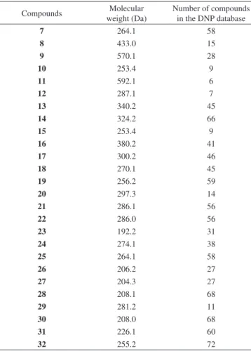
An example that illustrates the effectiveness of the rapid identification approach is the identification of sydowic acid (7). This metabolite was isolated from the culture medium of the
marine-derived strain A. sydowii DRG1M2. It has a molecular weight of 264 Da that matches other 58 fungal metabolites in the DNP database (Table 2). The identification of (7) was possible by analysis of LRMS and UV spectra together with analysis of its 1H NMR spectrum, which showed three methyl signals at δ 0.97 (s), δ 0.98 (s), and δ 1.50 (s), one trisubstituted benzene moiety with 1H signals at δ 7.28 (d, 1.7 Hz), δ 7.33 (dd, 8.0 and 1.7 Hz), δ 7.38 (d, 8.0 Hz), and a carboxylic acid 1H at δ 9.98 (s). Comparison of sydowic acid (7) 1H NMR data with the structures of compounds with the same molecular weight and similar UV absorption obtained in the DNP enabled its rapid identification,16 with no need for further NMR analyses. This approach defrayed the costs of time-consuming NMR experiments and provided rapid access to the structures of an extensive number of known fungal metabolites, many of which have been isolated several decades ago, when bioassays to evaluate such metabolites were still very limited.
Of the twenty-six compounds herein reported, the structures of (8) and (28) have not been previously reported in the literature. Compound (8) was identified as the opposite enantiomer of (+)-5,7-di-hydroxy-2-[1-(4-methoxy-6-oxo-6H -pyran-2-yl)-2-phenylethylami-no]-[1,4]naphthoquinone.17 It was isolated as a red amorphous solid and presented a [M+H]+ ion at m/z 434.0. Analysis of its 1H NMR spectrum indicated signals characteristic of a monosubstituted ben-zene ring at δ 7.30 (m), δ 7.28 (m), and δ 7.23 (m). Two hydrogens observed at δ 6.96 (d, 2.3 Hz) and δ 6.51 (d, 2.4 Hz) were assigned to a tetrasubstituted benzene moiety. Hydrogens characteristic of a pyrone group at δ 5.98 (d, 2.3 Hz) and δ 5.45 (d, 2.3 Hz) were de-tected, along with a protonat δ 5.59 (s) attached to a sp2 carbon, and two singlets at δ 12.91 (s) and at δ 3.77 (s) assigned to a hydroxyl and methoxyl groups, respectively. Finally, two diastereotopic hydrogens were observed at δ 3.31 (dd, 13.9; 6.0 Hz) and δ 6.21 (dd, 13.9; 8.2 Hz). Analysis of its 13C NMR spectrum confirmed the presence of a monosubstituted benzene ring at δ 132.1, 129.2 (2C), 128.6 (2C) and 127.1, the tetrasubstituted aromatic ring at δ 163.4, 161.0, 136.3, 108.9, 108.8, 107.9, the pyrone ring at δ 170.7, 163.3, 163.2, 101.2, 88.3, and a methoxyl group at δ 56.2. Two additional sp2 carbon signals were observed at δ 147.5, 100.4, a heteroatom-substituted
carbon at δ 55.2, two conjugated carbonyl groups δ 188.4 and at δ 180.7, and a methylene at δ 38.2.17
For compound (28), a query to the Dictionary of Natural Products database showed 68 additional fungal compounds with the same UV spectrum (λmax 240, 279 and 347 nm) and molecular weight (m/z 209.1 for the [M+H]+ ion). Analysis of the 1H, 13C and HSQC NMR spectra indicated the presence of a 1,2,4,5-tetrasubstituted benzene ring with 1H signals at δ 7.13 (s, CH-5) and δ 6.28 (s, CH-8). Two methyl groups represented by overlapping singlets at δ 1.40 (s, H3C-9 and H3C-10), one methylene group at δ 2.61 (s, CH2-3), one carbonyl group at δ 193.8 (C-4), as well as the presence of five quaternary carbons, including two hydroxylated aromatic carbons at δ 157.6 (C-7) and 141.7 (C-6) and one oxymethine group at δ 80.1 (C-2), were also observed. In the HMBC spectrum of (28), correlations from the methylene group at δ 2.61 (s) to C-2 (δC 80.1), C-9/C-10 (δC 26.8), C-4a (δC 113.2) and C-4 (δC 193.8), confirmed the structure and position of the pyranone ring in the molecule. HMBC correlations from CH-5 at δ 7.13 (s) to C-4a (δC 113.2) and C-8 (δC 104.6), and from CH-8 at δ 6.28 (s) to C-4a (δC 113.2) confirmed the substitu-tion pattern of the 1,2,4,5-tetrasubstituted benzene ring. Compound (28) was identified as 6,7-dihydroxy-2,2-dimethyl-4-chromanone, first synthesized in 1937.38 Related compounds displayed antioxi-dant activity and are used as inhibitors of self-oxidation of various organic substrates such as plastics, lubricants and rubber products often exposed to air during manufacturing processes.38,39 Production of (28) by Cochliobolus spp. is its first report from a natural source. Selected compounds were evaluated in antiviral assays. Compounds 8, 18, 21, harzianopyridone (29) and pyrenocine A (30)
displayed moderate to strong antiviral activity (Table 3). Compound (18) also showed moderate activity against strains of hepatitis C virus at a concentration of 2.5 µg mL-1.
In order to identify the stage of viral replication on which compounds (29) and (30) were active, three different treatments were performed. The first treatment consisted of infecting cells with viruses and incubating them for 1 h, followed by the addition of the compounds. The second treatment consisted of incubating the viruses with the compounds, in order to observe virus inacti-vation during the viral replication phase. Finally, the addition of compounds to cells prior to viral infection was performed in order to evaluate any effect on viral adsorption. Harzianopyridone (29) was active against HSV-1 during the viral inactivation phase (99% inhibition), with an EC50 of 0.2 µg mL-1, low toxicity (34 µg mL-1) and high selectivity against HSV-1. Pyrenocine A (30) presented 90% inhibition of AMPV growth during the viral replication phase, with an EC50 of 63.27 µg mL
-1, as well as 90% inhibition of HSV-1 during the viral adsorption phase, with an EC50 of 4.2 µg mL-1. Pyrenocine A also displayed both cytotoxic activity (~ 3 to 5 µg mL-1) and low selectivity. Both (29) and (30) displayed antiviral activity against HSV-1 KOS (acyclovir resistant HSV-1) virus strain (data not shown). Such result is particularly interesting, considering that the best drug for the systemic treatment of HSV-1 infection is acyclovir, which inhibits viral replication through competitive inhibition of the viral DNA polymerase and chain termination of viral DNA strains. HSV-1 resistance to acyclovir can result from mutations in either the viral TK gene or the viral DNA polymerase gene. This resistance is developed both in vitro and in vivo, and it
Table 1. Secondary metabolites identified by HPLC-UV-MS and 1H NMR analysis
Compounds Filamentous fungi (strain) Molecular formula UV (λmax nm) MS data (m/z)
Sydowic acid (7) A. sydowii (DRG1M2) C24H20NO7 212, 244, 299 247 [M-H2O]+
Naphthoquinoneimine (8) Aspergillus sp. (DLM38) C32H27O10 207, 270, 334 434.0 [M+H]+
Aurosperone A (9) Aspergillus sp. (DLM38) C32H26O10 228, 258, 277 571.1 [M+H]+
Pyranonigrin C (10) Aspergillus sp. (DLM38) C22H22N2O12 231, 290, 329 254.4 [M+H]
+
Aurosperone C (11) Aspergillus sp. (DLM38) C31 H29O12 233, 282, 334 593.1 [M+H]+
Pyrophen (12) Aspergillus sp. (DLM38) C16H17NO4 265 323.2 [M+Cl]
-Leucomelone (13) Aspergillus sp. (DLM38) C18H11O7 265 339.2 [M-H]
-Atromentin (14) Aspergillus sp. (DLM38) C18H11O6 266 323.2 [M-H]
-Pyranonigrin B (15) Aspergillus sp. (DLM38) C22H22N2O12 233, 291, 371 254.1 [M+H]
+
Penicitrinone A (16) P. glabrum (DLM218) C23H23O5 283, 307, 383 381.2 [M+H]+
Pinselin (17) P. glabrum (DLM218) C16H11O6 235, 263, 295, 384 323.1 [M+Na]+
Emodin (18) P. glabrum (DLM218) and
P. citrinum (DLM212)
C15H11O5 221, 266, 287 271.2 [M+H] +
Quinolactacin B (19) P. citrinum (DLM212) C15H16N2O2 213, 256, 315 257.2 [M+H]+
Penicillenol A (20) P. citrinum (DLM212) C16H27NO4 224, 283 298.3 [M+H]+
Citreorosein (21) P. citrinum (DLM212) C15H10O6 221, 287 287.1 [M+H]
+
Quinolactacin C (22) P. citrinum (DLM212) C16H18N2O3 218, 250, 316 287.0 [M+H]
+
5-methylmellein (23) Roussoella sp. (DLM33) C11H12O3 248, 322 193.2 [M+H]+
5,7-Dichloro-3-methyl-6-methoxy-8-hydroxy-3,4-dihy-droisocoumarin (24)
Roussoella sp. (DLM33) C12H15O3 259 207.2 [M+H]+
(+)-abscisic acid (25) Cochliobolus sp. (F56) C15H20O4 261 287.3 [M+Na]+
4-hydroxy-3-prenyl-benzoic acid (26) Cochliobolus sp. (F56) C12H14O3 259 207.2 [M+H]+
Anofinic acid (27). Cochliobolus sp. (F56) C12 H12O3 239 205.3 [M+H]+
6,7-dihydroxy-2,2-dimethyl-4-chromanone (28) Cochliobolus sp. (F56) C11H12O4 240, 279, 347 209.1 [M+H] +
Harzianopyridone (29) Trichoderma sp. (F254) C14H19NO5 203, 237, 269, 333 282.2 [M+H]
+
Pyrenocine A (30) P. paxilli (Ma(G)K) C11H12O4 228, 274 209.0 [M+H]+
Pyrenocine B (31) P. paxilli (Ma(G)K) C11H14O5 262 272.0 [M+H]+
is very significant in immunocompromised patients.10,40 Therefore, new, potent, and selective antiviral agents are necessary to provide alternative HSV-1 treatment choices, with (29) and (30) representing
attractive candidates for development as new antivirals. The structu-ral relatedness of (29) and (30) suggests that such structure scaffolds may be of interest for further synthesis and SAR investigations.
CONCLUSION
A strategy for the rapid identification of purified fungal meta-bolites by HPLC-UV-MS and 1H NMR analyses and, less often, by 13C NMR analysis, enabled the identification of 26 compounds produced by different marine-derived and endophytic fungal strains. Expensive and time-consuming 13C and two-dimensional NMR analyses for identification of known metabolites were avoided. An unreported enantiomer of (+)-5,7-dihydroxy-2-[1-(4-methoxy-6--oxo-6H-pyran-2-yl)-2-phenylethylamino]-[1,4]naphthoquinone, (-)-8, and 6,7-dihydroxy-2,2-dimethyl-4-chromanone (28), are herein reported as natural products for the first time. Evaluation in antiviral assays indicated that pyrenocine A (30) presents significant antiviral activity against HSV-1 in the adsorption phase, and against AMPV in the replication phase. Harzianopyridone (29) showed potent antiviral activity against HSV-1 during the viral inactivation phase coupled with a high selectivity, indicating that, although both compounds (29) and (30) present structural relatedness, their subtle structural differences have to be considered in order to better understand their antiviral mechanism-of-action against HSV-1. Since both pyrenocine
A and harzianopyridone can be obtained via fungal fermentation14,35,34 or by synthesis,41,42 these compounds clearly represent structural sca-ffolds worth of investigation for new antiviral leads in drug discovery. AKNOWLEDGMENTS
Financial support was provided by the FAPESP BIOTA/ BIOprospecTA grants (2010/50190-2 and 2013/50228-8) awarded to RGSB, and by FAPESP scholarships to LPI (2011/08064-2), MHK (2008/00331-9) and RPMU (2013/23153-7). Further financial support was provided by CNPq scholarships to LPI, ELFF and LKK, and by CAPES scholarships to MCFS, KJ, MSP and SR (BEX 4498-14-3). REFERENCES
1. Aly, A. H.; Debbab, A.; Proksch, P.; Fungal Diversity 2011, 50, 3. 2. Ratebab, M. E.; Ebel, R. Nat. Prod. Rep. 2011, 28, 290. 3. Nielsen, K. F.; Larsen, T. O.; Front. Microbiol. 2015, 6, 71. 4. Wolf, D.; Siems, K.; Chimia 2007, 61, 339.
5. El-Elimat, T.; Figueroa, M.; Ehrmann, B. M.; Cech, N. B.; Pearce, C. J.; Oberlies, N. H.; J. Nat. Prod. 2013, 76, 1709.
6. Klitgaard, A.; Iversen, A.; Andersen, M. R.; Larsen, T. O.; Frisvad, J. C.; Nielsen, K. F.; Anal. Bioanal. Chem. 2014, 406, 1933.
7. Wolfender, J.–L.; Marti, G.; Thomas, A.; Bertrand, S.; J. Chromatogr. A 2015, 1382, 136.
8. Ito, T.; Masubuchi, M.; J. Antibiot. 2014, 67, 353. 9. Gaudêncio, S. P.; Pereira, F.; Nat. Prod. Rep. 2015, 32, 779. 10. Antonelli, G.; Turriziani, O.; Int. J. Antimicrob. Agents 2012, 40, 95. 11. Hwang, Y.; Rowley, D.; Rhodes, D.; Gertsch, J.; Fenical, W.; Bushman,
F.; Mol. Pharm. 1999, 55, 1049.
12. Rowley, D. C.; Kelly, S.; Kauffman, C. A.; Jensen, P. R.; Fenical, W.; Bioorg. Med. Chem. 2003, 11, 4263.
13. Ióca, L. P.; Allard, P.-M.; Berlinck, R. G. S.; Nat. Prod. Rep. 2014, 31, 646.
14. Kossuga, M. H.; Romminger, S.; Xavier, C.; Milanetto, M. C.; Valle, M. Z.; Pimenta, E. F.; Morais, R. P.; Carvalho, E.; Mizuno, C. M.; Coradello, L. F. C.; Barroso, V. M.; Vacondio, B.; Seleghim, M. H. R.; Cavalcanti, B. C.; Pessoa, C.; Moraes, M. O.; Lima, B. A.; Gonçalves, R.; Bonugli-Santos, R. C.; Sette, L. D.; Berlinck, R. G. S.; Rev. Bras. Farmacogn. 2012, 22, 257.
15. Petrini, O. In Microbial Ecology of leaves; Andrews, J. H.; Hirano, S. S., eds.; Springer-Verlag: New York, 1991, pp. 179-197.
16. Hamasaki, T.; Sato, Y.; Hatsuda, Y.; Agr. Biol. Chem. 1975, 39, 2337. 17. Zhang, Y.; Li, X. M.; Wang, C. Y.; Wang, B. G.; Chinese Chem. Lett.
2007, 18, 951.
18. Priestap, H. A.; Tetrahedron 1984, 40, 3617.
19. Shaaban, M.; Shaaban, K. A.; Abdel-Aziz, M. S.; Org. Med. Chem. Lett.
2012, 2, 6.
20. Hiort, J.; Maksimenka, K.; Reichert, M.; Perovic-Ottstadt, S.; Lin, W. H.; Wray, V.; Steube, K.; Schaumann, K.; Weber, O. H.; Proksch, P.; Ebel, R.; Muller, W. E. G.; Bringmann, G.; J. Nat. Prod. 2004, 67, 1532. 21. Varoglu, M.; Crews, P.; J. Nat. Prod. 2000, 63, 41.
22. Gan, X.; Jiang, W.; Wang, W.; Hu, L.; Org. Lett. 2009, 11, 589. 23. Ye, Y. Q.; Koshino, H.; Abe, N.; Nakamura, T.; Hashizume, D.;
Taka-hashi, S.; Biosci. Biotechnol. Biochem. 2010, 74, 2342.
24. Wakana, D.; Hosoe, T.; Itabashi, T.; Okada, K.; Takaki, G. M. C.; Yagu-chi, T.; Fukushima, K.; Kawai, K.; J. Nat. Med. 2006, 60, 279. 25. Moppett, C. E.; J. Chem. Soc. D 1971, 423.
26. Danielsen, K.; Aksnes, D. W.; Francis, G. W.; Magn. Reson. Chem.
1992, 30, 359.
27. Kakinuma, N.; Iwai, H.; Takahashi, S.; Hamano, K.; Yanagisawa, T.; Nagai, K.; Tanaka, K.; Suzuki, K.; Kirikae, F.; Kirikae, T.; Nakagawa, A.; J. Antibiot. 2000, 53, 1247.
Table 2. Search outcome after taxonomy and HPLC-UV-MS data queries in the Dictionary of Natural Products database
Compounds Molecular
weight (Da)
Number of compounds in the DNP database
7 264.1 58
8 433.0 15
9 570.1 28
10 253.4 9
11 592.1 6
12 287.1 7
13 340.2 45
14 324.2 66
15 253.4 9
16 380.2 41
17 300.2 46
18 270.1 45
19 256.2 59
20 297.3 14
21 286.1 56
22 286.0 56
23 192.2 31
24 274.1 38
25 264.1 58
26 206.2 27
27 204.3 27
28 208.1 68
29 281.2 11
30 208.0 68
31 226.1 60
32 255.2 72
Table 3. Overall antiviral activity (% inhibition) of compounds 8, 18, 21,
29 and 30
Viral strain 8 18 21 29 30
Avian metapneumovirus (AMPV) i 68% 44% i 98%
Bovine diarrhea virus (BVDV) i 42% 39% i i
Herpes Simplex Virus Type 1 (HSV-1) 99% 44% 44% 99% 98%
28. Lin, Z. J.; Lu, Z. Y.; Zhu, T. J.; Fang, Y. C.; Gu, Q. Q.; Zhu, W. M.; Chem. Pharm. Bull. 2008, 56, 217.
29. Ren, H.; Tian, L.; Gu, Q.; Zhu, W.; Arch. Pharm. Res. 2006, 29, 59. 30. Bi, Y-M.; Bi, X-B.; Fang, A.; Zhao, Q-R.; Arch. Pharm. Res. 2007, 30,
267.
31. McGahren, W. J.; Mitscher, L. A.; J. Org. Chem. 1968, 33, 1577. 32. Assante, G.; Merlini, L.; Nasini, G.; Specialia 1977, 33, 1556. 33. Abraham, W. - R.; Arfmann, H, - A,; Phytochemistry 1990, 29, 2641. 34. Dickinson, J. M.; Hanson, J. R.; Hitchcock, P. B.; J. Chem. Soc. Perkin
Trans. I 1989, 11, 1885.
35. Sato, H.; Konoma, K.; Sakamura, S.; Agric. Biol. Chem. 1979, 43, 2409.
36. Liu, D.-Z.; Fei, W.; Liu, J.-K.; J. Nat. Prod. 2007, 70, 1503. 37. Bhat, H. B.; Vjznkataraman, K.; Tetrahedron 1963, 19, 77.
38. Toledo, T. R.; Dejani, N. N.; Silva Monnazzi, L. G.; Kossuga, M. H.; Berlinck, R. G. S.; Sette, L. D.; Medeiros, A. I.;Mediators Inflammation
2014, Article ID 767061, 11 pg.
39. Nishiyama, T.; Sugimoto, T.; Miyamoto, N.; Uezono, M.; Nakajima, Y.; Polym. Degrad. Stab. 2000, 70, 103.
40. Strasfeld, L.; Chou, S.; Infect. Dis. Clin. North Am. 2010, 24, 413. 41. Tréecourt, F.; Mallet, M.; Mongin, O.; Quéguiner, G.; J. Heterocyclic
Chem. 1995, 32, 1117.