r e v a s s o c m e d b r a s .2 0 1 3;5 9(5):411–412
Revista da
ASSOCIAÇÃO MÉDICA BRASILEIRA
w w w . r a m b . o r g . b r
Image in Medicine
Infectious cerebral embolism
Embolia cerebral infecciosa
Tsuneaki Kenzaka
∗, Yuki Ueda
Division of General Medicine, Center for Community Medicine, Jichi Medical University School of Medicine, Shimotsuke, Japan
An 82-year-old woman with a history of Alzheimer disease and hypertension was emergently transferred to our hos-pital with a fever of 37.4◦C, dysuria for three days, and
reduced level of consciousness. Her blood pressure was 88/55 mmHg and she had a Glasgow Coma Scale (GCS) score of E1V1M4; she presented no nuchal rigidity. Lumbar punc-ture was performed between the level of L4/L5. The cell counts of cerebrospinal fluid were 85/g; protein, 50 mg/dL
(standard value: 10-40 mg/dL); glucose, 40 mg/dL (blood glu-cose: 120 mg/dL). Considering the hypothesis of meningitis, antimicrobial agents were administered.
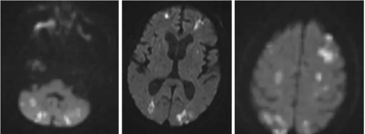
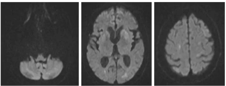
Fig. 1 – Diffusion-weighted image (DWI) showing multiple high signal-intensity lesions in the cerebral hemisphere (mainly watershed areas) and the cerebellar hemisphere.
∗ Corresponding author.
E-mail: [email protected] (T. Kenzaka).
Diffusion-weighted image (DWI) of brain magnetic res-onance image (MRI) showed multiple high signal-intensity lesions in cerebrum (mainly watershed areas) and cerebel-lum (Fig. 1). Trousseau syndrome, coagulation abnormalities including heparin-induced thrombocytopenia, eosinophilic meningoencephalitis, and fat embolism demonstrate images similar to the present patient’s.
However, bacteremia was suspected, based on acute course and examination of the cerebrospinal fluid. There were no findings in the transthoracic echocardiography to clearly demonstrate infective endocarditis in a visible range. Since
412
r e v a s s o c m e d b r a s .2 0 1 3;5 9(5):411–412Fig. 2 – MRI of the lumbar spine. Left image: T2-weighted image. Right image: Diffusion-weighted image. The white circles indicate the discitis image.
Fig. 3 – MRI of the head on the 15thday of hospitalization.
her general condition was poor, undergone transesophageal echocardiography in the acute phase was not possible. On the third day of hospitalization, her level of consciousness mildly improved, and she had a complaint of lumbago. MRI of the lumbar spine revealed a discitis image in the level of L2/L3 (Fig. 2). While Escherichia coli was detected in the urine, blood cultures, and cerebrospinal fluid, the suscepti-bility for the antimicrobial agents ofE. coliwas the same in
all the specimen materials. Therefore, the diagnosis of clini-cal infective endocarditis with infectious cerebral embolism and infectious discitis due to E. coli was made. Addition-ally, the MRI allowed for the diagnosis of infectious cerebral embolism.