08 artigo 546
ORIGINAL ARTICLE
1 – MSc and PhD in Orthopedics. Head of the Shoulder Group at the Orthopedic Hospital, Belo Horizonte Hospital and Lifecenter Hospital, Belo Horizonte, MG, Brazil. 2 – Attending Surgeon in the Shoulder Group at the Orthopedic Hospital and Lifecenter Hospital, Belo Horizonte, MG, Brazil.
3 – Attending Surgeon in the Shoulder Group at the Orthopedic Hospital, Belo Horizonte Hospital and Lifecenter Hospital, Belo Horizonte, MG, Brazil. 4 – Attending Surgeon in the Shoulder Group at the Orthopedic Hospital and Lifecenter Hospital, Belo Horizonte, MG, Brazil.
5 – Resident in the Shoulder Group at the Orthopedic Hospital, Belo Horizonte Hospital and Lifecenter Hospital, Belo Horizonte, MG, Brazil. Work performed at the Orthopedic Hospital, Belo Horizonte Hospital and Lifecenter Hospital, Belo Horizonte, MG, Brazil.
Correspondence: Hospital Ortopédico-BH, Rua Professor Otávio Coelho de Magalhães 111, Mangabeiras, 30210-300 Belo Horizonte, MG. E-mail: [email protected] Work received for publication: June 21, 2011; accepted for publication: August 19, 2011.
RESULTS FROM ARTHROSCOPIC REPAIR OF ISOLATED TEARS
OF THE SUBSCAPULARIS TENDON
Glaydson Gomes Godinho1, Flávio de Oliveira França2, José Márcio Alves Freita3, Flávio Márcio Lago Santos4, Ricardo Barreto Monteiro dos Santos5, Thiago Martins Taglietti5, Carlos Leonidas Escobar Guevara5
AbSTRACT
Objective: To evaluate the functional and clinical outcomes and identify prognostic factors in patients undergoing arthros-copic repair of isolated tears of the subscapularis tendon. Me-thods: Between January 2003 and May 2009, we identified 18 shoulders with isolated tears or deinsertions that were comple-te or affeccomple-ted at least one third of the subscapularis comple-tendon and underwent arthroscopic repair. Results: Three shoulders (17%) showed lesions in the upper third of the subscapularis; nine shoulders (50%) showed lesions in the upper two thirds; and six shoulders (33%) presented complete tears. In comparing the range of lateral rotation of the injured shoulder between before surgery and the time of the reevaluation, there was no statistical difference (p = 0.091). The LHBT was damaged in
INTRODUCTION
The tendon of the subscapularis muscle is located in the anterior portion of the rotator cuff. It is the main medial rotator of the humerus and contributes towards the dynamic anterior stability of the glenohumeral joint. It is the most important muscle not only for gle-nohumeral joint stability but also for function, since it contributes 52% of the shoulder abduction moment, while the supraspinatus contributes 14% and the in-fraspinatus and teres minor contribute 32%(1,2). Its
insertion extends from anteriorly in the lesser tubercle to posteriorly and laterally above the intertubercular sulcus, thus coalescing with the supraspinatus fibers, coracohumeral ligament and superior glenohumeral
The authors declare that there was no conflict of interest in conducting this work
This article is available online in Portuguese and English at the websites: www.rbo.org.br and www.scielo.br/rbort
11 shoulders (61%). According to the Constant score valida-tion, we had excellent and good results in 83% of the cases and 17% were reasonable. The reevaluations on three patients showed re-tearing on MRI. Acromioplasty was performed on ten patients and this procedure did not represent statistical differences in the final results (p = 0.57). Conclusions: There was no statistically significant difference in relation to preo-perative lateral rotation between the injured shoulder and the contralateral side. There was no significant loss of lateral ro-tation after surgery. The LHBT may be normal in deinsertions of the subscapularis tendon. Acromioplasty did not influence the results. The re-tearing rate for arthroscopic repair of the subscapularis tendon was 16.6%.
Keywords –Shoulder; Arthroscopy; Rotator Cuff
ligament to form the reflection pulley, which is an important stabilizer of the long head biceps tendon (LHBT)(3,4). Its amplitude is 7.3 cm, and it is the
stron-gest muscle of the rotator cuff(5). The upper two-thirds
of the craniocaudal extent of the subscapularis pre-sent a tendon insertion, while the lower one-third is inserted directly into the humerus by means of the muscle fibers, with a fine membranous structure(6).
In the tendinous portion of the subscapularis, 36% is intra-articular(7).
Rotator cuff injuries are common sources of shoul-der pain and dysfunction(8). Isolated deinsertion of
and these injuries are most frequent among men in their fifth decade of life(9-11). One of the principal
symptoms of a compromised subscapularis is pain that is more localized on the anterior face of the shoulder, in comparison with other rotator cuff tears(8).
Associa-ted involvement of the LHBT is observed in 31% to 56% of the cases and is generally a source of symp-toms(9,11,12). Arthroscopic viewing is the best means
of diagnosing subscapularis lesions, since more than 90% of such lesions start on the joint face and cephalic portion, close to the insertion in the lesser tubercle(13).
Our aim here was to evaluate the functional and clinical results and identify prognostic factors in pa-tients undergoing isolated deinsertion of the subsca-pular tendon arthroscopically.
MATERIAL AND METHODS
description of the sample and inclusion and ex-clusion criteria
Between January 2003 and May 2009, 2594 shoul-ders underwent arthroscopic repair of the rotator cuff at the Belo Horizonte Hospital, Lifecenter Hospital and Orthopedic Hospital. We retrospectively iden-tified 18 shoulders (0.7%) that underwent arthros-copic repair involving isolated of the subscapularis and fitted within the inclusion and exclusion criteria. Patients whose length of follow-up was less than one year, with partial tears only in the thickness of the subscapular tendon, without complete deinsertion of part or all of the tendon, or any associated lesions in other tendons of the rotator cuff. In addition, patients who underwent subscapularis repair by means of an open procedure, cases of rerupture after the opera-tion and cases of sequelae of infecopera-tion or advanced osteoarthrosis. The indication for arthroscopic repair was isolated tearing or deinsertion that was complete or affected at least one third of the subscapularis, was of degenerative or traumatic origin and was associated with functional deficit and pain.
Eighteen patients (18 shoulders) were evaluated: 13 men (72.2%) and five women (27.8%). Their mean age was 59 years (43-76 years). The length of follow--up ranged from 12 to 87 months, with a mean of 34.3 months. The right shoulder was affected in 12 patients and the left in six. The dominant side was affected in 12 cases (66.7%).
This study was authorized by the medical ethics
committees of the institutions involved. The patients agreed to participate through giving their free and informed consent, as approved by the institutions’ ethics committees.
Nine of the patients had suffered traumatic dein-sertion: eight through a fall mechanism involving hyperextension with external rotation; and one due to a car accident. In the other nine cases, we did not identify any traumatic event in their clinical histories, and thus these cases were classified as presenting degenerative etiology. The mean time that elapsed between the start of symptoms and the surgical pro-cedure was 5.8 months (1-36 months).
variables analyzed
Preoperatively, the patients were evaluated using range of motion (ROM) measurements and by means of sufficiency tests for the subscapularis: the lift-off and belly-press tests. The latter was used only when the patient presented limitations of passive Internal rotation(12). The lift-off test is based on observation of
the weakness of Internal rotation, which is most easily demonstrated with the upper arm in hyperextension with Internal rotation, and with the dorsum of the hand positioned at the L3 level, which is when the subscapularis is at its maximum amplitude of contrac-tion. Patients with subscapularis tears are incapable of moving the dorsum of the hand away from the back. Gerber et al(11) described an alternative maneuver, the
belly-press test, for identifying subscapularis tears in situations when the lift-off test could not be perfor-med due to pain or limitation of ROM. In the belly--press test, subscapularis function is tested by asking the patient to press his hand onto his abdomen. If the elbow remains in front of the trunk with the wrist ex-tended, the subscapularis is functional. If the wrist is flexed, thereby compensating for the Internal rotation with the strength of the posterior deltoid, the test will be positive(12). Both tests, when positive, indicate
dys-function of the subscapularis. Before the operation, all the patients underwent magnetic resonance imaging to assess the degree of deinsertion of the subscapular tendon (Figure 1). This was classified based on the magnetic resonance images as follows: complete tear affecting the upper third of its craniocaudal extent; complete tear affecting two thirds of the extent; and total tear (Figures 2 and 3).
the subscapularis(12), and the results were scored
accor-ding to the Constant and Murley method(14). The scores
thus obtained were grouped into categories according to the methodological criteria described by Boehm: 91-100 points, excellent; 81-90, good; 71-80, satisfactory; 61-70, adequate; less than 60 points, weak(15).
surgical technique
After administration of general anesthesia in association with regional blockade of the brachial plexus, the patients were positioned in lateral decubitus
with the arm abducted at 20 to 30° and flexed at 20°. Arthroscopy was started through the standard posterior portal, using a 30° optic device, followed by assessment of the joint. In this, the conditions of the LHBT and the subscapularis tendon were assessed. The stability of the LHBT in the intertubercular sulcus was evaluated dynamically, by asking the attending surgeon to perform medial and external rotation on the limb. In situations in which limb instability was observed, tenotomy or tenodesis was then performed. In situations of partial lesions of the biceps, debridement was performed if the lesion affected less than 50% of the thickness. The anterosuperior portal (1 cm anterior to the anterolateral angle of the acromion) was used for debridement, tenotomy or tenodesis of the LHBT, if necessary and subsequently was used to help in suturing the subscapularis. The anteroinferior portal, which was constructed slightly laterally to the lateral edge of the coracoid process, was used for surgical instrumentation.
Acromioplasty was performed when bursal inspec-tion showed the presence of signs of impact on the anteroinferior acromion or subacromial spur.
After the surgery, the patients had to use a Velpeau sling for three weeks if the deinsertion was partial, or for six weeks if it was complete. Following the immobilization period, a physiotherapy protocol for improving the range of motion and for strengthening was started.
Figure 1 – Magnetic arthro-resonance showing subacromial--subdeltoid bursa filled with paramagnetic contrast, due to com-plete tearing of the subscapular tendon. The LHBT was seen to be subluxated medially.
Figure 2 – Representative diagram showing the three types of com-plete tearing of the subscapular tendon: undamaged (A); affecting the upper third of the craniocaudal extent (B); affecting two thirds of the extent (C); and total (D).
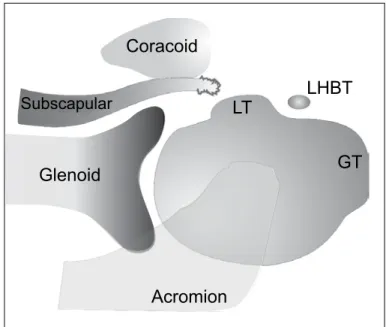
Figure 3 – Schematic drawing in axial view showing complete te-aring of the total thickness of the subscapularis tendon. LT: lesser tubercle; GT: greater tubercle; LHBT: long head biceps tendon.
Acromion Glenoid
Coracoid
Subscapular LT
STATISTICAL ANALYSIS
For descriptive statistical analysis, the mean and the minimum and maximum values were used. For analysis on the continuous variables, the nonparame-tric Wilcoxon text was used, while the t test was used for independent variables. The significance level was taken to be p < 0.05. For the statistical calculations, the resources of the R statistical software (version 2.11.1) were used.
RESULTS
Three shoulders (17%) presented deinsertion of the upper third of the subscapularis; nine shoulders (50%), the upper two thirds; and six shoulders (33%), complete deinsertion. In the preoperative evaluation, the mean degree of external rotation of the affected limb was 90º (range: 60º to 110º), while the mean for the unaffected side was 85º (range: 60º to 90º), without any statistical difference (p = 0.073). Com-parison of the amplitude of external rotation of the affected limb between before the operation and the time of the reassessment showed that there was no statistical difference (p = 0.091) (Table 1).
To attach the subscapularis to the lesser tubercle, a mean of two anchors was used (range: 0-4 anchors). In one case, the tear in the subscapular tendon occurred in the substance of the tendon, and this was repaired with three side-to-side stitches. Coracoplasty was not done in any of the cases. Tenodesis of the LHBT was performed on three shoulders (37.5%) and was included in suturing the subscapularis.
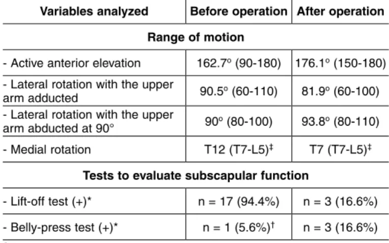
The degree of active anterior elevation before the operation ranged from 90 to 180º, with a mean of 162º, while the range was from 150 to 180º after the operation, with a mean of 176º (p = 0.61). In the pre-operative evaluation, 17 patients presented a positive lift-off test. There was only a single patient on whom this test could not be done, and this was because of limited Internal rotation (level of the gluteus). The belly-press test on this patient was positive. Three pa-tients who presented positive lift-off tests at the time of the reassessment had magnetic resonance images showing rerupture (Table 2).
During the arthroscopic evaluation, the LHBT was seen to be affected in 11 shoulders (61%; 95% confidence interval: 38.6-83.6). In eight cases, the LHBT was found to be dislocated or subluxated; in
two cases, it was torn; and in one case, it was stable but more than 50% of its thickness was compromised. In these cases, tenotomy was performed in six cases and tenodesis in three cases. In the other seven cases, no lesion or instability of the LHBT was observed. Comparison of the Constant and Murley score results between the group in which the LHBT was shown to be compromised and the group in which it was not affected did not show any statistical difference (p = 0.083) (Table 3).
Table 1 – Evaluation on the range of motion of lateral rotation. External rotation Mean (min-max) p Preoperative external rotation of the
unaffected side 85° (60°-90°)
} p = 0.073*
} p = 0.091* Preoperative external rotation of the
affected side 90.5° (60°-110°) Postoperative external rotation of
the affected side 81.9° (60°-100°)
Source: research data. * Wilcoxon test.
Table 2 – Physical examination on the affected limb, before and after the operation.
Variables analyzed before operation After operation Range of motion
- Active anterior elevation 162.7o (90-180) 176.1o (150-180)
- Lateral rotation with the upper
arm adducted 90.5o (60-110) 81.9o (60-100) - Lateral rotation with the upper
arm abducted at 90° 90o (80-100) 93.8o (80-110) - Medial rotation T12 (T7-L5)‡ T7 (T7-L5)‡
Tests to evaluate subscapular function
- Lift-off test (+)* n = 17 (94.4%) n = 3 (16.6%) - Belly-press test (+)* n = 1 (5.6%)† n = 3 (16.6%) Source: research data.
* Indicates a positive test for functional insufficiency of the subscapularis.
† Patient with limitation of medial rotation on the gluteus; it was not possible to perform the lift-off test. ‡ Values corresponding to vertebral level.
Table 3 – Arthroscopic evaluation of the LHBT, n (%).
Inspection/procedure n (%) p
Arthroscopic inspection of the LHbT n = 18
LHBT stable and unaffected n = 7 (39%)
} p = 0.083* LHBT affected IC (38.6 a 83.6)n = 11 (61%) †
- Dislocated or subluxated n = 8 (44%)
- Tear n = 2 (11%)
- Stable, with lesion greater than 50% n = 1 (6%)
Procedure performed on the LHbT n = 9
- Tenotomy n = 6 (67%)
- Tenodesis n = 3 (33%)
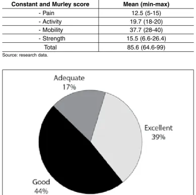
Application of the Constant and Murley score to evaluate the results after the operation showed that the range of scores was from 64.6 to 99 points, with a mean of 85.6 points (Table 4).
Using the validation of the Constant score descri-bed by Boehm(15), we found that 39% of the patients
had excellent results; 44%, good; and 17%, adequate (Figure 4).
Acromioplasty was performed in 10 cases. All of these cases presented signs of subacromial friction on inspection. Statistically, no influence from this pro-cedure was seen in the results (p = 0.57) (Table 5).
They observed only 26% ± 11% of the tendon area and concluded that the arthroscopic viewing was in-complete. Burkhart and Brady(17) recommended that a
70-degree optic device should be used for evaluating the subscapularis.
We did not find any difficulty in identifying the lesions of the subscapularis and performing the repair solely using a 30-degree optic device, by doing this in association with the maneuver of rotating the upper arm medially in order to improve the viewing of the cephalic portion of the subscapularis.
Associations with tenosynovitis, tendinosis and su-bluxation of the LHBT occur frequently and are an important cause of pain. In our study, the frequency of patients with biceps involvement was 61% (95% confidence interval: 38.6-83.6%). Comparison of the interval in this study with results in the literature, which show involvement in 31 to 56% of the cases, indicates that our results were similar. However, the first interval presented greater amplitude because of the small number of samples(9,11,12).
Bennett advocated repairing the subscapularis in association with reconstruction of the medial ligament complex of the LHBT, with the aim of recovering its stability in the intertubercular sulcus, given that repair of the subscapularis alone was insufficient to prevent its subluxation(18). Burkhart and Tehrany(9)
recommended that tenotomy or tenodesis of the LHBT should always be done when it was found to be unstable during the arthroscopic procedure. These authors believed that the arthroscopic procedure for reconstructing the ligament complex was flawed and was responsible for compromising the fixation of the subscapularis. We agree with Burkhart, and we belie-ve that situations in which the LHBT remains unstable or degenerated are an important reason for lack of success, because the painful condition is perpetuated. Provocative test results for the biceps (Speed and Yer-gason) are common findings in these patients(19,20).
Despite reports in the literature that patients with subscapular tears present increased amplitude of pas-sive external rotation, there was no statistical diffe-rence in our sample between the affected limb and the contralateral limb. This may be related to the percentage of the patients who undergo operations with deinsertions that are only partial. It is known that patients with partial ruptures may such increases that are less perceptible on clinical examination(18).
Table 4 – Postoperative results assessed by means of the Constant and Murley score.
Constant and Murley score Mean (min-max)
- Pain 12.5 (5-15)
- Activity 19.7 (18-20) - Mobility 37.7 (28-40) - Strength 15.5 (6.6-26.4)
Total 85.6 (64.6-99)
Source: research data.
Table 5 – Evaluation of the scores relating to the Constant variable according to acromioplasty procedure group.
Acromioplasty
procedure Cases (%)
Constant
Mean (min-max) p
No 8 45% 87.22 (64.6-99) 0.571* Yes 10 55% 84.39 (73-97)
Source: research data. * t test for independent variables.
Figure 4 – Results expressed as percentages obtained through Boehm’s validation(15).
DISCUSSION
Identification and arthroscopic repair of lesions of the subscapularis has received attention recently. Wright et al(16) evaluated six shoulders of fresh
Subcoracoid impact has been suggested to be a possible cause of partial or total tearing of the subsca-pularis and the rotator cuff, as well as a factor trigge-ring anterior pain in the shoulder. Some studies have suggested that the subscapular tendon is subjected to high tension on its posterior (convex) face when it crosses the stenosed subcoracoid space, thereby ini-tially leading to rupture of the joint fibers (Figure 1). Such lesions have been named tensile undersurface fiber failure (TUFF)(21,22). The normal
coracohume-ral distance is 8.7 to 11 mm(23,24). We do not believe
that subcoracoid impact is the primary etiology of subscapular lesions. There are large divergences of opinion in the literature regarding whether this type of lesion exists. Because of the lack of scientific evi-dence, we did not go ahead with coracoplasty in any of our cases.
All of the patients in our series underwent arthros-copic inspection of the subacromial space in order to investigate any lesions associated with other tendons. The cases in which signs of impact and/or subacro-mial spurs were identified underwent acromioplasty. This procedure was performed on 10 patients (56% of the cases) and there was no statistically significant difference in comparison with the group that did not undergo decompression.
Burkhart and Tehrany(9) reported that 92% of
the results from arthroscopic repair of subscapular lesions were excellent, among 25 shoulders in pa-tients of mean age 61 years and mean follow-up of 10.7 months.
Bennett(18) evaluated the results from eight patients
with isolated tears of the subscapularis that were re-paired arthroscopically, with a minimum follow-up of two years and a maximum of four years. They ob-served satisfactory results over both the short and the long term. Their surgical technique used not only the standard portals but also an accessory anterolateral portal and a portal through the supraspinatus in order to reconstruct the reflection pulley.
Kim et al(25) evaluated 29 patients of mean age 54
years with isolated partial joint lesions of the subsca-pularis who underwent arthroscopic repair. The mean length of follow-up was 2.3 years, with 18 excellent results, 10 good results and one failure.
Edwards et al(26) evaluated the results from
arthros-copic debridement of isolated lesions of the subsca-pularis and tenotomy of the biceps in 11 patients with
injuries that were considered to be arthroscopically irreparable or patients who did not choose to follow the entire rehabilitation protocol, if the lesion was repaired. These authors demonstrated good results and high satisfaction among the patients.
Lafosse et al(27) published prospective results
from 17 patients of mean age 47 years with isolated tears of the subscapularis who underwent arthros-copic repair. The mean follow-up was 2.4 years; 12 patients were very satisfied, four were satisfied and one was dissatisfied.
Adams et al(28) assessed the medium-term results
(three years of follow-up) from 40 patients with tears of the subscapularis. There were only seven patients with an isolated tear of the subscapularis. Coraco-plasty was performed in 46% of the cases, and was indicated when the coracohumeral interval was less than 6 mm. These authors reported that 80% of the results were excellent and good.
Balsini et al(29) retrospectively evaluated the
re-sults from 12 patients who underwent arthroscopic repair of isolated complete lesions of the subsca-pular tendon. After a minimum follow-up of one year, there were satisfactory results in 91.67% of the cases. These authors performed coracoplasty in 33% of the cases, which was indicated when the coracohumeral space seen during arthroscopy was less than 6 mm.
In our sample, occurrences of rerupture of the subscapularis were confirmed through magnetic re-sonance imaging in three cases. In clinical examina-tions on these patients, they presented positive tests for subscapular insufficiency (lift-off and belly-press). However, in assessing the Constant score, as validated by Boehm, we found that these patients presented adequate results.
In our sample, we observed that 83% of the results were good and excellent, while 17% were adequate. The LHBT was frequently affected (61% of the ca-ses), thus proving its close relationship with subsca-pular lesions.
CONCLUSIONS
There was no statistically significant difference in preoperative external rotation between the affected and contralateral sides (p = 0.073);
The LHBT may have a normal presentation in deinsertions of the subscapular tendon, provided that the reflection pulley is preserved;
The lesions of the LHBT did not influence the final result, according to the Constant and Murley index (p = 0.083);
Acromioplasty did not have any statistical influence
on the results (p = 0.57);
The arthroscopic repair on the deinsertions of the subscapular tendon presented a high rate of favorable results (83% of the results were excellent and good); and
The rerupture rate among the arthroscopic repairs on the subscapular tendon was 16.6%.
REFERENCES
1. Keating JF, Waterworth P, Shaw-Dunn J, Crossan J. The relative strengths of the rotator cuff muscles. A cadaver study. J Bone Joint Surg Br. 1993;75(1):137-40.
2. Warner JJ. Management of massive irreparable rotator cuff tears: the role of tendon transfer. Instr Course Lect. 2001;50:63-71.
3. Hunt SA, Kwon YW, Zuckerman JD. The rotator interval: anatomy, pathology, and strategies for treatment. J Am Acad Orthop Surg. 2007;15(4):218-27.
4. Jost B, Koch PP, Gerber C. Anatomy and functional aspects of the rotator interval. J Shoulder Elbow Surg. 2000;9(4):336-41.
5. Gerber C, Hersche O. Tendon transfers for the treatment of irreparable rotator cuff defects. Orthop Clin North Am. 1997;28(2):195-203.
6. Arai R, Sugaya H, Mochizuki T, Nimura A, Moriishi J, Akita K. Subscapu-laris tendon tear: an anatomic and clinical investigation. Arthroscopy. 2008;24(9):997-1004.
7. Pearsall AWt, Holovacs TF, Speer KP. The intra-articular component of the subscapularis tendon: anatomic and histological correlation in reference to surgical release in patients with frozen-shoulder syndrome. Arthroscopy. 2000;16(3):236-42.
8. Lyons RP, Green A. Subscapularis tendon tears. J Am Acad Orthop Surg. 2005;13(5):353-63.
9. Burkhart SS, Tehrany AM. Arthroscopic subscapularis tendon repair: Technique and preliminary results. Arthroscopy. 2002;18(5):454-63.
10. Bennett WF. Arthroscopic repair of anterosuperior (supraspinatus/subscapu-laris) rotator cuff tears: a prospective cohort with 2- to 4-year follow-up. Clas-sification of biceps subluxation/instability. Arthroscopy. 2003;19(1):21-33.
11. Gerber C, Krushell RJ. Isolated rupture of the tendon of the subscapularis muscle. Clinical features in 16 cases. J Bone Joint Surg Br. 1991;73(3):389-94.
12. Gerber C, Hersche O, Farron A. Isolated rupture of the subscapularis tendon. J Bone Joint Surg Am. 1996;78(7):1015-23.
13. Adams CR, Schoolfield JD, Burkhart SS. Accuracy of preoperative magnetic resonance imaging in predicting a subscapularis tendon tear based on arthros-copy. Arthrosarthros-copy. 2010;26(11):1427-33.
14. Constant CR, Murley AH. A clinical method of functional assessment of the shoulder. Clin Orthop Relat Res. 1987;(214):160-4.
15. Boehm D. Scores. In: Gohlke F, editor. Schulter: das Standardwerk für Klinik und Praxis. New York: Thieme; 2002. p. 98-104.
16. Wright JM, Heavrin B, Hawkins RJ, Noonan T. Arthroscopic visualization of the subscapularis tendon. Arthroscopy. 2001;17(7):677-84.
17. Burkhart SS, Brady PC. Arthroscopic subscapularis repair: surgical tips and pearls A to Z. Arthroscopy. 2006;22(9):1014-27.
18. Bennett WF. Arthroscopic repair of isolated subscapularis tears: A prospective cohort with 2- to 4-year follow-up. Arthroscopy. 2003;19(2):131-43.
19. Yoshikawa GI, Hori K, Kaneko H, Matsusue Y, Murakami M. Acute subscapu-laris tendon rupture caused by throwing: a case report. J Shoulder Elbow Surg. 2005;14(2):218-20.
20. Deutsch A, Altchek DW, Veltri DM, Potter HG, Warren RF. Traumatic tears of the subscapularis tendon. Clinical diagnosis, magnetic resonance imaging findings, and operative treatment. Am J Sports Med. 1997;25(1):13-22.
21. Richards DP, Burkhart SS, Campbell SE. Relation between narrowed coraco-humeral distance and subscapularis tears. Arthroscopy. 2005;21(10):1223-8.
22. Lo IK, Burkhart SS. The etiology and assessment of subscapularis tendon tears: a case for subcoracoid impingement, the roller-wringer effect, and TUFF lesions of the subscapularis. Arthroscopy. 2003;19(10):1142-50.
23. Friedman RJ, Bonutti PM, Genez B. Cine magnetic resonance imaging of the subcoracoid region. Orthopedics. 1998;21(5):545-8.
24. Gerber C, Terrier F, Zehnder R, Ganz R. The subcoracoid space. An anatomic study. Clin Orthop Relat Res. 1987;(215):132-8.
25. Kim SH, Oh I, Park JS, Shin SK, Jeong WK. Intra-articular repair of an isolated partial articular-surface tear of the subscapularis tendon. Am J Sports Med. 2005;33(12):1825-30.
26. Edwards TB, Walch G, Nove-Josserand L, Boulahia A, Neyton L, O’Connor DP, et al. Arthroscopic debridement in the treatment of patients with isolated tears of the subscapularis. Arthroscopy. 2006;22(9):941-6.
27. Lafosse L, Jost B, Reiland Y, Audebert S, Toussaint B, Gobezie R. Structural integrity and clinical outcomes after arthroscopic repair of isolated subscapularis tears. J Bone Joint Surg Am. 2007;89(6):1184-93.
28. Adams CR, Schoolfield JD, Burkhart SS. The results of arthroscopic subscapu-laris tendon repairs. Arthroscopy. 2008;24(12):1381-9.