SOCIEDADE BRASILEIRA DE ORTOPEDIA E TRAUMATOLOGIA
w w w . r b o . o r g . b r
Case
report
Osteoid
osteoma
of
the
distal
clavicle
夽
Bernardo
Barcellos
Terra
∗,
Leandro
Marano
Rodrigues,
David
Victoria
Hoffmann
Padua,
Tannous
Jorge
Sassine,
José
Maria
Cavatte,
Anderson
De
Nadai
SantaCasadeMisericórdiadeVitória,DepartamentodeOrtopediaeTraumatologia,Vitória,ES,Brazil
a
r
t
i
c
l
e
i
n
f
o
Articlehistory:
Received23November2015
Accepted29March2016
Availableonline17February2017
Keywords:
Clavicle
Osteoidosteoma
Shoulder
a
b
s
t
r
a
c
t
Theosteoid osteomais a bonetumor thataccounts for10%ofbenigntumors. Itwas
describedin1935byJaffe,asatumorthataffectstheyoungadultpopulation,witha
pre-dominanceofmales.Thisstudyaimstopresentacaseoflatediagnosisofapatientwith
osteoidosteomaofthedistalclavicleregion.Femalepatient,44yearsold,non-professional
volleyballplayer, reportedpainintheanterior andsuperiorregionoftheshoulder
gir-dle,specificallyinthe acromioclavicular joint,whichworsenedatnight andhadbeen
treatedforninemonthsastendinitisoftherotatorcuffandacromioclavicularjoint
arthri-tis.Afterconfirmingthediagnosis,thepatientunderwentopensurgerywithresectionof
thedistalclavicle.Attwoyearsoffollow-up,thepatientpresentswithoutlocalpain.In
theradiographicevaluation,coracoclaviculardistanceispreservedandtherearenosigns
ofrecurrence.Tumorsoftheshouldergirdlearerareandareoftendiagnosedlate.Ahigh
degreeofsuspicionforthediagnosisoftumorsoftheshouldergirdleisneededinorderto
avoidlatediagnosis.
©2016SociedadeBrasileiradeOrtopediaeTraumatologia.PublishedbyElsevierEditora
Ltda.ThisisanopenaccessarticleundertheCCBY-NC-NDlicense(http://
creativecommons.org/licenses/by-nc-nd/4.0/).
Osteoma
osteóide
da
clavícula
distal
Palavras-chave:
Clavícula
Osteomaosteóide
Ombro
r
e
s
u
m
o
Oosteomaosteóideéumtumorósseoquecorrespondea10%dostumoresbenignos.Foi
descritoem1935porJaffe,comoumtumorqueacometeapopulac¸ãoadultajovem,com
predominâncianosexomasculino.Oobjetivodotrabalhoéapresentarumcasode
diag-nósticotardiodeumapacientecomosteomaosteóidedaregiãodaclavículadistalerelatar
seutratamento.Pacientede44anos,jogadoradevôleinãoprofissional,comdoresnaregião
anterioresuperiordacinturaescapular,maisespecificamentenaarticulac¸ão
acromioclav-icular,asquaispioravamanoiteequeeratratadahavianovemesescomoumatendinitedo
manguitorotadoreartritedaarticulac¸ãoacromioclavicular.Apósconfirmac¸ãodiagnóstica,
apacientefoisubmetidaaotratamentocirúrgicoabertocomressecc¸ãodaclavículadistal.
夽
StudyconductedattheSantaCasadeMisericórdiadeVitória,DepartamentodeOrtopediaeTraumatologia,Vitória,ES,Brazil.
∗ Correspondingauthor.
E-mail:bernardomed@hotmail.com(B.B.Terra).
http://dx.doi.org/10.1016/j.rboe.2017.01.006
2255-4971/©2016SociedadeBrasileiradeOrtopediaeTraumatologia.PublishedbyElsevierEditoraLtda.Thisisanopenaccessarticle
Atualmenteapacienteencontra-secomdoisanosdeevoluc¸ãosemdorlocal.Naavaliac¸ão
radiográfica,adistânciacoracoclavicularencontra-sepreservadaenãohásinaisderecidiva.
Tumoresósseosdacinturaescapularsãorarosefrequentementesãodiagnosticados
tardia-mente.Deve-seterumaltograudesuspeic¸ãoparaodiagnósticodeneoplasiasdacintura
escapular,afimdeevitarodiagnósticotardio.
©2016SociedadeBrasileiradeOrtopediaeTraumatologia.PublicadoporElsevier
EditoraLtda.Este ´eumartigoOpenAccesssobumalicenc¸aCCBY-NC-ND(http://
creativecommons.org/licenses/by-nc-nd/4.0/).
Introduction
Osteoidosteomaisabenignbonetumorthataccountsfor10%
ofbenigntumors,representingthethirdmostcommonbone
tumor.Ithasapreferenceforthediaphysisoflongbones,such
asthetibiaandfemur.Itwasdescribedin1935andlaterin
1953byJaffe;itisatumorthataffectstheyoungadult
popu-lation,inthesecondandthirddecadeoflife,predominantly
inmales.1–3
Theclinical presentationcomprises mildtoseverepain,
predominantlyatnight,whichisusuallyrelievedbytheuse
ofsalicylates.Itcanbelocatedinanyboneregion,buthalfof
casesinvolvethefemurandthetibia.
Itspresenceintheregion ofthescapula and clavicleis
extremelyrare,withfewreportsintheliterature.Aliterature
searchretrievednocasesdescribed atthedistalendofthe
clavicle.
Thisstudyaimedtopresentacaseoflatediagnosisofan
osteoidosteomaofthedistalclavicleregionandtoreportits
treatment.
Case
report
A44-year-oldfemale, recreativevolleyballplayer,presented
withanteriorandsuperiorscapulargirdlepain,more
specifi-callyattheacromioclavicularjoint,whichworsenedatnight;
thisconditionhad beentreatedforninemonthsasrotator
cufftendinitisandacromioclavicularjointarthritis.Painwas
partiallyalleviatedbysalicylates.Patientdeniedahistoryof
previoustraumaorfall.
Onphysicalexamination,noedema,deformities,or
atro-phies in the region of the shoulder girdle were observed.
Passiveandactiverangeofmotionwerenormal,exceptfor
thefactthatforcedadductionwaspainfulattheextremeend
ofthemovement.
O’Brientestwas positiveinthesemiologicalmaneuvers
andatthepalpationoftheacromioclavicularjoint.Othertests
forrotatorcuffandinstabilitywerenegative.
Patientunderwentcomplementaryexaminations,through
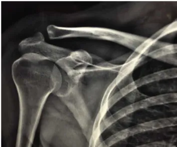
whichtheosteoidosteomawasevidenced,withitspeculiar
characteristicsatthedistalendoftheclavicleatthe
acromio-clavicularjoint(Figs.1–3).
Patient underwent open resection of the distal end of
the clavicle (approximately 1.5cm) in a way that did not
compromisetheinsertionofthecoracoclavicularligaments;
electrocoagulation withradiofrequencywas performeddue
to bone bleeding (Figs. 4 and 5). Tissue sample was sent
forhistopathological analysis and the diagnosis of osteoid
osteomawasconfirmed(AppendixA1,inadditionalmaterial).
Fig.1–Anteroposteriorviewradiographshowinganarea
ofsclerosisinthelateraldistalregionoftheclavicle.
Fig.2–Magneticresonanceimaging(MRI)incoronal
sectionshowingimageofthetumortogetherwithintense
Fig.3–MRIinsagittalsectionshowingtheimageofthe
nidusalongwithintenseedemainthelateraldistalregion
oftheclavicle.
Fig.4–Photographofthesuperioraccessrouteoverthe
claviclewiththeopenedacromioclavicularjointanda
curvedKellyforcepspointingtowardtheosteoidosteoma
region.
Intwomonths,patientevolvedfromavisualanalogscale
(VAS)scoreof9inthepreoperativeperiodto1.
Currently,at24monthsoffollow-up,shepresentsnolocal
pain.Radiographicevaluationshowsthatthe
coracoclavicu-lardistanceispreservedandtherearenosignsofrecurrence
(Fig.6).
Discussion
The clavicle is a rare location for tumors.4–6 Therefore,
orthopedistshavelittleexperienceinthediagnosisand
man-agementoftumorsandneoplasticconditionsofthisbone.The
Fig.5–Photographshowingtheosteotomyofthelateral
distalregionoftheclavicle.
oncologicalcharacteristicsofclavicletumorsresemblethose
offlatboneswhencomparedwiththoseoflongbones.Tumors
oftheclaviclearemostlymalignant;diagnosisisoftenlatedue
tothelowdegreeofsuspicionofthispathology.7
Osteoidosteomaaccountsfor10%ofbenignbonetumors,
being more common in menand inthe second and third
decadeoflife.8Inthesuperiorcingulate,themostcommon
siteofinvolvementistheproximalhumerus.Among
imag-ing studies, osteoid osteoma presents in radiographs as a
radiolucent imagewith centralcalcification. Lesionsin the
spongy boneusuallypresentas asmallarea ofrarefaction
with an area surrounded bysclerosis. Computed
tomogra-phyaidsinthelocalizationofthetumor.Magneticresonance
imagingpresentstumorchangesasalowintensitysignalat
T1andsignsofvariableintensityatT2-weighted.Although
tomographyisthestudyofchoice,theedemasurroundingthe
tumorandthebonemarrowalterationsarebestobservedwith
magneticresonanceimaging,asdemonstratedinthepresent
case.
Kapooretal.2reportedacaseseriesof12tumorsonthe
clavicle,the mostcommon ofwhichwasEwing’s sarcoma;
treatmentrangedfrompartialclaviculectomyto
chemother-apy. In theirseries, those authors did notreport acaseof
osteoid osteoma, despite reporting of a case with a rare
periostealdesmoidtumor.
Only 1% of all bone tumors affect the clavicle.9–11 The
mostcommonsiteistheacromialend,whichisinagreement
withthepresentcasereport.Miyasakietal.12reportedacase
ofosteoidosteoma intheacromionsimulatingpain inthe
acromioclavicularjoint,whichwasresectedarthroscopically
andassociatedwithanacromioplasty;patienthadacomplete
recovery ofrange ofmotionand no signsof recurrencein
sevenyearsoffollow-up.Degreefetal.11alsoreportedacase
inwhich theacromion wasresectedthrough theMumford
arthroscopic procedure.Inthe presentcase,openresection
was chosen due to the fact that this was a tumor, albeit
benign,andthismethodfacilitatesthecollectionofmaterial
Fig.6–Radiographyonanteroposteriorandprofileviews,inwhichexcisionofthedistalfragmentoftheclaviclewiththe
osteoidosteomaisnoted.
Glanzmannetal.13reportedacaseofosteoidosteomaof
thecoracoidsimulatingadhesivecapsulitis,whichwasalso
resectedbyarthroscopyandelectrocauterizationthroughthe
rotatorinterval.
The natural course of osteoma may be spontaneous
resolution over time; however, residual pain and
persis-tentsymptomsareindicativeofsurgery.Multipletreatment
optionsfor this tumorare available, suchasdrug therapy,
percutaneous radiofrequency ablation, and surgical
proce-duresinvolvingcompleteremovalofthenidus,whichcanbe
obtainedbycurettage, enbloc resectionand,morerecently,
arthroscopy,withgoodresults.Minimallyinvasivetreatments,
suchasradiofrequencythermocoagulationandpercutaneous
excisionalcorebiopsy,arethetreatmentsofchoiceinmany
centers,avoidingthecomplicationsofopensurgery.Themain
advantagesofpercutaneoustechniquesarefasterreturnto
activities,lowermorbidity,and,incasesofosteoidosteoma
ofthespine,maintenanceofstability.However,orthopedists
shouldbeawareofpossiblethermallesionstoneurological
structuresand,incasesofexcisionalbiopsy,ofincomplete
removalofthetumor,whichcouldleadtorecurrenceof
symp-tomsandlesion.4,14–17
Inordertoavoidlatediagnosis,scapulargirdleneoplasms
shouldbeconsideredinthediagnosisofrefractorypaininthe
superiorcingulateregion.
Conclusion
Theauthorsdescribedararecaseofosteoidosteomaatthe
distalendoftheclavicle.Despitetherarity,itisnecessaryto
includeneoplasmsinthedifferentialdiagnosesofpathologies
oftheshoulder.
Conflicts
of
interest
Theauthorsdeclarenoconflictsofinterest.
Appendix
A.
Supplementary
data
Supplementarydataassociatedwiththisarticlecanbefound,
intheonlineversion,atdoi:10.1016/j.rboe.2017.01.006.
r
e
f
e
r
e
n
c
e
s
1.JaffeHL.Osteoid-osteoma.ProcRSocMed. 1953;46(12):1007–12.
2.KapoorS,TiwariA,KapoorS.Primarytumoursandtumorous lesionsofclavicle.IntOrthop.2008;32(6):829–34.
3.MobergE.Thenaturalcourseofosteoidosteoma.JBoneJoint SurgAm.1951;33(1):166–70.
4.ReimanHM,DahlinDC.Cartilage-andbone-formingtumors ofthesofttissues.SeminDiagnPathol.1986;3(4):288–305.
5.KleinMH,ShankmanS.Osteoidosteoma:radiologicand pathologiccorrelation.SkeletalRadiol.1992;21(1):23–31.
6.KleinMJ,LusskinR,BeckerMH,AntopolSC.Osteoidosteoma oftheclavicle.ClinOrthopRelatRes.1979;(143):162–4.
7.OgoseA,SimFH,O’ConnorMI,UnniKK.Bonetumorsofthe coracoidprocessofthescapula.ClinOrthopRelatRes. 1999;(358):205–14.
8.SamilsonRL,MorrisJM,ThompsonRW.Tumorsofthe scapula.Areviewoftheliteratureandananalysisof31cases. ClinOrthopRelatRes.1968;(58):105–15.
9.IshikawaY,OkadaK,MiyakoshiN,TakahashiS,ShimadaY, ItoiE,etal.Osteoidosteomaofthescapulaassociatedwith synovitisoftheshoulder.JShoulderElbowSurg.
10.CohenMD,HarringtonTM,GinsburgWW.Osteoidosteoma: 95casesandareviewoftheliterature.SeminArthritis Rheum.1983;12(3):265–81.
11.DegreefI,VerduycktJ,DebeerP,DeSmetL.Anunusualcause ofshoulderpain:osteoidosteomaoftheacromion–acase report.JShoulderElbowSurg.2005;14(6):643–4.
12.MiyazakiAN,FregonezeM,SantosPD,daSilvaLA,doValSella G,NetoDL,etal.Osteoidosteomaoftheacromionsimulating acromioclavicularpain.RevBrasOrtop.2014;49(1):82–5.
13.GlanzmannMC,HinterwimmerS,WoertlerK,ImhoffAB. Osteoidosteomaofthecoracoidmaskedaslocalized capsulitisoftheshoulder.JShoulderElbowSurg. 2011;20(8):e4–7.
14.WoodsER,MartelW,MandellSH,CrabbeJP.Reactive
soft-tissuemassassociatedwithosteoidosteoma:correlation ofMRimagingfeatureswithpathologicfindings.Radiology. 1993;186(1):221–5.
15.ShaffreyCI,MoskalJT,ShaffreyME.Osteoidosteomaofthe clavicle.JShoulderElbowSurg.1997;6(4):396–9.
16.BednarMS,WeilandAJ,LightTR.Osteoidosteomaofthe upperextremity.HandClin.1995;11(2):211–21.


