*Correspondence: R. M. Mainardes. Departamento de Farmácia, Universidade Estadual do Centro-Oeste/UNICENTRO. Rua Simeão Camargo Varela de Sá 03, 85040-080 - Guarapuava - PR, Brasil. E-mail: [email protected]
A
vol. 49, n. 1, jan./mar., 2013
Development and validation of an HPLC method for the
determination of fluorouracil in polymeric nanoparticles
Ana Cristina de Mattos, Najeh Maissar Khalil, Rubiana Mara Mainardes
*Department of Pharmacy, Midwestern State University/UNICENTRO, Guarapuava, Paraná, Brasil
The objective of this work was to develop and validate a rapid high performance liquid chromatography
(HPLC) method for the quantitative analysis of luorouracil (5-FU) in polymeric nanoparticles.
Chromatographic analyses were performed on an RP C18 column with a mobile phase consisting of
acetonitrile and water (10:90, v/v) at a low rate of 1 mL/min. The 5-FU was detected and quantitated using a photodiode array detector at a wavelength of 265 nm. The method was shown to be speciic and linear in the range of 0.1-10 μg/mL (r = 0.9997). The precision (intra- and inter-day) was demonstrated
because the maximum relative standard deviation was 3.51%. The method is robust relative to
changes in low rate, column and temperature. The limits of detection and quantitation were 10.86 and 32.78 ng/mL, respectively. The method fulilled the requirements for reliability and feasibility for
application to the quantitative analysis of 5-FU in polymeric nanoparticles.
Uniterms: Fluorouracil/determination. Nanoparticles. High performance liquid chromatography/ quantitative analysis/method validation.
O objetivo deste trabalho foi desenvolver e validar um método rápido de cromatograia líquida de alta eiciência (CLAE) para análise quantitativa de luorouracila (5-FU) em nanopartículas poliméricas. Corridas cromatográicas foram realizadas sob uma coluna RP C18 com uma fase móvel consistindo de acetonitrila e água (10:90, v/v) a um luxo de 1 mL/min. O 5-FU foi detectado e quantiicado através de um detector de fotodiodos em um comprimento de onda de 265 nm. O método demonstrou ser especíico e linear na faixa de 0,1-10 μg/mL (r =0.9997). As precisões (intra e inter dia) revelaram um desvio padrão relativo máximo de 3,51%. O método é robusto considerando mudanças realizadas no luxo da fase móvel, temperatura e marca da coluna. Os limites de detecção e quantiicação foram de 10,86 e 32,78 ng/mL, respectivamente. O método cumpriu os requisitos para ser considerado coniável e viável para aplicação na análise quantitativa de 5-FU em nanopartículas poliméricas.
Unitermos: Fluorouracila/determinação. Nanopartículas. Cromatograia líquida de alta eiciências/
análise quantitativa/validação de método.
INTRODUCTION
The luorouracil (5-FU) is an anticancer agent used
in the treatment of solid tumors. This drug, an analog of the natural pyrimidine uracil, must be converted to the
nucleo-tide to exert its effect. The drug is rapidly metabolized after administration, giving cytotoxic luoronucleotides
with well-known antineoplastic properties (Pinedo, Peters, 1988). One explanation for this phenomenon is the
possi-bility that 5-FU pharmacomodulation markedly increases
the antitumor eficacy of this antimetabolite (Peters, Van
Groeningein, 1991).
The mechanism of 5-FU cytotoxicity is complex because the drug is activated through different pathways
leading to at least three cytotoxic compounds: luorode -oxyuridine monophosphate, which inhibits thymidylate
synthase and subsequent DNA synthesis; luorouridine triphosphate, which is directly incorporated into RNA; and luorodeoxyuridine triphosphate, for which incorporation into DNA has been suggested (Grem, 1990).
pharma-cokinetics than other anticancer drugs. For these reasons, some inter-patient differences in terms of toxicity and
eficacy can be expected based on individual pharmaco -kinetic parameters, especially in the area under the time
vs. concentration curve (AUC) (Gamelin, Boisdron-Celle,
1999; Gamelin et al., 1999; Casale et al., 2004). Another
problem with 5-FU therapy is its toxicity to the bone mar-row and the gastrointestinal tract (Tanaka et al., 2000; Lai,
Guo, 2011). Therefore, 5-FU represents an interesting drug model to be improved by nanotechnology.
Numerous investigators have shown that the
biologi-cal distribution of drugs, proteins or DNA can be modi
-ied, both at the cellular and organ levels, using micro/
nanoparticle delivery systems (Moghimi et al., 2001;
Panyam, Labhasetwar, 2003; Labhasetwar, Prabha, 2004; Akagi et al., 2005). The nanoencapsulation of drugs has many advantages for the protection from premature deg-radation and interaction with the biological environment, enhancement of absorption into a selected tissue, bioavail-ability, retention time and improvement of intracellular penetration. However, polymeric nanoparticles, when used intravenously, are removed from systemic circulation by the cells of the mononuclear phagocyte system (MPS). Several methods are used to modify the surface of the nanoparticles to avoid recognition and capture by the cells of the MPS and promote a long plasma circulating time
and improved pharmacokinetics. Among these methods,
coating nanoparticles with hydrophilic polymers such as polyethylene glycol (PEG) is the most popular because the
long chains of PEG prevent opsonization and phagocytosis by steric hindrance (Alexis et al., 2008).
To characterize the delivery systems such as poly -meric nanoparticles fully, suitable and validated quantita-tion methods are required to assess pharmaceutical param-eters such as drug content. Several methods are described in the literature for the determination of 5-FU in samples of biological matrices using gas chromatography/mass
spec-trometry (GC/MS) (Anderson et al., 1997), spectrometry
(Badea et al., 2002), high performance liquid
chromatog-raphy (HPLC) (Escoriaza et al., 1999), hydrophilic
inter-action liquid chromatography-APCI-mass spectrometry
(Pisano et al., 2005) or liquid chromatography tandem
mass spectrometry (LC–MS/MS) (Licea-Perez, Wang, Bowen, 2009; Liu et al., 2010). The analytical determi-nation of 5-FU in pharmaceutical dosage forms such as nanoparticles has been performed by several authors using
spectrophotometry (Bozkir, Saka, 2005; Liu et al., 2006;
Zhu et al., 2009; Lai, Guo, 2011; Li et al., 2011; Rejinold et al., 2011a; Rejinold et al., 2011b; Zhang et al., 2011), but few studies report the use of HPLC methods for this
deter-mination. Arbós, Campanero and Irache (2002) described
an HPLC method for the quantitation of 5-luorouridine in nanoparticles and veriied the possible degradation of
5-FU, but the investigators used a C8 column. Zheng et al.
(2007) described an HPLC method using as mobile phase
a mixture of methanol and 3.6% acetic acid (80:20, v/v), but details such as the retention time, peak characteristics and validation data were not described.
The objective of this work was therefore to develop
and validate a fast, simple and optimized HPLC method to determine the encapsulation eficiency of 5-FU incor
-porated in poly(lactic acid) (PLA) and PLA-PEG blended
nanoparticles.
MATERIAL AND METHODS
Materials
Fluorouracil (99% TLC), poly(lactic acid) (PLA)
(MW 85-160 kDa), polyethylene glycol (10 kDa) and
polyvinyl alcohol (PVA, 31 KDa, 88% hydrolyzed) were purchased from Sigma-Aldrich (St. Louis, MO, USA).
Methylene chloride was purchased from FMaia® (Brazil).
HPLC-grade acetonitrile was purchased from JTBaker®
(USA). Water was puriied in a Milli-Q Plus system (Mil -lipore®), and its resistivity was 18.2 MƱ cm. All other
solvents and chemicals were analytical or HPLC grade.
Instrumentation
A Waters 2695 Alliance HPLC system (Milford, MA, USA) was used for method development. The HPLC
system was equipped with a column compartment with temperature control, an on-line degasser, a quaternary
pump, an auto sampler and a photodiode array (PDA) wavelength detector (Waters 2998). Data acquisition,
analysis, and reporting were performed using Empower
chromatography software (Milford, MA, USA). HPLC
analysis was conducted using a RP C18 column (Xterra
Waters®), with 5 µm particle size, 4.6 mm internal diam
-eter and 250 mm length.
Preparation of standard and sample solutions
A stock standard solution of 500 µg/mL of 5-FU
was prepared in water and subsequent dilutions were car-ried out to obtain eight standard solutions (0.1, 0.5, 1.0,
2.0, 4.0, 6.0, 8.0 and 10.0 μg/mL). Similarly, six standard
solutions were obtained by serial dilutions of a 5-FU
stan-dard solution (1.0 μg/mL) with water (50.0, 75.0, 100.0,
125.0, 150.0, and 200.0 ng/mL) to determine the limit of
method. The samples were appropriately diluted in water.
The standards and samples had previously been iltered through a 0.22 μm pore size ilter (Millipore, Bedford, USA) prior to injection.
Chromatographic conditions
Chromatographic analysis was performed in the isocratic mode. The mobile phase consisted of a mixture of acetonitrile and water (10:90, v/v), which was pumped
at a low rate of 1.0 mL/min. The sample injection volume was 100 µL, and the PDA detection wavelength was 265
nm. The method run time was 4 min, and all experiments were performed at 25°C.
Method validation
The HPLC method was validated according to the
International Conference on Harmonization (ICH) guide -lines (2005). The following characteristics were
consid-ered for validation: speciicity, linearity, range, accuracy, precision, LOD, LOQ and robustness.
The specificity was evaluated by comparing the representative chromatograms of samples containing pos-sible interfering substances and samples containing 5-FU.
Additionally, speciicity was demonstrated by performing
stress studies (i.e., light stability, pH variation, temperature and oxidation).
Linearity was determined by calculating a regression line from the plot of peak area vs. concentration for the eight standard solutions in water (i.e., 0.1, 0.5, 1.0, 2.0,
4.0, 6.0, 8.0 and 10.0 μg/mL) using the linear least squares
methodology.
The accuracy was tested by calculating the percent recovery of the mean concentration of 5-FU at three differ-ent concdiffer-entration levels, and the relative standard devia-tion (RSD) was determined. The mean concentradevia-tion value obtained for each level was compared to the theoretical value, which was considered to be 100%.
Precision was assessed at two levels: repeatabil-ity or intra-day variabilrepeatabil-ity and intermediate precision or inter-day variability. The repeatability was assessed by testing three different standard solutions on the same day and using a standard solution with concentration of 1.0
μg/mL analyzed 10 times in succession. The results were
reported as RSD. The intermediate precision was
evalu-ated by analyzing three different standard samples on two
different days. The results were reported as the standard deviation (SD) and RSD.
The LOD and LOQ were determined from the spe
-ciic calibration curve obtained using six standard solu
-tions (50.0, 75.0, 100.0, 125.0, 150.0, and 200.0 ng/mL) that were the closest to the LOQ. The following equations
(1 and 2) were used according to ICH (2005):
LOD=3.3/σ.S Eq. 1 LOQ=10/σ.S Eq. 2
where σ is the standard deviation of the response, and S is
the slope of the calibration curve.
Robustness was evaluated by deliberately varying
the temperature of the analytical column (35 °C), the low
rate (0.9 and 1.1 mL/min) and using a similar C18 column
(5 μm particle size, 4.6 mm internal diameter, and 250 mm length; Vertical Chromatography Co.®).
Method applicability
Preparation of 5-FU-loaded PLA or PLA-PEG blended nanoparticles
The nanoparticles were obtained using the double emulsion solvent-evaporation technique (Zabaux et al.,
1998). Initially, PLA was dissolved in methylene chloride
either with or without PEG at room temperature. This
solution was poured rapidly into a PVA aqueous solution containing 5-FU and emulsiied by means of sonication for 1 min (35% of 500W, Unique® Ultrasonic Mixing, mod.
DES 500, equipped with a 4 mm probe, Unique Group,
Brazil). The resulting water-in-oil (W/O) emulsion was further emulsiied with PVA aqueous solution by sonica -tion for 5 min, resulting in a water-in-oil-in-water emulsion
(W/O/W). Next, the organic solvent was rapidly eliminated by evaporation under vacuum at 37 ºC. The particles were then recovered by ultracentrifugation (19,975 g, 30 min,
4 °C, Cientec CT-15000R centrifuge, Brazil) and washed
twice with water to remove the surfactant. The nanoparticles were dispersed in the cryoprotectant sucrose, and the
result-ing nanosuspension was subsequently cooled to −18 °C and freeze-dried (Terroni, Brazil). All details presented here are under patent, as requested in Brazil, and must be protected according to the Brazilian agency regulation.
The mean particle size, size distribution and poly -dispersity index were determined by dynamic light scat-tering (BIC 90 plus, Brookhaven Instruments Corp.). The analyses were performed at a scattering angle of 90° and a temperature of 25 °C. For each sample, the mean particle diameter, polydispersity and standard deviation for ten determinations were calculated.
Determination of the 5-FU encapsulation
nanopar-ticles was determined indirectly (Das Neves et al., 2010). The analyte was the supernatant, which contained free 5-FU separated from solid nanoparticles by ultracentrifugation.
After appropriate dilutions in water, 100 μL of the
sample was injected into the HPLC system, and the drug concentration in the supernatant was obtained by compar-ing the concentration obtained to a previously constructed
analytical curve. Before injection, all solutions were il
-tered through a membrane ilter (0.22-μm pore size, Mil -lipore). The amount of 5-FU entrapped in the nanoparticles was obtained by subtracting the amount in the supernatant from the total amount used during the preparation. These analyses were performed in triplicate.
RESULTS AND DISCUSSION
Method development
Due to the high aqueous solubility of 5-FU,
hydro-philic solvents were used to reduce the afinity of the drug
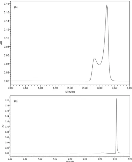
for the column and thus obtain short retention times. Initially, the methodology described in the United States Pharmacopoeia USP 34 (2010) was tested, describing ultrapure water as mobile phase, with a rate of 1.0 mL/min. However, for samples, irregular peaks were observed
us-ing low resolution chromatography (Figure 1A), possibly
because of instrumental or column differences.
Acetonitrile was added to the mobile phase to
improve the resolution of chromatographic peaks of
5-FU. Analyses were performed using acetonitrile and
water in many proportions, in isocratic mode and with the proportions of acetonitrile:water ranging from 10:90
(v/v) to 70:30 (v/v). Noticeable tailing and an irregular
shape of the 5-FU peak were observed when the propor-tion of acetonitrile was higher than the water. Increas-ing the water proportion in the mobile phase, the 5-FU peak became more regular, and, when the proportion of acetonitrile:water of 10:90 (v/v) was used, a regular and
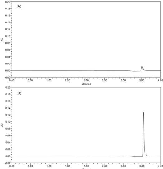
FIGURE 2 - Representative HPLC chromatograms of the PVA aqueous solution (1% (w/v)) (A) and supernatant from the production of 5-FU nanoparticles (B).
symmetric peak was observed. Under these conditions, the 5-FU peak was detected in approximately 3.5 min (Figure 1B).
Method validation
Specificity
The PVA aqueous solution (i.e., the main component
of supernatant obtained by ultracentrifugation of an
aque-ous dispersion of nanoparticles) was analyzed using the HPLC method described (Figure 2A) and compared with
the chromatogram of the 5-FU standard (Figure 1B) and
5-FU sample (5-FU in supernatant) (Figure 2B). A peak at
the 5-FU retention time was observed in the chromatogram
of the PVA aqueous solution, but the peak is so small that
it does not interfere with the quantitative determination of
5-FU from the formulation components. To conirm this observation, a 5-FU standard solution prepared in PVA aqueous solution was analyzed and compared to the 5-FU
standard solution in water (as usual). The percent recovery
of 5-FU in PVA aqueous solution was 99.89%.
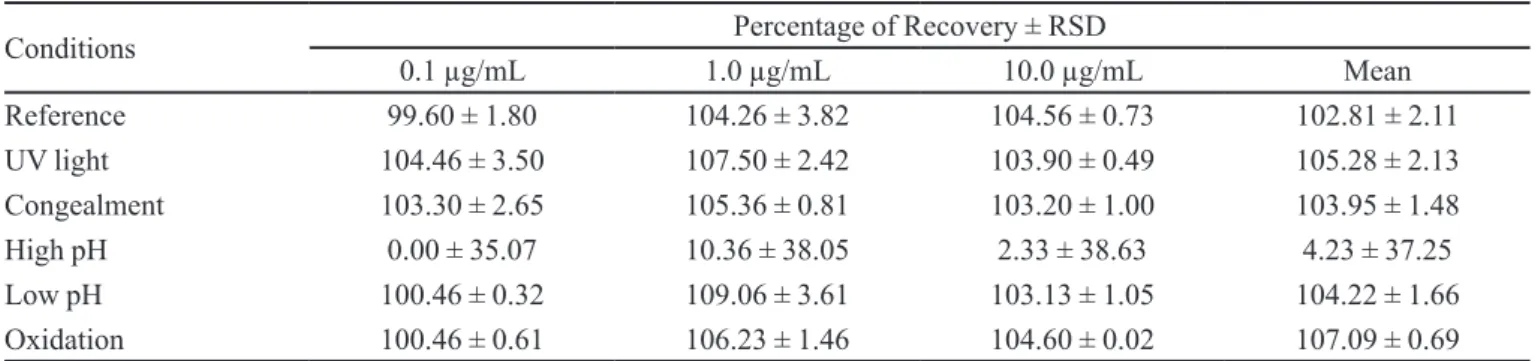
Tests were also performed under four stress condi-tions (i.e., temperature, visible light, pH and oxidation) to detect the occurrence of possible interfering peaks at 265 nm resulting from the degradation of 5-FU. These tests are regarded as helpful tools in establishing degradation pathways and the inherent stability of the molecule and help validate the power of the method for studying the drug stability (Das Neves et al., 2010). The percent recovery under stress conditions also revealed that 5-FU was not affected, except at high pH values (4.23%). The results obtained are presented in Table I, showing no alterations in 5-FU retention time.
Linearity
Linearity was evaluated at eight concentration levels
the method of least squares:
Y = 0.041259 × A − 4.66.104 Eq. 3
r = 0.9997
where Y is the peak area and A is the standard solution concentration in μg/mL. The r-value near 1 indicates linearity in the proposed range.
The validity of the assay was conirmed by an analy -sis of variance, which showed that the linear regression
was signiicant and the deviation from linearity was not signiicant (p <0.01).
Accuracy
Accuracy was assessed by calculating the percent
recovery and the RSD of the mean concentration of the analyte at three different concentrations. Three standard
solutions (0.1, 1 and 10 μg/mL) were carefully prepared in triplicate and analyzed using the proposed method.
Detailed results for these three tested concentration levels are presented in Table II. The mean percent recovery of
5-FU from the samples was 99.95% (RSD = 1.49%, n = 9).
The results show agreement between experimental and theoretical values.
Precision
The precision is a measure of the relative errors of the method, expressed as the RSD for repeatability and
intermediate precision. Three concentrations of 5-FU (0.1,
1 and 10 μg/mL) were prepared in triplicate and analyzed
on one day or two different days to evaluate intra-day or inter-day variation, respectively. The RSDs of responses were calculated in each case and are shown in Table III, indicating that precision was obtained because the maxi-mal RSD obtained was 3.51%.
The instrumental precision is a measure of the rela-tive errors inherent in the equipment and is expressed as the
RSD for repeatability. One standard of 5-FU (1.0 μg/mL) was prepared and analyzed in one day (n=10). The results indicate the instrument precision (percent recovery = 98.57%) because the maximum RSD obtained was 0.13%.
Limits of quantification and detection
The lowest concentration at which an analyte can
TABLE I - Results of exposure of 5-FU standards solutions to stress conditions (n=3)
Conditions Percentage of Recovery ± RSD
0.1 µg/mL 1.0 µg/mL 10.0 µg/mL Mean
Reference 99.60 ± 1.80 104.26 ± 3.82 104.56 ± 0.73 102.81 ± 2.11
UV light 104.46 ± 3.50 107.50 ± 2.42 103.90 ± 0.49 105.28 ± 2.13
Congealment 103.30 ± 2.65 105.36 ± 0.81 103.20 ± 1.00 103.95 ± 1.48
High pH 0.00 ± 35.07 10.36 ± 38.05 2.33 ± 38.63 4.23 ± 37.25
Low pH 100.46 ± 0.32 109.06 ± 3.61 103.13 ± 1.05 104.22 ± 1.66
Oxidation 100.46 ± 0.61 106.23 ± 1.46 104.60 ± 0.02 107.09 ± 0.69
TABLE II - Accuracy results for 5-FU concentrations in the standard solutions
Standard solution
(µg/mL)* Recovery (%) RSD (%)
0.1 99.55 1.87
1.0 98.57 0.17
10.0 99.71 1.27
*n=3
TABLE III - Precision results for different levels of 5-FU in the standard solutions
Standard solution (µg/mL)
Measured concentration ± SD
(µg/mL)
RSD (%)
Analysis repeatability (n=3)
0.1 0.09 ± 0.02 1.87
1.0 0.98 ± 0.00 0.17
10.0 9.97 ± 0.13 1.27
Intermediate precision (n=3)
Day 1
0.1 0.09 ± 0.00 1.46
1.0 0.95 ± 0.02 2.11
10.0 10.14 ± 0.07 0.70
Day 2
0.1 0.09 ± 0.00 1.66
1.0 1.04 ± 0.04 3.51
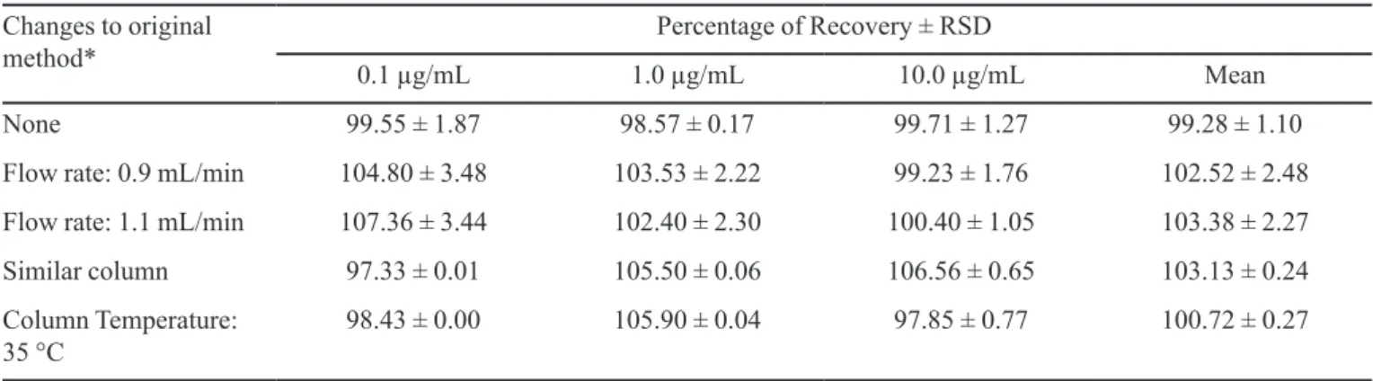
TABLE IV - Robustness results for different low rate, column brand and temperature
Changes to original method*
Percentage of Recovery ± RSD
0.1 µg/mL 1.0 µg/mL 10.0 µg/mL Mean
None 99.55 ± 1.87 98.57 ± 0.17 99.71 ± 1.27 99.28 ± 1.10
Flow rate: 0.9 mL/min 104.80 ± 3.48 103.53 ± 2.22 99.23 ± 1.76 102.52 ± 2.48
Flow rate: 1.1 mL/min 107.36 ± 3.44 102.40 ± 2.30 100.40 ± 1.05 103.38 ± 2.27
Similar column 97.33 ± 0.01 105.50 ± 0.06 106.56 ± 0.65 103.13 ± 0.24
Column Temperature: 35 °C
98.43 ± 0.00 105.90 ± 0.04 97.85 ± 0.77 100.72 ± 0.27
* 1.0mL/min, column at 25 °C and mobile phase (acetonitrile:water 10:90, v/v).
be detected (LOD) or quantiied (LOQ) with acceptable
precision and accuracy, in the present study, was calcu-lated from the SD of the response and the slope obtained from linear regression of a specific calibration curve (50-200 ng/mL) in the low-end region of the proposed range (ICH, 2005). The method was linear in this range
because the r-value was 0.993. The LOD and LOQ were found to be 10.86 and 32.78 ng/mL, respectively.
Range
The working range of the method, defined as the range that possesses the required linearity, accuracy and
precision, was between the LOQ and 10 μg/mL. Samples
containing these concentration levels may therefore be assayed using the proposed HPLC method.
Robustness
Robustness is a measure of the inluence of small
changes in the analytical procedures/parameters on the response. The evaluation of robustness was based on the percent recovery and RSD values obtained using
differ-ent parameters for low rate of mobile phase and column
temperature and using a similar C18 column. The method is robust concerning these alterations in chromatographic parameters (Table IV). The maximum RSD obtained was 3.48%.
Method applicability
The proposed analytical method was used to
evalu-ate the content of 5-FU in PLA and PLA-PEG blended nanoparticles. An indirect method, which separated the
supernatant containing free 5-FU from the solid nanopar-ticles by ultracentrifugation, was used. The supernatant
was analyzed using HPLC. As shown in the speciicity
test, no alterations to the chromatograms or unusual peaks
were observed during the drug quantiication.
The method of double emulsion-solvent evaporation was adequate to obtain the nanoparticles containing 5-FU. The main characteristics of the nanoparticles are described
in Table V. The presence of PEG did not inluence the en
-capsulation eficiency because both nanoparticles showed the same eficiency in loading the 5-FU. Considering the particle size, the PEG provided a slight increase in particle size compared with PLA nanoparticles (p<0.05), but both
formulations presented a bimodal distribution profile. Nanoparticles can be considered potential carriers for 5-FU delivery.
Our objective was to develop a fast, simple and
ef-fective HPLC-PDA method for quantitative analyses of 5-FU in PLA and PLA-PEG blended nanoparticles. The
literature describes mainly spectrophotometric methods
for 5-FU quantitation in nanoformulations (Bozkir, Saka,
TABLE V - Nanoparticle characteristics
Nanoparticle Mean size (nm)ª Polydispersity indexª Size distributionª eficiency (%)ªEncapsulation
PLA 282.87 ± 3.64 0.116 195-233 nm (54%) and 398-476 nm (46%) 51.23 ± 7.82
PLA-PEG 305.80 ± 3.62 0.173 171-208 nm (39%) and 432-525 nm (61%) 55.53 ± 9.21
2005; Liu et al., 2006; Zhu et al., 2009; Lai, Guo, 2011;
Li et al., 2011; Rejinold et al., 2011a; Rejinold et al.,
2011b; Zhang et al., 2011), but these methods are not as convenient as HPLC methods considering the sensitivity. The only example in the literature of an HPLC-UV method applied to 5-FU determination in nanoparticles was the method cited by Zheng et al. (2007), but the authors cited
only the mobile phase (methanol:3.6% acetic acid - 80:20, v/v) and did not give any information about validation data
and retention time of 5-FU. Arbós, Campanero and Irache
(2002) developed an HPLC-UV method for quantitation of
5-luorouridine in nanoparticles and, in addition to other parameters, veriied whether this drug degrades in 5-FU.
The mobile phase was comprised of 0.05 M ammonium acetate (pH 6.5), and the results showed a retention time
of 6.7 min for 5-luorouridine and 4.5 min for 5-FU. The
method was sensitive and could be applied to 5-FU, but a C8 column was used, so the method does not correspond
to our chromatographic conditions. Thus, the HPLC-PDA
method developed and validated in this work represents an alternative to spectrophotometric methods for the
analysis of 5-FU in nanoparticles and fulills the require
-ment for detailed data in the literature for analyzing 5-FU in nanoparticles via HPLC-PDA detection. The proposed
method could be applied not only for the determination
of encapsulation eficiency of 5-FU in nanoparticles but
also for other assays, such as determining the in vitro 5-FU
release proile and stability studies.
CONCLUSION
A fast, simple and reliable HPLC method using
photodiode array detection for determining the
encapsu-lation eficiency of 5-FU in PLA and PLA-PEG blended
nanoparticles has been developed and validated according
to the ICH guidelines. The method fulilled the require -ments to be considered a reliable and feasible method, including specificity, linearity, precision, accuracy,
robustness, LOD and LOQ. The analytical procedure
has a chromatographic run time of 4 min, which allows
analyzing a large number of samples in a short period of
time. The low percentage of acetonitrile used is important because it reduces the cost of solvent and the damage to the environment. This method was found to be suitable for
determining 5-FU encapsulation eficiency in polymeric
nanoparticles.
ACKNOWLEDGMENTS
This study was supported by CAPES (scholarship).
REFERENCES
AKAGI, T.; KANEKO, T.; KIDA, T.; AKASHI, M. Preparation and characterization of biodegradable nanoparticles based
on poly(g-glutamic acid) with l-phenylalanine as a protein
carrier. J. Control. Release, v.108, n.2-3, p.226-236, 2005.
ALEXIS F.; PRIDGEN E.;MOLNAR, L.K.;FAROKHZAD,
O.C. Factors affecting the clearance and biodistribution of
polymeric nanoparticles. Mol. Pharm., v.5, n.4, p.505-515,
2008.
ANDERSON, D.; KERR, D.J.; BLESING, C.; SEYMOUR, L.W. Simultaneous gas chromatographic-mass spectrophotometric determination of a-luoro-P-alanine
and 5-luorouracil in plasma. J. Chromatogr. B, v.688, n.1,
p.87-93, 1997.
ARBÓS, P.; CAMPANERO, M.A.; IRACHE, J.M. RP-LC
determination of 5-fluorouridine in nanoparticulate
formulations. J. Pharm. Biomed. Anal., v.28, n.5,
p.857-866, 2002.
BADEA, I.; MOJA, D.; TUDOSE, A.; STOICESCU, D. Determination of the 5-luorouraciland N1(2?-furanidyl) uracil in the presence of tegafur by zero-crossing first
derivative spectrometry. J. Pharm. Biomed. Anal., v.30,
n.4, p.1371-1378, 2002.
BOZKIR, A.; SAKA, O.M. Formulation and investigation of
5-FU nanoparticles with factorial design-based studies.
Farmaco, v.60, n.10, p.840-846, 2005.
CASALE, F., CANAPARO, R; SERPE, L.; MUNTONI, E.; PEPA, C.D; COSTA, M.; MAIRONE, L.; ZARA, G.P.; FORNARI, G.; EANDI, M. Plasma concentrations of
5-luorouracil and its metabolites in colon cancer patients.
Pharmacol. Res., v.50, n.2, p.173-179, 2004.
DAS NEVES, J.; SARMENTO, B.; AMIJI, M.M.; BAHIA,
M.F. Development and validation of a rapid reversed-phase HPLC method for the determination of the non-nucleoside reverse transcriptase inhibitor dapivirine from polymeric
nanoparticles, J. Pharm. Biomed. Anal., v.52, n.2,
p.167-172, 2010.
ESCORIAZA, J.; ALDAZ, A.; CALVO, E.;GIRÁLDEZ, J.
Simple and sensitive determination of 5-fluorouracil in plasma by high-performance liquid chromatography.
Application to clinical pharmacokinetic studies. J.
GAMELIN, E., BOISDRON-CELLE, M. Dose monitoring of 5-luorouracil in patients with colorectal or head and neck
cancer - status of the art. Crit. Rev. Oncol. Hemat., v.30,
n.1, p.71-79, 1999.
GAMELIN, E.; BOISDRON-CELLE, M.; GUÉRIN-MEYER, V.; DELVA, R.; LORTHOLARY, A.; GENEVIEVE, F.
Correlation between uracil and dihydrouracil plasma ratio, fluorouracil (5-FU) pharmacokinetic parameters, and tolerance in patients with advanced colorectal cancer: a potential interest for predicting 5-FU toxicity and
determining optimal 5-FU dosage. J. Clin. Oncol., v.17,
n.4, p.1105-1110, 1999.
GREM, J.L. Fluorinated pyrimidines. In: CHABNER, B.A.;
COLLINS, J.M. (Eds.) Cancer chemotherapy: principles
and practice. Philadelphia: J.B. Lippincott, 1990. p.180-224.
INTERNATIONAL CONFERENCE ON HARMONISATION O F T E C H N I C A L R E Q U I R E M E N T S F O R REGISTRATION OF PHARMACEUTICALS FOR HUMAN USE. ICH HARMONISED TRIPARTITE GUIDELINE: Validation of Analytical procedures: text and methodology Q2(R1), 2005, p. 1-13.
LABHASETWAR, V.; PRABHA, S. Nanoparticle-mediated
wild-type p53 gene delivery results in sustained antiproliferative activity in breast cancer cells. Mol. Pharm.,
v.1, n.3, p.211-219, 2004.
LAI, L.F.; GUO, H.X. Preparation of new 5-luorouracil-loaded
zein nanoparticles for liver targeting. Int. J. Pharm., v.404,
n.1-2, p.317-323, 2011.
LI, P.; WANG, Y.; PENG, Z.; SHE, F.; KONG, L. Development
of chitosan nanoparticles as drug delivery systems for
5-luorouracil and leucovorin blends. Carbohyd. Polym.,
v.85, n.3, p.698-704, 2011.
LICEA-PEREZ, H.; WANG, S.; BOWEN, C. Development
of a sensitive and selective LC MS/MS method for the
determination of α-fluoro-β-alanine, 5-fluorouracil and
capecitabine in human plasma. J. Chromatogr. B, v.877,
n.11-12, p.1040-1046, 2009.
LIU, L.; JIN, P.; CHENG, M.; ZHANG, G. ;ZHANG, F.
5-Fluorouracil-loaded self assembled ph-sensitive nanoparticles as novel drug carrier for treatment of
malignant tumors. Chinese J. Chem. Eng., v.14, n.3,
p.377-382, 2006.
LIU, K.; ZHONG, D.; ZOU, H.; CHEN, X. Determination
of tegafur, 5-fluorouracil, gimeracil and oxonic acid in human plasma using liquid chromatography-tandem mass
spectrometry. J. Pharm. Biomed. Anal., v.52, n.4,
p.550-556, 2010.
MOGHIMI, S.M.; HUNTER, A.C.; MURRAY, J.C.
Long-circulating and target specific nanoparticles: theory to
practice. Pharmacol. Rev., v.53, p.283-318, 2001.
PANYAM, J.; LABHASETWAR, V. Biodegradable
nanoparticles for drug and gene delivery to cells and tissue.
Adv. Drug Deliver. Rev., v.55, n.3, p.329-347, 2003.
PETERS, G.J.; VAN GROENINGEN, C.J. Clinical relevance
of biochemical modulation of 5-luorouracil. Ann. Oncol.,
v.2, n.7, p.469-480, 1991.
PINEDO, H.M.; PETERS, G.F. Fluorouracil: biochemistry
and pharmacology. J. Clin. Oncol., v.6, n.10, p.1653-1664,
1988.
PISANO, R.; BREDA, M.; GRASSI, S.; JAMES, C.A. Hydrophilic interaction liquid chromatography-APCI-mass spectrometry determination of 5-luorouracil in plasma and
tissues. J. Pharm. Biomed. Anal., v.38, n.4, p.738-745, 2005.
REJINOLD, N.S.; CHENNAZHI, K.P.; NAIR, S.V.; TAMURA, H.; JAYAKUMAR, R. Biodegradable and thermo-sensitive
chitosan-g-poly(N-vinylcaprolactam) nanoparticles as
5-luorouracil Carrier. Carbohyd. Polym., v.83, n.2,
p.776-786, 2011a.
REJINOLD, N.S.; MUTHUNARAYANAN, M.; CHENNAZHI, K.P.; NAIR, S.V.; JAYAKUMAR, R. 5-Fluorouracil loaded ibrinogen nanoparticles for cancer drug delivery
application. Int. J. Biol. Macromol., v.48, n.1, p.98-105,
2011b.
TANAKA, F.; FUKUSE, T.; WADA, H.; FUKUSHIMA, M. The history, mechanism and clinical use of oral 5-luorouracil
derivative chemotherapeutic agents. Curr. Pharm. Biotech.,
v.1, n.2, p.137-164, 2000.
UNITED STATES PHARMACOPEIA. USP 34: The National
formulary: NF 29: by authority of the United States Pharmacopeia Convention prepared by the council of experts and its expert committees. 29.ed. Rockville: United
ZHANG, Y.; LI, J.; LANG, M.; TANG, X.; LI, L.; SHEN, X. Folate-functionalized nanoparticles for controlled
5-Fluorouracil delivery. J. Colloid Interf. Sci., v.354, n.1,
p.202-209, 2011.
ZHENG, Y.; YANG, W.; WANG, C.; HU, J.; FU, S.; DONG, L.; WU, L.; SHEN, X. Nanoparticles based on the complex
of chitosan and polyaspartic acid sodium salt: Preparation,
characterization and the use for 5-luorouracil delivery. Eur.
J. Pharm. Biopharm., v.67, n.3, p.621-631, 2007.
ZHU, L.; MA, J.; JIA, N.; ZHAO, Y.; SHEN, H.
Chitosan-coated magnetic nanoparticles as carriers of 5-Fluorouracil:
Preparation, characterization and cytotoxicity studies. Coll.
Surf. B, v.68, n.1, p.1-6, 2009.
ZAMBAUX, M.F.; BONNEAUX, F.; GREF, R.; MAINCENT, P.; DELLACHERIE, E.; ALONSO, M.J.; LABRUDE, P.;
VIGNERON, C. Influence of experimental parameters on the characteristics of poly(lactic acid) nanoparticles
prepared by a double emulsion method. J. Control. Release,
v.50, n.1-3, p.31-40, 1998.
Received for publication on 28th May 2012