Ectopic development of skeletal muscle
induced by subcutaneous transplant of
rat satellite cells
Departamentos de 1Farmacologia and 2Neurologia e Neurocirurgia,
Universidade Federal de São Paulo, Escola Paulista de Medicina, São Paulo, SP, Brasil
M.G. Fukushima1,
I. Furlan1, T. Chiavegatti1,
B.H. Kiyomoto2
and R.O. Godinho1
Abstract
The present study analyzes the ectopic development of the rat skeletal muscle originated from transplanted satellite cells. Satellite cells (106
cells) obtained from hindlimb muscles of newborn female 2BAW Wistar rats were injected subcutaneously into the dorsal area of adult male rats. After 3, 7, and 14 days, the transplanted tissues (N = 4-5) were processed for histochemical analysis of peripheral nerves, inac-tive X-chromosome and acetylcholinesterase. Nicotinic acetylcholine receptors (nAChRs) were also labeled with tetramethylrhodamine-labeled α-bungarotoxin. The development of ectopic muscles was
successful in 86% of the implantation sites. By day 3, the transplanted cells were organized as multinucleated fibers containing multiple clusters of nAChRs (N = 2-4), resembling those from non-innervated cultured skeletal muscle fibers. After 7 days, the transplanted cells appeared as a highly vascularized tissue formed by bundles of fibers containing peripheral nuclei. The presence of X chromatin body indicated that subcutaneously developed fibers originated from fe-male donor satellite cells. Differently from the extensor digitorum longus muscle of adult male rat (87.9 ± 1.0 µm; N = 213), the diameter of ectopic fibers (59.1 µm; N = 213) did not obey a Gaussian distribution and had a higher coefficient of variation. After 7 and 14 days, the organization of the nAChR clusters was similar to that of clusters from adult innervated extensor digitorum longus muscle. These findings indicate the histocompatibility of rats from 2BAW colony and that satellite cells transplanted into the subcutaneous space of adult animals are able to develop and fuse to form differentiated skeletal muscle fibers.
Correspondence
R.O. Godinho
Departamento de Farmacologia EPM, UNIFESP
Rua 3 de maio, 100 04044-020 São Paulo, SP Brasil
Fax: +55-11-5576-4499 E-mail: [email protected]
Research supported by FAPESP (No. 01/01417-5). M.G. Fukushima was recipient of a PIBIC/CNPq fellowship. T. Chiavegatti and I. Furlan were recipients of CNPq and FAPESP MS fellowships, respectively.
Received June 21, 2004 Accepted November 23, 2004
Key words
•Skeletal muscle •Satellite cells •Stem cells •Transplantation •Myogenesis •2BAW rats
Introduction
The development of skeletal muscle fibers initiates during embryogenesis, when mono-nucleated myoblasts derived from pluripotent mesodermic cells fuse to form multinucleated myotubes. Subsequently, these fusiform cells differentiate into skeletal muscle fibers with
myo-genic precursor cells (satellite cells) located under the basal lamina of individual postnatal and adult myofibers can be activated, leading to complete regeneration of skeletal muscle. Once activated, these cells proliferate and fuse to form new myofibers morphologically and functionally indistinguishable from undam-aged ones (2,3). In fact, recent studies indicate that satellite cells represent a unique popula-tion of progenitor cells committed to the myo-genic lineage and derived from multipotent muscle-derived stem cells (4).
The demonstration that intramuscular in-jection of myoblasts induces fusion between host and donor myoblasts/satellite cells in skel-etal muscle grafts (5,6) initiated a series of studies focusing on the transplantation of myo-genic cells as a potential therapeutic strategy for treatment of skeletal muscle disorders, including Duchenne muscular dystrophy. More recently, the therapeutic benefits of transplan-tation of skeletal muscle precursors (myo-blasts/satellite cells) extended to treatment of myocardial diseases, such as infarction and end-stage heart failure (for a review, see Ref. 7). In different animal species, it has been reported that injection of myoblasts/satellite cells into the heart muscle improves cardiac contraction (8-10). Interestingly, at this site, the implanted cells acquire some of the con-tractile properties of cardiomyocytes (8,11).
Assuming that differentiation of multi-potent cells occurs in response to signals provided by their host tissue (12), in the present study we investigated whether freshly isolated myogenic precursor cells from new-born 2BAW rats were able to differentiate into skeletal muscle fibers in the subcutane-ous region, an environment free from skel-etal muscle cells.
Material and Methods
The experiments were conducted on adult male (3 months old) and newborn female Wistar 2BAW rats. The adult rats were kept on 12-h light-dark cycles with free access to
tap water and food. All protocols were in accordance with the Guide for the Care and Use of Laboratory Animals (National Insti-tutes of Health, 1986).
Isolation and implantation of satellite cells
Satellite cells were obtained from hind-limb muscles of newborn female Wistar rats. Briefly, the animals were killed under CO2
anesthesia, the muscles were removed and the cells dissociated for 3 h in Hanks’ bal-anced salt solution (HBSS), pH 7.4, contain-ing collagenase type IA (200 U/ml) as de-scribed by da Costa et al. (13). After me-chanical dissociation, the cell suspension was centrifuged at 500 g for 5 min and the resulting pellet was resuspended in Dul-becco’s Modified Eagle’s Medium (DMEM, Gibco-BRL, Life Technologies, Grand Is-land, NY, USA) containing 40 mg/l gen-tamicin. To avoid fibroblast contamination, the cells were preincubated in culture flasks for 30 min at 37ºC in a humidified atmos-phere of 90% air and 10% CO2.
Non-at-tached mononucleated satellite cells were rinsed three times with HBSS and injected subcutaneously (106 cells/200 µl) into the
dorsal area of adult male Wistar rats using a 0.5 ml syringe with a 26-gauge needle.
In another set of experiments, 106
satel-lite cells were diluted in 2 ml DMEM supple-mented with 15% fetal calf serum, seeded onto 35-mm collagen-coated culture dishes and maintained at 37ºC in a humidified at-mosphere of 90% air and 10% CO2 (14). On
the third day and every other day thereafter the medium was replaced with DMEM supplemented with 10% horse serum (HS) and 2% fetal calf serum. On day 7, myotubes were multinucleated and contracting cells indicated an appropriate maturation of the muscle fibers in vitro.
Processing transplanted tissue
were sacrificed by section of the abdominal aorta under ether anesthesia. The skin from the dorsal area was removed and transferred to a Petri dish containing phosphate-buff-ered saline (PBS), pH 7.4. The transplanted tissues were dissected, weighed, covered with tissue freezing medium (Triangle Bio-medical Sciences, Durham, NC, USA) and immediately frozen in liquid nitrogen. The tissues were sectioned on a cryostat at a thickness of 8-10 µm, mounted on slides and kept frozen at -80ºC until the time for histo-logical analysis. The extensor digitorum lon-gus (EDL) muscle from adult rats was used as control.
Determination of fiber diameter
The sections were stained with hema-toxylin and eosin, dehydrated by successive immersions in 70%, 95% and absolute etha-nol, and dried. The slides were then im-mersed in xylene, mounted with Entellan®neu
(Merck, Darmstadt, Germany) and exam-ined under a light microscope. At least three sections from each implant were photo-graphed with 40 and 60X objectives and the diameter of up to 213 fibers was determined using public domain NIH-Image 1.61 soft-ware. Frequency histograms of the fiber di-ameter were constructed and compared to that obtained from the EDL muscle of adult male rats.
Acetylcholinesterase staining
Sections were fixed in 2% paraformalde-hyde for 1 h, rinsed with PBS and incubated in 100 mM acetate buffer, pH 6.0, contain-ing 2 mM acetylthiocholine iodide, 10 mM sodium citrate, 3 mM copper sulfate, and 0.5 mM potassium ferricyanide at 25ºC, accord-ing to the method of Karnovsky and Roots (15). After 1-2 h, the sections were rinsed in PBS, counterstained with hematoxylin and mounted with 9:1 glycerin/PBS solution, pH 7.4.
Peripheral nerve staining
In order to analyze the possible innerva-tion of muscle fibers, the peripheral neurons were stained with the Bielschowsky silver stain modified by Mirra et al. (16). After AChE staining, tissue sections were incu-bated with 20% silver nitrate solution for 15 min at 37ºC. The slides were then rinsed three times with water and incubated with ammoniacal silver solution at 37ºC. After 10 min, the sections were incubated with devel-oper solution containing 0.5% formaldehyde, 50 µM HNO3, 160 µM citric acid, and 0.2%
ammonium hydroxide for 1-2 min, and rinsed successively with 0.2% ammonium hydrox-ide and distilled water. The reaction was stopped with 5% sodium thiosulfate and the slides were finally washed again in water, dehydrated and mounted with Entellan®neu.
Nicotinic acetylcholine receptor staining
Sections were rinsed for 10 min with DMEM containing 10% HS (DMEM-HS) and incubated with tetramethylrhodamine-labeled α-bungarotoxin (TRICT-α-BTX; 1
µg/ml in PBS; Molecular Probes, Eugene, OR, USA) and Hoescht dye 33258 (0.5 µg/ ml; Molecular Probes) at 25ºC, in order to stain the nAChRs and nuclei, respectively. After 1 h, the sections were washed three times with DMEM-HS and once with PBS and covered with an anti-quenching gel mounting solution (Biomeda Corp., Foster City, CA, USA).
Inactive X chromosome staining
(GraphPad Software, Inc., San Diego, CA, USA). Differences between means were an-alyzed by the Student t-test, with the level of significance set at P < 0.05.
Results
Figure 1a,b shows the subcutaneous layer from host rats 7 days after satellite cell trans-plantation. The transplanted cells appear as a highly vascularized and well-defined tis-sue weighing 23.4 ± 2.8 mg (N = 5). HE staining of transplant sections showed
multi-Figure 1. Ectopic development of skeletal muscle induced by subcutaneous transplantation of rat satellite cells. Satellite cells (106 cells) obtained from hindlimb muscles of newborn female rats were injected subcutaneously into the dorsal region of adult male rats. After 7 days, the transplants were removed, processed and stained with hematoxylin and eosin (HE) (a,b; Bars, 1 cm). A tissue mass formed under the skin of adult male rat after transplantation of satellite cells. Cross-sections (c,d,g) or longitudinal sections (e,f) of transplanted tissue (c-f) or extensor digitorum longus muscle from an adult male rat (g), stained with HE (Bars, 50 µm). The arrows indicate the presence of vascular formation (a,b) and the central nuclei (c,d).
were immersed in 5 M HCl for 10 min for acid hydrolysis and rinsed in distilled water. The samples were then incubated in 0.1% cresyl violet solution for 30 min, rinsed five times with water and mounted with Entellan®neu.
Statistical analysis
Data are reported as the means ± SEM of at least three implants. The distribution of fiber diameter was submitted to a normality test using GraphPad Prism v.3 software
nucleated cells (Figure 1c-f) that resemble those from adult skeletal muscle (EDL, Fig-ure 1g). Although most of the ectopic fibers displayed characteristics of mature muscle fibers, such as cross-striations and peripher-al nuclei, myotubes with centrperipher-ally located nuclei were also found (Figure 1c,d, arrows), indicating the presence of fibers in interme-diate stages of differentiation.
The presence of X inactive chromatin, identified as a dark stained body in close association with the nuclear membrane, en-sured that subcutaneous muscle fibers were derived from female donor satellite cells (Figure 2a,b). The X chromosome positive nuclei were also observed in EDL from adult female rats (Figure 2c,d) but not from male rats (Figure 2e,f).
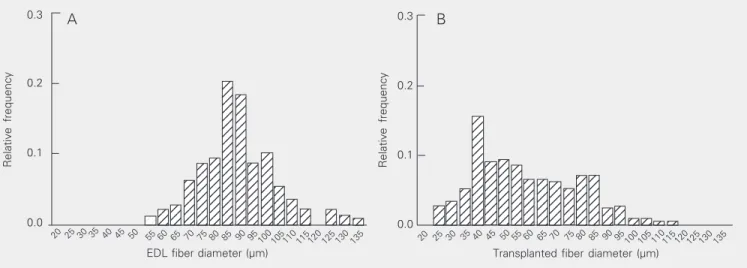
The transplanted tissue also exhibited distinct features when compared to the EDL sections, such as a higher amount of connec-tive tissue (Figure 1c,d) and high variability of fiber sizes. Figure 3 shows the frequency histogram of fiber diameters 7 days after cell transplantation in comparison to the EDL from adult male rats. The mean diameter of fibers from transplants was smaller (59.1 µm; N = 213) than that of fibers from EDL
Figure 3. Frequency histograms of skeletal muscle fiber diameter. Sections obtained from adult male rat extensor digitorum longus (EDL) muscle (A) or from ectopic muscle 7 days after transplantation (B) were stained with hematoxylin and eosin and photographed with 40X objectives and the fiber diameter was measured using public domain NIH-Image 1.61 software.
(87.9 ± 1.0 µm, N = 213) and did not obey a normal distribution. By day 7, the coeffi-cient of variation of fiber diameters was 33.6% whereas that from adult EDL fiber was only 16%.
In order to analyze the developmental expression of nAChRs, receptors were stained with TRICT-α-BTX. Three days
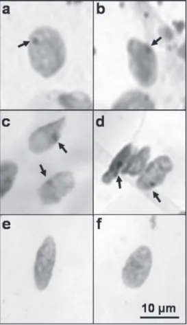
af-ter transplantation, ectopic fibers expressed multiple clusters of nAChRs (Figure 4a) that resembled those from non-innervated cul-tured skeletal muscle fibers (Figure 4b). By days 7 and 14, however, the nAChRs were distributed in a single cluster per fiber (Fig-ure 5a-d), an arrangement similar to that observed in innervated EDL from adult rats (Figure 5e,f).
At this stage, the transplanted fibers also exhibited localized expression of AChE (data not shown). Since synaptic contact is re-sponsible for the organization of nAChRs and AChE clusters in vivo, we investigated the possible innervation of ectopic skeletal muscle fibers. As shown in Figure 6a-d, black-stained axons and intramuscular nerve branches were detected after silver impreg-nation, indicating the innervation of ectopic muscle fibers.
Relative frequency
0.3
0.2
0.1
EDL fiber diameter (µm) 0.0
30 40 50 60 70 75 85 95 105
Transplanted fiber diameter (µm)
20 25 35 45 55 65 80 90 100 110115 120 125 130 135 30 40 50 60 70 75 85 95 105
20 25 35 45 55 65 80 90 100 110115 120 125 130 135
Figure 5. Nicotinic acetylcholine receptor (nAChR) clusters ex-pressed in ectopic muscle and in the extensor digitorum longus (EDL) endplate from adult male rats. Tissue sections from 7-day (a,b) or 14-day (c,d) ectopic skel-etal muscle fibers were labeled with tetramethylrhodamine-la-beled α-bungarotoxin and the distribution of nAChRs was compared to that observed in the neuromuscular junction of EDL from an adult male rat (e,f).
Figure 6. Detection of a periph-eral nerve at the site of ectopic skeletal muscle fibers. Fourteen days after transplantation of sat-ellite cells, the allografts were submitted to peripheral nerve staining (stained in black, a-d).
rats. The actual study was based on a similar report that used a murine animal model and
in vitro-expanded primary myoblast cultures, after 2-5 passages (17). It is important to emphasize that, differently from the previ-ous report, our study was performed on a 2BAW rat colony (18) whose isogenicity has not been established. Besides, the ectopic growth of skeletal muscle shown here was obtained after subcutaneous transplantation of freshly dissociated myoblasts, avoiding possible artifacts induced by ex vivo expan-sion of a muscle-derived stem cell popula-tion. In fact, it has been recently demon-strated that the passage procedure reduces the percentage of cells with myogenic mark-ers and consequently their potential for dif-ferentiation into muscle fibers (19).
In order to eliminate the possible fusion of transplanted satellite cells with host muscle fibers or satellite cells, which could modify the development of implants, the subcutane-ous space, a skeletal muscle-free environ-ment, was used as the recipient location. In addition, the detection of an inactive X chro-mosome in the nuclei demonstrated unam-biguously that ectopic muscle fibers origi-nated from implanted cells of female donor rats.
By day 7, the transplanted cells formed a vascularized tissue mass consisting of bundles of multinucleated skeletal muscle fibers surrounded by connective tissue. The well-defined alignment and the fusiform ap-pearance of transplanted cells indicate that, following the injection of the cell suspen-sion into the subcutaneous space, rearrange-ment of satellite cells took place prior to cell
Figure 4. Nicotinic acetylcho-line receptor clusters in ec-topic muscle and in cultured muscle fibers. Tissue sections from 3-day ectopic skeletal muscle fibers (a) or from cul-tured rat skeletal muscle fi-bers (b) were labeled with tet-ramethylrhodamine-labeled α -bungarotoxin.
Discussion
fusion. These results support the idea that muscle fiber phenotype is determined, at least in part, by intrinsic influences that are not dependent on the environment (20). How-ever, the reorientation of the satellite cells cannot explain the discrepancies observed in the diameter of ectopic muscle fibers. Actu-ally, during embryogenesis, the myoblasts align and fuse synchronously to form pri-mary myotubes, providing a suitable scaf-fold for secondary myoblast fusion and sub-sequent formation of a uniform muscle fiber population (21). The lack of this environ-ment in the subcutaneous space may be re-sponsible for the random satellite cell fu-sion, leading to a higher variation of fiber size.
Another interesting aspect of the trans-plants reported here was the detection of AChE and nAChR clustering, feature pro-teins from the neuromuscular synapse, indi-cating a high stage of muscle fiber matura-tion. Some aspects of nAChR expression in transplanted tissue resembled those observed during the formation of the neuromuscular synapse. From day 3 to day 14, the ectopic fibers expressed high-density nAChR aggre-gates. As aneural cultured muscle fibers, by day 3, the ectopic fibers expressed multiple plaque-shaped nAChR clusters, whereas by days 7 and 14, the distribution of nAChRs was similar to that observed in adult inner-vated muscle. Although we showed the ex-istence of peripheral nerves in close proxim-ity to ectopic muscle fibers, we cannot as-sure that the developed fibers were vated by motor neurons. Besides, the inner-vation of muscle fibers originated from ec-topic or intramuscular injection of myogenic progenitor cells has only been reported after 4 weeks of implantation (17,22). More
re-cently, an elegant study has demonstrated the formation of pretzel-shaped aggregates of nAChRs on muscle fibers by nerve-inde-pendent mechanisms. According to Kummer et al. (23), both laminin and fibronectin are able to induce the rearrangement of nAChR aggregates in aneural-cultured muscle fibers, resembling those found in the adult neuro-muscular junction. Since subcutaneous space is an environment rich in laminin, fibronec-tin and other adhesion molecules (24), it is possible that these molecules contribute to the correct arrangement of nAChR clusters at the ectopic site.
Our results show that, when transplanted into the subcutaneous space, satellite cells obtained from neonatal 2BAW rats are able to differentiate into multinucleated muscle fibers containing AChE and nAChR-rich domains, similar to those seen in adult skel-etal muscle fibers. Since multipotent satel-lite cells can easily grow, fuse and differen-tiate into skeletal muscle fibers, the subcuta-neous transplantation of freshly isolated sat-ellite cells becomes an accessible approach to study the molecular mechanisms involved in the myogenesis and differentiation of muscle-derived stem cells in vivo. In addi-tion, our results show histocompatibility of rats from the Wistar 2BAW colony. Although isogenicity of the Wistar 2BAW colony, an animal model used by many Brazilian re-searchers, has not been proved definitively, the recipient rats did not reject the myogenic allografts for up to 14 days, indicating that the 2BAW lineage became homogeneous during the uninterrupted inbreeding of com-mon ancestor rats for more than 5 decades. These data also support the use of this rat lineage in future allograft studies.
References
1. Sanes JR & Lichtman JW (1999). Development of the vertebrate neuromuscular junction. Annual Review of Neuroscience, 22: 389-442.
3. Asakura A (2003). Stem cells in adult skeletal muscle. Trends in Cardiovascular Medicine, 13: 123-128.
4. Deasy BM, Jankowski RJ & Huard J (2001). Muscle-derived stem cells: characterization and potential for cell-mediated therapy. Blood Cells, Molecules, and Diseases, 27: 924-933.
5. Partridge TA, Grounds M & Sloper JC (1978). Evidence of fusion between host and donor myoblasts in skeletal muscle grafts. Na-ture, 273: 306-308.
6. Snow MH (1978). An autoradiographic study of satellite cell differ-entiation into regenerating myotubes following transplantation of muscles in young rats. Cell and Tissue Research, 186: 535-540. 7. Taylor DA (2001). Cellular cardiomyoplasty with autologous skeletal
myoblasts for ischemic heart disease and heart failure. Current Controlled Trials in Cardiovascular Medicine, 2: 208-210.
8. Taylor DA, Atkins BZ, Hungspreugs P, Jones TR, Reedy MC, Hutcheson KA, Glower DD & Kraus WE (1998). Regenerating func-tional myocardium: improved performance after skeletal myoblast transplantation. Nature Medicine, 4: 929-933.
9. Pouzet B, Vilquin JT, Hagege AA, Scorsin M, Messas E, Fiszman M, Schwartz K & Menasche P (2000). Intramyocardial transplantation of autologous myoblasts: can tissue processing be optimized? Cir-culation, 102: 210-III-215-III.
10. Leobon B, Garcin I, Menasche P, Vilquin JT, Audinat E & Charpak S (2003). Myoblasts transplanted into rat infarcted myocardium are functionally isolated from their host. Proceedings of the National Academy of Sciences, USA, 100: 7808-7811.
11. Dorfman J, Duong M, Zibaitis A, Pelletier MP, Shum-Tim D, Li C & Chiu RC (1998). Myocardial tissue engineering with autologous myoblast implantation. Journal of Thoracic and Cardiovascular Sur-gery, 116: 744-751.
12. Seale P, Asakura A & Rudnicki MA (2001). The potential of muscle stem cells. Developmental Cell, 1: 333-342.
13. da Costa VL, Lapa AJ & Godinho RO (2001). Short- and long-term
influences of calcitonin gene-related peptide on the synthesis of acetylcholinesterase in mammalian myotubes. British Journal of Pharmacology, 133: 229-236.
14. Godinho RO & Costa-Jr VL (2003). Regulation of intracellular cyclic AMP in skeletal muscle cells involves the efflux of cyclic nucleotide to the extracellular compartment. British Journal of Pharmacology, 138: 995-1003.
15. Karnovsky MJ & Roots L (1961). Direct-coloring thiocholine method for cholinesterases. Journal of Histochemistry and Cytochemistry, 12: 219-220.
16. Mirra SS, Hart MN & Terry RD (1993). Making the diagnosis of Alzheimer’s disease. A primer for practicing pathologists. Archives of Pathology and Laboratory Medicine, 117: 132-144.
17. Irintchev A, Rosenblatt JD, Cullen MJ, Zweyer M & Wernig A (1998). Ectopic skeletal muscles derived from myoblasts implanted under the skin. Journal of Cell Science, 111 (Part 22): 3287-3297. 18. Valle JR (1949). Colônia de ratos 2BAW. Ciência e Cultura, 1: 156. 19. Machida S, Spangenburg EE & Booth FW (2004). Primary rat muscle
progenitor cells have decreased proliferation and myotube forma-tion during passages. Cell Proliferation, 37: 267-277.
20. Stockdale FE (1992). Myogenic cell lineages. Developmental Biol-ogy, 154: 284-298.
21. Ontell M & Kozeka K (1984). Organogenesis of the mouse extensor digitorum longus muscle: a quantitative study. American Journal of Anatomy, 171: 149-161.
22. Pin CL & Merrifield PA (1997). Developmental potential of rat L6 myoblasts in vivo following injection into regenerating muscles.
Developmental Biology, 188: 147-166.
23. Kummer TT, Misgeld T, Lichtman JW & Sanes JR (2004). Nerve-independent formation of a topologically complex postsynaptic ap-paratus. Journal of Cell Biology, 164: 1077-1087.