Known Viruses in Species Human Adenovirus D in Which
Serology and Genomics Do Not Correlate
Elizabeth B. Liu1., Debra A. Wadford2., Jason Seto1
, Maria Vu2, Nolan Ryan Hudson3, Lisa Thrasher3, Sarah Torres3, David W. Dyer4, James Chodosh5, Donald Seto1, Morris S. Jones1*
1School of Systems Biology, George Mason University, Manassas, Virginia, United States of America,2Viral and Rickettsial Disease Laboratory, California Department of Public Health, Richmond, California, United States of America,3Clinical Investigation Facility, David Grant USAF Medical Center, Travis AFB, Fairfield, California, United States of America,4Department of Microbiology and Immunology, University of Oklahoma Health Sciences Center, Oklahoma City, Oklahoma, United States of America,
5Howe Laboratory, Massachusetts Eye and Ear Infirmary, Department of Ophthalmology, Harvard Medical School, Boston, Massachusetts, United States of America
Abstract
In November of 2007 a human adenovirus (HAdV) was isolated from a bronchoalveolar lavage (BAL) sample recovered from a biopsy of an AIDS patient who presented with fever, cough, tachycardia, and expiratory wheezes. To better understand the isolated virus, the genome was sequenced and analyzed using bioinformatic and phylogenomic analysis. The results suggest that this novel virus, which is provisionally named HAdV-D59, may have been created from multiple recombination events. Specifically, the penton, hexon, and fiber genes have high nucleotide identity to D19C, D25, and HAdV-D56, respectively. Serological results demonstrated that HAdV-D59 has a neutralization profile that is similar yet not identical to that of D25. Furthermore, we observed a two-fold difference between the ability of D15 and HAdV-D25 to be neutralized by reciprocal antiserum indicating that the two hexon proteins may be more similar in epitopic conformation than previously assumed. In contrast, hexon loops 1 and 2 of HAdV-D15 and HAdV-D25 share 79.13 and 92.56 percent nucleotide identity, respectively. These data suggest that serology and genomics do not always correlate.
Citation:Liu EB, Wadford DA, Seto J, Vu M, Hudson NR, et al. (2012) Computational and Serologic Analysis of Novel and Known Viruses in Species Human Adenovirus D in Which Serology and Genomics Do Not Correlate. PLoS ONE 7(3): e33212. doi:10.1371/journal.pone.0033212
Editor:Michael A. Barry, Mayo Clinic, United States of America
ReceivedJuly 23, 2011;AcceptedFebruary 12, 2012;PublishedMarch 13, 2012
Copyright:ß2012 Liu et al. This is an open-access article distributed under the terms of the Creative Commons Attribution License, which permits unrestricted use, distribution, and reproduction in any medium, provided the original author and source are credited.
Funding:This research was financially supported by grant R01EY013124 (DWD, DS, MSJ and JC) and P30EY014104 (JC). LS, NRH, and MSJ were supported by United States Air Force Surgeon General grant FDG20040024E. JC is also funded by an unrestricted grant to the Department of Ophthalmology, Harvard Medical School, from Research to Prevent Blindness, Inc. The funders had no role in study design, data collection and analysis, decision to publish, or preparation of the manuscript.
Competing Interests:The authors have declared that no competing interests exist.
* E-mail: [email protected]
.These authors contributed equally to this work.
Introduction
The first human adenoviruses (HAdVs) were isolated in 1953 from a military basic trainee and the adenoid tissue of a pediatric patient as respiratory pathogens [1]. Presently there are greater than 60 types that have been isolated and characterized either with serological methods or, more recently, genomic methods [2,3,4,5,6]. HAdVs are classified into the Mastadenovirus genus and are further parsed into seven species (A–G) [3,4,5,6,7,8]. Originally, HAdV types were identified, characterized and classified based on serum neutralization and hemagglutination inhibition assays among other biological attributes [9]; however recently, bioinformatics and genomic analysis of the whole genome have replaced serology-based methods for typing novel HAdVs [3,4,5,6,8,10]. At the nucleotide level, the members of each adenovirus species are highly similar to each other, and do not commonly recombine with members of other species. The species groupings, in part, may reflect the cell tropism of the viruses, as well as the resulting symptoms and diseases caused by the individual HAdV types. For example, species HAdV-B1 viruses are known to cause respiratory infections of the lower lung [11] whereas viruses in species HAdV-D can cause ocular disease, including epidemic keratoconjunctivitis [12], and gastrointestinal disease [3,13].
In this report, an adenovirus isolated from a bronchoalveolar lavage (BAL) sample that was biopsied from an AIDS patient who presented with fever, cough, tachycardia and expiratory wheezes is examined using genomics and bioinformatics. Based upon the whole genome analysis and supported by limited serological data, this adenovirus belongs to species HAdV-D, and is a ‘never seen before’ novel virus, to be given the name of HAdV-D59.
Materials and Methods
Ethics Statement
The work reported herein was performed under United States Air Force Surgeon General-approved Clinical Investigation No. FDG20040024E, by the Institutional Review Board at the David Grant USAF Medical Center. Informed Consent was not required, because we did not use clinical samples.
Viruses, cells, and serum neutralization assay
antisera raised against reference stock adenoviruses types 1–49 from the collection maintained by the Viral and Rickettsial Disease Laboratory of the California Department of Public Health, Richmond, CA. Reference viruses were originally obtained from the reporting investigators: the Research Reference Reagents Branch, National Institutes of Health; the Respiratory Viral Disease Unit, Centers for Disease Control and Prevention; or the American Type Culture Collection. Stock virus cultures were passaged in A549 cells (American Type Culture Collection, Rockville, MD), the cells were disrupted by vortexing, and cell-free supernatant fluid was then frozen at270uC.
Equal volumes of diluted virus and immune serum were mixed and incubated for one hour in 5% CO2at 37uC. Thereafter A549
cells were added, mixed, and incubated at 37uC in 5% CO2for 7
days. Each assay contained a back titration of the virus used. Living cells were distinguished from dead cells by measuring the amount of Finter’s Neutral Red [15] present as indicated by absorbance at 550 nm using a microplate spectrophotometer (Bio-Tek Instruments, Winooski, VT). Virus neutralization titers were determined by equating cell death to virus growth (no virus neutralization). Neutralization was plotted as a percentage of cell control absorbance, to determine endpoint virus and serum titers. Three independent experiments were run yielding similar results.
Nucleotide sequence accession numbers
The HAdV-D59 genome sequence and its annotation are deposited in GenBank and retrievable as accession number JF799911. In addition, the following HAdV genomes (GenBank accession numbers) were used for comparative computational analyses: D8 (AB448767), D9 (AJ854486), HAdV-D15 (AB562586), HAdV-D17 (AF108105), HAdV-D19C (EF121005), HAdV-D22 (FJ404771), HAdV-D25 (unpublished), HAdV-D26 (EF153474), HAdV-D28 (FJ824826), HAdV-D36 (GQ384080), HAdV-D37 (DQ900900), HAdV-D46 (AY875648), HAdV-D48 (EF153473), HAdV-D49 (DQ393829), HAdV-D53 (FJ169625), HAdV-D54 (AB333801), HAdV-D56 (HM770721), and HAdV-D58 (HQ883276).
Fiber coding sequences used for analysis were as follows: HAdV-D8 (AB448767), HAdV-D9 (AJ854486), HAdV-D10 (AB369368), HAdV-D19C (AB448774), HAdV-D19a
(CS301726), HAdV-D22 (FJ619037), HAdV-D26 (EF153474), HAdV-D28 (FJ824826), HAdV-D29 (AB562587), HAdV-D36 (GQ384080), HAdV-D37 (DQ900900), HAdV-D46 (AY875648), HAdV-D48 (EF153473), HAdV-D49 (DQ393829), HAdV-D54 (AB333801), HAdV-D56 (HM770721), and HAdV-D58 (HQ883276). Fiber genes from species HAdV-D genomes were aligned using ClustalW [16]. For this analysis, the default gap opening and gap extension penalties were 15.0 and 6.66.
Amplification and DNA sequencing of the HAdV-D59 genome
To amplify regions of HAdV-D59 using the polymerase chain reaction (PCR) protocol, conserved adenovirus sequences in species HAdV-D were used to design primers. All amplicons were then sequenced on an ABI 3130xl using a primer walking strategy. The HAdV-D59 genome was sequenced to 8-fold coverage following PCR amplification, with both strands represented.
Bioinformatics
The HAdV-D59 genome was compared against a select number of viral genomes from the HAdV-D group based on its GC content, which is indicative of HAdV species. The selection of which genomes was based on initial overall high nucleotide identity to HAdV-D59. The data presented are final iterations of analyses that initially included all of the sequenced genomes in species HAdV-D.
Recombination analysis
Whole genome sequences of HAdV-D59 and members of species HAdV-D were first aligned with kalign (http://www.ebi. ac.uk/Tools/msa/kalign/) for a broad perspective of the genome. SimPlot [17] was then used to construct a Bootscan analysis of the aligned sequences. The window size and step size were set to 1000 and 200 respectively.
Following this, to provide a detailed close inspection of recombination events, the penton base gene, hexon gene, E3 coding region and fiber gene from the HAdV-D genomes were aligned to their counterparts using ClustalW [16]. This was also followed by recombination analysis using SimPlot with the window size and step size set to 250 and 50, respectively.
Figure 1. Genome organization of HAdV-D59.The HAdV-D59 genome is represented by a black horizontal line marked at 5-kbp intervals. Protein encoding regions are shown as arrows indicating transcriptional orientation above and below the genome. Spliced genes are indicated by V-shaped lines.
doi:10.1371/journal.pone.0033212.g001
Computational and Serologic Analysis of Adenovirus
Percent Identity
Whole genome, penton base gene, hexon gene, E3 coding region and fiber gene nucleotide sequences of HAdV-D59, along with members of species HAdV-D, were aligned using kalign; these were then compared to each other based on percent identity values calculated with Chimera [18].
Phylogenomic analysis of HAdV-D59
Sequence alignments for phylogenomic analysis were generated using the kalign method noted earlier. Phylogenetic trees were constructed from these aligned sequences using Molecular Genetic Analysis Software (MEGA 4.1; http://www.megasoftware.net), via neighbor-joining methods and bootstrap test of phylogeny with replicates set to 1000.
Results
Clinical Investigation
In November 2007, an AIDS patient was admitted to San Francisco General Hospital, presenting with fever. The patient also complained of a cough productive of yellow sputum and blood. Clinical examination revealed a body temperature of 101uF, tachycardia and expiratory wheezes. During the hospital stay, a CT scan displayed results suggestive of a cavitary lung lesion. This prompted a bronchoalveolar lavage (BAL) for a diagnostic specimen (via bronchoscopy). A virus was cultured from the BAL sample and sent to the California Department of Public Health (CDPH) for further analysis and identification. No other pathogens were isolated from this patient.
The virus was propagated at the Viral and Rickettsial Disease Laboratory at the California Department of Public Health, and was identified as an unknown adenovirus by serum neutralization assay. Initial sequence analysis of amplicons derived from the hexon and the fiber genes revealed similarity to gene sequences from HAdV-D25 and HAdV-D56, respectively. The possibility
that this virus might represent a novel, recombinant pathogen provoked whole genome analysis in order to characterize this isolate more thoroughly.
Amplification, sequencing, and genetic characteristics of the novel adenovirus
To elucidate the genetic characteristics of this pathogen (HAdV-D59), we sequenced and analyzed the entire genome. The genome length of HAdV-D59 is 35,072 base pairs (Figure 1), with a base composition of 22.4% A, 20.4% T, 28.5% G, 28.7% C. The GC content of 57.2% is consistent with the values found for members of species HAdV-D (mean of 57.0%). The organization of the 36 open reading frames (ORFs) that were annotated had a genome organization similar to other mastadenoviruses (Fig. 1). The inverted terminal repeat (ITR) sequences for HAdV-D59 were determined to be 151 bp in length. Within species HAdV-D, HAdV-D59 has a genome percent identity ranging from a low of 92.06% (HAdV-D8) to 96.48% (HAdV-D9).
Genome Analysis
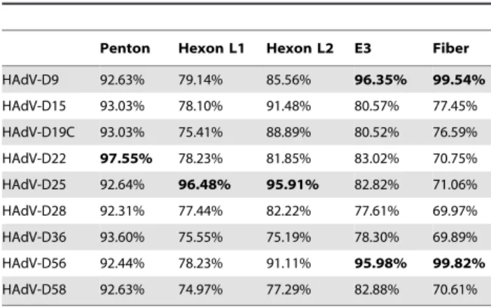
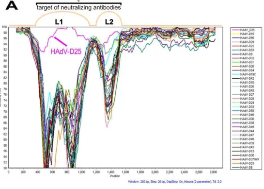
Since initial DNA sequencing suggested evidence of recombi-nation, we analyzed the HAdV-D59 genome using zPicture, a dynamic blastz alignment visualization program designed for comparative analysis (http://zpicture.dcode.org/). Comparisons of the HAdV-D59 genome with the whole genome sequences of HAdV-D9, -D22, -D19C, -D25, -D28, -D36, -D56 and -D58 were performed. Consistent with other previously reported viruses in species HAdV-D [4,19], HAdV-D59 shows heterogeneity in the penton base, hexon, E3, and fiber coding sequences (Fig. 2A). Pairwise alignment suggested that these regions had highest nucleotide identities with sequences from D9, HAdV-D19C, HAdV-D25, HAdV-D28, and HAdV-D56 (Table 1).
Comprehensive phylogenomic analyses of whole genome HAdVs were performed. Using sequences available in GenBank as well as the unpublished sequence of HAdV-D25, the whole genome phylogenetic tree analysis resulted in a subclade that includes HAdV-D59, HAdV-D9, and HAdV-D56 with a high confidence bootstrap value of 97 (Fig. 2B).
Penton Base Gene Analysis
Recently it was shown that two coding sequences for the external hypervariable loops in the penton base gene contain hotspots for recombination in species HAdV-D [20]. Analysis of the primary amino acid sequences in species HAdV-D showed that the most similar loop1 sequences (nucleotides 200–600) to that of HAdV-D59 were HAdV-D28 and HAdV-D36 (80.95%). In addition, the most similar RGD loop (nucleotides 650–1150) to HAdV-D59 was HAdV-D22 with 100% amino acid identity. Bootscan analysis [17] with penton base sequences from species HAdV-D confirmed the aforementioned relationships (Fig. 3). Phylogenetic analysis of the HAdV-D59 penton base hypervari-able loop 1 also confirmed that it is a close relative of HAdV-D28 and HAdV-D36 with a robust bootstrap value of 91 (Fig. 3B). Phylogenetic analysis also demonstrated that the HAdV-D59
Figure 2. Comparative genomic analysis.(A) Pairwise nucleotide comparison of selected HAdV-D genomes to HAdV-D59 using zPicture. The arrows above the x-axis demarcate the positions of penton base, hexon, E3 region, and fiber coding sequences in the genome of HAdV-D59. The y-axis notes the percent identity. HAdV-D9, HAdV-D22, HAdV-D19C, HAdV-D25, HAdV-D28, HAdV-D36, and HAdV-D56 were used for comparison to HAdV-D59 because they share high nucleotide identity to the aforementioned virus in different sections of their genomes. (B) Whole genome phylogenetic analysis. The phylogenetic tree was constructed from aligned sequences using MEGA, via the neighbor-joining methods and a bootstrap test of phylogeny. Bootstrap values shown at the branching points indicate the percentages of 1000 replications produced the clade. A Bootstrap value of 70 and above is considered to be robust.
doi:10.1371/journal.pone.0033212.g002
Table 1.Percent identities of the nucleotide coding sequences of selected HAdV-D59 coding regions to
homologous sequences from other viruses in species HAdV-D.
Penton Hexon L1 Hexon L2 E3 Fiber
HAdV-D9 92.63% 79.14% 85.56% 96.35% 99.54%
HAdV-D15 93.03% 78.10% 91.48% 80.57% 77.45%
HAdV-D19C 93.03% 75.41% 88.89% 80.52% 76.59%
HAdV-D22 97.55% 78.23% 81.85% 83.02% 70.75% HAdV-D25 92.64% 96.48% 95.91% 82.82% 71.06%
HAdV-D28 92.31% 77.44% 82.22% 77.61% 69.97%
HAdV-D36 93.60% 75.55% 75.19% 78.30% 69.89% HAdV-D56 92.44% 78.23% 91.11% 95.98% 99.82%
HAdV-D58 92.63% 74.97% 77.29% 82.88% 70.61%
doi:10.1371/journal.pone.0033212.t001
Computational and Serologic Analysis of Adenovirus
RGD loop segregates with the subclade that includes HAdV-D19C and HAdV-D22 with a bootstrap value of 92 (Fig. 3C).
Hexon Gene Analysis
A previous study showed that recombination in HAdVs can occur within the hexon gene of viruses in species HAdV-D [8]. This has also been demonstrated in other species of HAdVs [4,5]. To determine whether or not the HAdV-D59 hexon coding region was either novel or the result of a recombination event, SimPlot analysis was performed. SimPlot results were not consistent with a recent recombination event in the hexon of HAdV-D59 (Fig. 4A). In addition, the nucleotide percent identity to other hexon coding sequences in species D was determined (Table 1). HAdV-D59 loops 1 and 2 (L1 and L2) were 96.48% and 95.19% identical to L1 and L2 of HAdV-D25, respectively (Table 1). Phylogenetic analysis demonstrated that L1 of HAdV-D59 and HAdV-D25 are as distantly related as the following HAdV pairs which were shown to be distinct via serology [21]: HAdV-D39 and HAdV-D43; HAdV-D37 and HAdV-D13; HAdV-D15 and HAdV-D30; HAdV-D29 and HAdV-D30; HAdV-D9 and HAdV-D32, as well as HAdV-D45 and HAdV-D26 (Fig. 4B). Furthermore, the same phenomenon was observed for L2 with the exception of the HAdV-D45/D26 pair (Fig. 4C).
The difference in nucleotide identity between HAdV-D59 and HAdV-D25 (the nearest phylogenetic relative to HAdV-D59) in the L1 and L2 domains are greater than 2.5% (3.52% and 4.81%, respectively). Madisch et al stated that percent nucleotide identity differences greater than 2.4% and 2.5% in L1 and L2, respectively, strongly suggests identification of a novel HAdV [22]. Therefore the percent of nucleotide identity differences in L1 and L2 of HAdV-D59 further suggests that the aforementioned virus is novel.
E3 Genome Region Analysis
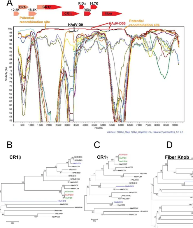
SimPlot and Bootscan results suggest that a large portion of the E3 transcription region (genes encoding for the CR1b, 18.4 k, CR1c, RIDa, RIDb, and 14.7 k proteins) in HAdV-D59 may have originated from a recombination event between either HAdV-D56 or HAdV-D9 and another yet to be described HAdV (Fig. 5). Interestingly, the SimPlot results suggest that a recombination event took place within the open reading frame of the CR1bgene (Fig. 5A). We also examined other E3 genes in species HAdV-D and did not detect common recombination loci (data not shown).
Phylogenetic analyses of two genes (CR1band CR1cgenes) in the E3 region demonstrate that the coding regions for CR1band CR1cin HAdV-D59 are closely related to those of HAdV-D9 and HAdV-D56 (Fig. 5B, 5C).
Fiber Gene Analysis
SimPlot analyses of the HAdV-D59 fiber was performed on fiber sequences extracted from GenBank. The results suggested that the fiber gene of HAdV-D59 is nearly identical with sequences from both HAdV-D56 and HAdV-D9 (Fig. 5A). Furthermore, the fiber of HAdV-D59 had 99.54% and 99.84% nucleotide identities with HAdV-D9 and HAdV-D56 (Table 1), respectively. Phylogenetic analysis of the fiber genes in species HAdV-D confirms that the fiber of HAdV-D59 was closest in
sequence to the corresponding sequences in HAdV-D56 and HAdV-D9 (Fig. 5D).
Serum Neutralization
D59 was neutralized by both D25 and HAdV-D15 antiserum, yet not by HAdV-D9 antiserum (Table 2). Interestingly, HAdV-D25 antiserum showed at least a two-fold higher neutralization titer to HAdV-D59 (greater than 1:4096) than it did against its cognate antigen HAdV-D25 (1:2048) (Table 2). These results demonstrate that the antigenic profile of HAdV-D59 differs from that of HAdV-D25 (Table 2). HAdV-D15 antiserum demonstrated only a two-fold increase in its ability to neutralize its cognate HAdV-D15 (1:1024) relative to the heterologous HAdV-D25 antigen (1:512). Similar reciprocal results were obtained with HAdV-D25 antiserum against HAdV-D15 (1:1024) and HAdV-D25 (1:2048). When tested with rabbit serum our results indicate that HAdV-D15 and HAdV-D25 are not as serologically distinct as previously reported [21].
Discussion
Our results demonstrated that HAdV-D25 antiserum was more effective at neutralizing HAdV-D59 than HAdV-D25 (Table 2). Since L1 and L2 protrude from the surface of HAdVs [23], it is not surprising that there is a difference between the ability of HAdV-D25 antiserum to neutralize the different viruses. One possibility for the differences in neutralization may be that the few differences in the primary amino acid structure present the HAdV-D59 hexon three-dimensional structure in such a way that the neutralizing epitopes are enhanced, thus making the virus easier to neutralize. Interestingly, we also observed a two-fold difference between the ability of HAdV-D15 and HAdV-D25 to be neutralized by reciprocal antiserum. This contradicts one study that showed antiserum to HAdV-D15 and HAdV-D25 did not cross-react in reciprocal neutralization experiments [21]. Howev-er, our data are consistent with the original characterization of HAdV-D15 and HAdV-D25 (previously called BP-1) [24], thus we conclude that HAdV-D15 and HAdV-D25 are not separate serotypes according to the traditional methods used for differen-tiating serotypes. These results also demonstrate that using neutralization as a criterion to type novel adenoviruses is complicated by non-standard serology methods and reagents that may yield interlaboratory variability of neutralization results. In contrast, using genomics as a method for typing HAdVs is consistent regardless of which laboratory generates the results.
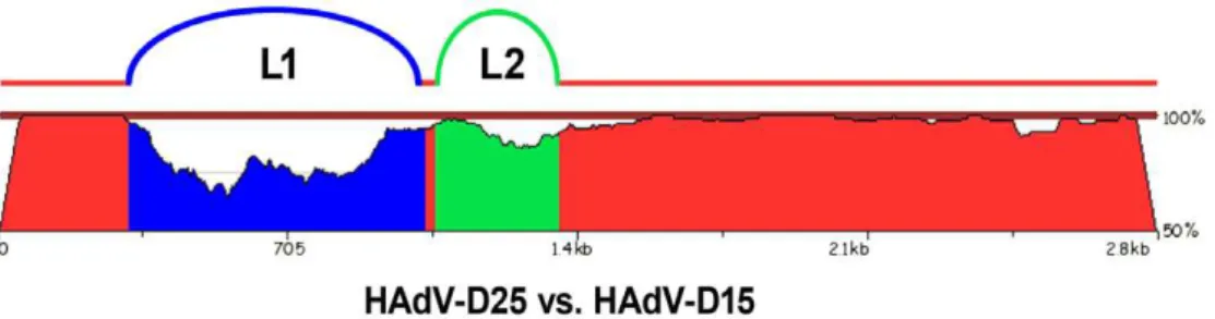
Even though HAdV-D15 and HAdV-D25 showed only a two-fold difference via serum neutralization, they were recognized as different serotypes by Rosen et al, because they had different fiber proteins [24]. HAdV-D15 and HAdV-D25 share 79.13 and 92.56 percent nucleotide identity in L1 and L2, respectively. Thus, bioinformatic analysis demonstrates that they are actually different types using the criteria established by Madisch et al. which states that the nucleotide identity of L2 must differ by greater than 2.5 percent to type a novel virus [22]. Furthermore, pairwise nucleotide comparison of the hexon coding sequences for HAdV-D15 and HAdV-D25 show how genetically divergent they are (Fig. 6). Neutralization assays measure the overall effect of various antibodies that bind to multiple epitopes and may yield
Figure 3. Computational analysis of the penton base gene.(A) Bootscan analysis of the HAdV-D59 penton base gene with fully sequenced penton genes in species HAdV-D using a window size of 250 bp and step size of 50 bp. (B) Phylogenetic analysis of the hypervariable loop 1 penton base gene sequences in species HAdV-D. (C) Phylogenetic analysis of the RGD motif and surrounding variable region of sequences in species HAdV-D. The phylogenetic trees were generated from aligned sequences using MEGA, via the neighbor-joining method and a bootstrap test of phylogeny. doi:10.1371/journal.pone.0033212.g003
Computational and Serologic Analysis of Adenovirus
representatives from the other species. Phylogenetic trees were generated from aligned sequences using MEGA, via the neighbor-joining method and a bootstrap test of phylogeny.
doi:10.1371/journal.pone.0033212.g004
Figure 5. Computational analysis of the E3 and Fiber regions.(A) SimPlot analysis of the E3 and fiber region of HAdV-D59 compared to fully sequenced E3 and fiber regions from species HAdV-D. The arrows over the Bootscan demarcate the approximate positions of the E3 coding sequences. Phylogenetic analysis of (B) HAdV-D59 CR1b(C) HAdV-D59 CR1c, and (D) fiber knob. The phylogenetic trees were generated from aligned sequences using MEGA, via the neighbor-joining method and a bootstrap test of phylogeny.
doi:10.1371/journal.pone.0033212.g005
Computational and Serologic Analysis of Adenovirus
variable interlaboratory results due to non-standard methods and reagents. Genomic analyses measure genetic differences in the genome that have the potential to affect the pathogenicity of the virus and can be independently verified by most laboratories. The contrasting serology and genomics results for HAdV-D15 and HAdV-D25 demonstrate that these two methods do not always yield concordant results.
SimPlot analysis of the HAdV-D59 L1 and L2 regions demonstrates high nucleotide identity between the hexons of HAdV-D25 and HAdV-D59 and suggests that they may be derived from a yet undiscovered common ancestor. If L1 and L2 of HAdV-D25 and HAdV-D59 are from a common ancestor, the recombination event may be ancient, as evidenced by 3.52 and 4.81 percent nucleotide differences in the L1 and L2 sequences, respectively. If the recombination events were recent, SimPlot analysis would illustrate near 100% nucleotide identity, which was shown for HAdV-D53 and HAdV-D56 [4,5,19] With a distant past recombination event the hexon genes from HAdV-D25 and HAdV-D59 would have mutated over many replication cycles, after the initial recombination, to result in the variation we detected.
Multiple studies have shown that HAdVs in species HAdV-D recombine with one another in the penton base and hexon genes [4,8,20]. In this paper, we demonstrate that recombination may have occurred in the E3 region of HAdV-D59; however, after
examining all of the sequenced E3 genes in species HAdV-D, we found that there was not a predictable pattern of recombination (data not shown). Viruses in species HAdV-D show variability in their cell tropism ranging from growth in ocular tissues to gastrointestinal and/or respiratory tissues [4,25,26]. Given that the fiber knob is an important determinant of cell tropism, it may be concluded that recombination is an important molecular evolution pathway for the diversity observed within species HAdV-D.
The section of the HAdV-D59, -D56, and –D9 genomes that encodes for CR1b, CR1c, RIDa, RIDb, 14.7K, and fiber show high nucleotide identity (Fig. 5A). From the nucleotide data, it is impossible to tell whether or not the 39end of HAdV-D59 came from HAdV-D56 or HAdV-D9. Although HAdV-D9 was discovered in 1957 [25], there has been no disease associated with this virus. In contrast, prior serological evidence suggests that HAdV-D56, an ocular and respiratory pathogen (with hexon and fiber coding sequences similar to HAdV-D15 and HAdV-D9, respectively) [12,26] has been implicated in human disease as early as 1960 [27] and at other points in time as well [27,28,29,30,31,32]. HAdV-D59 may have also existed prior to our current description yet had gone undetected during the same time periods. Thus, it is impossible to say with absolute certainty which of the aforementioned viruses existed first and/or whether they evolved from a common ancestor. Future genomic analysis of known and unknown adenoviruses is needed to elucidate further the evolutionary history of HAdVs.
Acknowledgments
The views expressed in this material are those of the authors, and do not reflect the official policy or position of the U.S. Government, the Department of Defense, or the Department of the Air Force. We thank Drs. Michael Walsh and Shoaleh Dehghan for advice, critical discussion and preliminary analyses.
Author Contributions
Conceived and designed the experiments: MSJ DS DAW. Performed the experiments: EBL JS MV NRH LT ST. Analyzed the data: EBL DAW JS DWD JC DS MSJ. Contributed reagents/materials/analysis tools: DWD JC DS MSJ. Wrote the paper: EBL DAW DWD JC DS MSJ.
References
1. Rowe WP, Huebner RJ, Gilmore LK, Parrott RH, Ward TG (1953) Isolation of a cytopathogenic agent from human adenoids undergoing spontaneous degenera-tion in tissue culture. Proceedings of the Society for Experimental Biology and Medicine Society for Experimental Biology and Medicine 84: 570–573. 2. Ishiko H, Aoki K (2009) Spread of epidemic keratoconjunctivitis due to a novel
serotype of human adenovirus in Japan. J Clin Microbiol 47: 2678–2679.
3. Jones MS, 2nd, Harrach B, Ganac RD, Gozum MM, Dela Cruz WP, et al. (2007) New adenovirus species found in a patient presenting with gastroenteritis. J Virol 81: 5978–5984.
4. Robinson CM, Singh G, Henquell C, Walsh MP, Peigue-Lafeuille H, et al. (2011) Computational analysis and identification of an emergent human adenovirus pathogen implicated in a respiratory fatality. Virology 409: 141–147. Table 2.Neutralization of HAdV-D59 with hyper immune
serum.
Antiserum
aHAdV-D9 aHAdV-D15 aHAdV-D25
HAdV-D9 1:128 ,8*
,8*
HAdV-D15 ,8* 1:1024 1:1024
HAdV-D25 ,8* 1:512 1:2048
HAdV-D59 ,8* 1:128 .1:4096
*No neutralization.
doi:10.1371/journal.pone.0033212.t002
Figure 6. Pairwise nucleotide comparison of the hexon genes of HAdV-D15 and HAdV-D25.The blastz program zPicture was used to compare the hexon genes of HAdV-D15 and HAdV-D25. The blue region represents L1 and the green region represents L2.
5. Walsh MP, Seto J, Jones MS, Chodosh J, Xu W, et al. (2010) Computational analysis identifies human adenovirus type 55 as a re-emergent acute respiratory disease pathogen. J Clin Microbiol 48: 991–993.
6. Walsh MP, Seto J, Liu EB, Dehghan S, Hudson NR, et al. (2011) Computational analysis of two species C human adenoviruses provides evidence of a novel virus. J Clin Microbiol 49: 3482–3490.
7. Liu EB, Ferreyra L, Fischer SL, Pavan JV, Nates SV, et al. (2011) Genetic analysis of a novel human adenovirus with a serologically unique hexon and a recombinant fiber gene. PLoS One 6: e24491.
8. Walsh MP, Chintakuntlawar A, Robinson CM, Madisch I, Harrach B, et al. (2009) Evidence of molecular evolution driven by recombination events influencing tropism in a novel human adenovirus that causes epidemic keratoconjunctivitis. PLoS One 4: e5635.
9. Rosen L (1960) A hemagglutination-inhibition technique for typing adenovi-ruses. Am J Hyg 71: 120–128.
10. Seto D, Chodosh J, Brister JR, Jones MS (2011) Using the whole-genome sequence to characterize and name human adenoviruses. J Virol 85: 5701–5702. 11. Metzgar D, Gibbins C, Hudson NR, Jones MS (2010) Evaluation of multiplex type-specific real-time PCR assays using the LightCycler and joint biological agent identification and diagnostic system platforms for detection and quantitation of adult human respiratory adenoviruses. J Clin Microbiol 48: 1397–1403.
12. Robinson CM, Shariati F, Zaitshik J, Gillaspy AF, Dyer DW, et al. (2009) Human adenovirus type 19: genomic and bioinformatics analysis of a keratoconjunctivitis isolate. Virus Res 139: 122–126.
13. Echavarria M In Principles and Practice of Clinical Virology: John Wiley & Sons.
14. Crawford-Miksza LK, Schnurr DP (1994) Quantitative colorimetric microneu-tralization assay for characterization of adenoviruses. Journal of clinical microbiology 32: 2331–2334.
15. Montefiori DC, Robinson WE, Jr., Schuffman SS, Mitchell WM (1988) Evaluation of antiviral drugs and neutralizing antibodies to human immuno-deficiency virus by a rapid and sensitive microtiter infection assay. Journal of clinical microbiology 26: 231–235.
16. Larkin MA, Blackshields G, Brown NP, Chenna R, McGettigan PA, et al. (2007) Clustal W and Clustal X version 2.0. Bioinformatics 23: 2947–2948. 17. Lole KS, Bollinger RC, Paranjape RS, Gadkari D, Kulkarni SS, et al. (1999)
Full-length human immunodeficiency virus type 1 genomes from subtype C-infected seroconverters in India, with evidence of intersubtype recombination. J Virol 73: 152–160.
18. Pettersen EF, Goddard TD, Huang CC, Couch GS, Greenblatt DM, et al. (2004) UCSF Chimera–a visualization system for exploratory research and analysis. J Comput Chem 25: 1605–1612.
19. Robinson CM, Seto D, Jones MS, Dyer DW, Chodosh J (2011) Molecular evolution of human species D adenoviruses. Infect Genet Evol 11: 1208–1217. 20. Robinson CM, Rajaiya J, Walsh MP, Seto D, Dyer DW, et al. (2009) Computational analysis of human adenovirus type 22 provides evidence for recombination among species D human adenoviruses in the penton base gene. J Virol 83: 8980–8985.
21. Hierholzer JC, Stone YO, Broderson JR (1991) Antigenic relationships among the 47 human adenoviruses determined in reference horse antisera. Archives of virology 121: 179–197.
22. Madisch I, Harste G, Pommer H, Heim A (2005) Phylogenetic analysis of the main neutralization and hemagglutination determinants of all human adeno-virus prototypes as a basis for molecular classification and taxonomy. J Virol 79: 15265–15276.
23. Rux JJ, Burnett RM (2004) Adenovirus structure. Hum Gene Ther 15: 1167–1176.
24. Rosen L, Baron S, Bell JA (1961) Four newly recognized adenoviruses. Proc Soc Exp Biol Med 107: 434–437.
25. Kibrick S, Melendez L, Enders JF (1957) Clinical associations of enteric viruses with particular reference to agents exhibiting properties of the ECHO group. Ann N Y Acad Sci 67: 311–325.
26. Henquell C, Boeuf B, Mirand A, Bacher C, Traore O, et al. (2009) Fatal adenovirus infection in a neonate and transmission to health-care workers. J Clin Virol 45: 345–348.
27. Cramblett HG, Kasel JA, Langmack M, Wilken FD (1960) Illnesses in children infected with an adenovirus antigenically related to types 9 and 15. Pediatrics 25: 822–828.
28. Adrian T, Bastian B, Benoist W, Hierholzer JC, Wigand R (1985) Characterization of adenovirus 15/H9 intermediate strains. Intervirology 23: 15–22.
29. Adrian T, Bastian B, Wagner V (1989) Restriction site mapping of adenovirus types 9 and 15 and genome types of intermediate adenovirus 15/H9. Intervirology 30: 169–176.
30. Hierholzer JC, Rodriguez FH, Jr. (1981) Antigenically intermediate human adenovirus strain associated with conjunctivitis. J Clin Microbiol 13: 395–397. 31. Wigand R, Fliedner D (1968) Serologically intermediate adenovirus strains: a regular feature of group II adenoviruses. Arch Gesamte Virusforsch 24: 245–256.
32. Wigand R (1987) Pitfalls in the identification of adenoviruses. Journal of virological methods 16: 161–169.
Computational and Serologic Analysis of Adenovirus