(1) Unidad de Biología Molecular y Grupo de Micología, Corporación para Investigaciones Biológicas (CIB), Medellín, Colombia. (2) Facultad de Medicina Veterinaria y Zootecnia, Universidad de Caldas, Manizales, Colombia.
Correspondence to: Dr. J.G. McEwen, Corporación para Investigaciones Biológicas (CIB), Carrera 72A- # 78B-141, Medellín, Colombia. Telephone (574) 441 0855; FAX (574) 441-5514, Email: [email protected]
PCR WITH Paracoccidioides brasiliensis SPECIFIC PRIMERS:
POTENTIAL USE IN ECOLOGICAL STUDIES
S. DÍEZ(1), E.A. GARCIA(1), P.A. PINO(1), S. BOTERO(1), G.G. CORREDOR(2), L.A. PERALTA(2), J.H. CASTAÑO(2), A. RESTREPO(1) & J.G. McEWEN(1)
SUMMARY
The precise microenvironment of Paracoccidioides brasiliensis has not yet been discovered perhaps because the methods used are not sensitive enough. We applied to this purpose the polymerase chain reaction (PCR) using three sets of specific primers corresponding to two P. brasiliensis genes. This fungus as well as several other fungi, were grown and their DNA obtained by
mechanical disruption and a phenol chloroform isoamylalcohol-based purification method. The DNA served for a PCR reaction that employed specific primers from two P. brasiliensis genes that codify for antigenic proteins, namely, the 27 kDa and the 43 kDa. The
lowest detection range for the 27 kDa gene was 3 pg. The amplification for both genes was positive only with DNA from P. brasiliensis;
additionally, the mRNA for the 27 kDa gene was present only in P. brasiliensis, as indicated by the Northern analysis. The standardization
of PCR technology permitted the amplification of P. brasiliensis DNA in artificially contaminated soils and in tissues of armadillos
naturally infected with the fungus. These results indicate that PCR technology could play an important role in the search for P. brasiliensis’ habitat and could also be used in other ecological studies.
KEYWORDS: P. brasiliensis; PCR detection; Specific primers; Soil and animal detection.
INTRODUCTION
Paracoccidioides brasiliensis is the dimorphic fungus responsible
for paracoccidioidomycosis (PCM), a disease highly prevalent in Latin America6. Skin testing studies carried out in areas of endemicity have shown a relative high frequency of reactivity (≥12%) in healthy persons. However, only a few of these will develop the disease later on. The absence of epidemic outbreaks and the long periods of latency of the mycosis hinder determination of the source of the primary contact6. PCM endemic areas have certain ecological characteristics which suggest that the following factors favor the presence of P. brasiliensis:temperatures between 17 to 24 °C, high humidity with annual pluviosities ranging from 500 to 2,500 mm, abundant forests and watercourses, short winters and rainy summers12.
The precise microenvironment of P. brasiliensis has not yet been discovered, although its habitat is considered to be exogenous to man24. The fungus has been rarely isolated from natural sources, such as soils; as an example, it was isolated once from Argentinean soil22, twice from the same farm in Venezuela1, and once from a coffee growing area in Brazil27. Additionally the fungus has been isolated from dog food11, bats and penguin excreta14,15; nonetheless, numerous attempts to repeat these observations have failed24.
In the past, the disease had rarely been informed in animals; more recently, however, the presence of the fungus has been repeatedly reported in armadillos (Dasypus novemcinctus). These isolations have been
confirmed in different paracoccidioidomycosis endemic areas of Brazil. The armadillos become infected in nature and as such, they are enzootic hosts to P. brasiliensis3,9,21.
The search for the natural habitat of P. brasiliensis using conventional
techniques has been difficult. The application of molecular biology procedures, specifically, the polymerase chain reaction (PCR) technique may prove more appropriate to detect this pathogenic fungus, as it has high sensitivity and specificity16. Additionally, it can also be applied to detection of environmental microorganisms17,31; when used to amplify DNA in soil samples, the procedures may be hindered by the presence of natural contaminants such as polysaccharides and/or humic acids, two powerful PCR inhibitors, which interfere with the reaction2,4,28.
An essential step for the development of PCR for P. brasiliensis is
the designing of specific primers. We have approached this step by means of two genes, one that codifies for the 43 kDa glycoprotein, an immunodominant antigen8 and the other codifying for the 27 kDa protein, also an antigenic protein. The latter has been the second P. brasiliensis
We report here on the attemp to standardize a PCR technique using primers designed to match two specific sequences of P. brasiliensis, for
amplification in artificially contaminated soils as well as in the tissues of naturally infected armadillos, captured in an endemic area for paracoccidioidomycosis in Colombia.
MATERIALS AND METHODS
The different fungi that were studied were grown and their total RNA and genomic DNA extracted and analyzed by Northern-blot and PCR, respectively. Additionally, DNA was extracted both from negative soil samples artificially contaminated with P. brasiliensis and tissues
obtained from armadillos captured in an endemic area. These materials were used as the template for a PCR reaction that used P. brasiliensis
specific primers (Table 1).
Culture conditions
P. brasiliensis isolates B 339 (ATCC 32069) and Gar (ATCC 60855),
Candida albicans (ATCC 90028), Saccharomyces cerevisae (ATCC
9763), Cryptococcus neoformans (ATCC 90112), Histoplasma capsulatum var capsulatum Hc1980, Sporothrix schenkii CIB37405, Aspergillus fumigatus CIB 214 were obtained from the culture collection
of the CIB, and Blastomyces dermatitidis was obtained from the MAMH
3559, Alberta University, Canada. P. brasiliensis and H. capsulatum were
grown for 10-12 days in a gyratory shaker set at 120 rpm (Gallenkamp, Leicester, England) both in the mycelial and yeast phases, in the modified liquid synthetic medium of McVeigh and Morton25, at their optimal growth temperatures (26 °C and 36 °C, respectively). C. albicans and A. fumigatus were grown in Sabouraud liquid medium supplemented with
L-asparagine (0.14%) and thiamine (0.01%), at 22 °C (± 4 °C) in a gyratory shaker for 8 days. B. dermatitidis, S. schenkii,S. cerevisae, and C. neoformans, were grown in the same medium but at 37 °C .
Armadillos
As previously described9 the area where the armadillos were captured was selected, on the basis of the records corresponding to patients with paracoccidioidomycosis, in the Department of Caldas, Colombia. Par-ticular attention was given to those cases that have lived and worked within the same locality; we selected a coffee growing terrain. This place has ecological conditions favorable to the growth of P. brasiliensis and
it corresponds simultaneously, to the habitat of the nine-banded armadillo9. Permission for capture of armadillos was obtained from the wildlife preservation authorities (Instituto Colombiano Agropecuario -ICA, Corpocaldas branch of ICA). Four male adults and one female adult D. novemcinctus were captured within a period of 6 months. The
animals were sacrificed by prolonged anesthesia9. Samples from the liver, the spleen, the lungs, and the mesentheric lymph nodes were cultured, used for animal inoculation and PCR.
Processing of tissues from armadillos for culture and inoculation
The technique used for the study of animal tissues can be summarized as follows9: biopsies were minced and/or homogenized in tissue grinders and splited in three portions, one of which was then plated in Sabouraud dextrose agar and Mycosel agar (BBL, Beckton Dickinson Microbiology
Systems, Cockeysville, MD, USA) and incubated under appropriate conditions. When growth occurred, microscopic observations were carried out and the mycelial growth sub-cultured at 36 ºC in Sabouraud glucose agar with thiamine and asparagine, both at 0.1%, with incubation at 36 °C, in order to facilitate conversion to the yeast phase. Colonies developing here were subjected to microscopic observation. Other portion of the homogenized tissues was used for the intraperitoneal inoculation of adult, male BALB/c mice, 4 per tissue. These mice were sacrificed after 8 weeks and the lungs, spleen, liver and mesenteric lymph nodes, cultured as indicated above9. The remaining tissue homogenates were subjected to molecular biology procedures, as described below.
Artificial contamination of soils with P. brasiliensis
Fresh garden soil (1g) was contaminated with different quantities of
P. brasiliensis isolate B339 in the mycelial phase (1.5x10⁵-20x106 cells). The quantification of the inoculum was done as described by ESPINEL-INGROFF & KERKERING10. The inoculated soils were resuspended in 6 volumes of EDTA 0.125 M and 6 volumes of extraction buffer (2% triton x-100, 1% SDS, 100 mM NaCl, 10 mM Tris-HCl [pH 8.0], 1mM EDTA). Prior to DNA extraction, these soils were shaken at 100 rpm overnight (Gallenkamp, Leicester, England) at room temperature.
Northern blot analysis of the p27 gene
Total RNA was extracted using Trizol TM (GIBCO BRL, Gaithersburg, MD), from the fungi described above, except for A. fumigatus and S. schenckii which were not used in the Northern blot analysis. These RNAs
were run in agarose gel, transferred to nylon membranes and visualized with ethidium bromide. The probe corresponding to a 536 bp from the gene’s sequence that codifies for the 27 kDa protein19 was obtained by PCR, using primers LO/UP (Table 1) and labelled using the Fluorescein Gene ImagesTM from Amersham (Buckinghamshire, UK). The probes were then exposed in the nylon membrane (Probe to X-ray film) for 2 hours26.
DNA Fungal extraction
Table 1
Primers used for PCR amplifications
Primers Sequence Amplification conditions Specific amplification
Temperature(ºC) & time (m,s) products (bp) 1 cycle at 95 x 5 m
ITS 1 5´TCCGTAGGTGAACCTGCGG 95 x 50 s
ITS 4 5´TCCTCCGCTTATTGATATGC 30 cycles at 50 x 50 s 620
72 x 50 s 1 cycle at 72 x 7 m 1 cycle at 95 x 5 m
gp43-1 5´ATGAATTTTAGTTCTCTTAACCTGGCTCTT 95 x 1 m
gp43-2 5´CCTGCATCCACCATACTTCCTAGCCCA 30 cycles at 40 x 1 m 1303
72 x 2 m 1 cycle at 72 x 7 m 1 cycle at 95 x 5 m
gp2 5´TCACCTGTCGACACCATACTTCCTAGCCCAAATC 95 x 1 m
gpM 5´GCCATGGTCGACAATGGGCGTGGCATAGGTTCG 30 cycles at 55 x 1 m 300
(nested) 72 x 1 m
1 cycle at 72 x 7 m 1 cycle at 95 x 5 m
LO 5´CACTCTTGGCTTTGGTTGAAG 95 x 1 m
UP 5´CTGTTGTTTCCGTCCTTGCGC 30 cycles at 55 x 1 m 536
72 x 1 m 1 cycle at 72 x 7 m
Extraction and purification of P. brasiliensis DNA from artificially contaminated soils
After overnight shaking the corresponding supernatants were transferred to different tubes. These were centrifuged at 7,000 x g for 20 min. DNA from these pellets was extracted using the same procedure described above. In order to eliminate soil inhibitors, we employed the procedure reported by MOREIRA with some modifications20, as follows: the pellet containing DNA was resuspended in TE buffer and 1 volume of melted 1.6% low melting point agarose (GIBCO BRL, Grand Island, N.Y., USA) was added and mixed. The mixture was poured into sterile 200 µL moulds prior to solidification. The agarose blocks were dialyzed against 15 ml of TE buffer for 24 hours. This procedure was repeated using new TE buffer. PCR was done using the 2 µL obtained from melted (60 ºC) agarose blocks.
Extraction of P. brasiliensis DNA from animal tissues
Homogenized tissues (lymph nodes, liver, spleen) from both the armadillos and the mice inoculated with their organs were subjected to PCR5. The DNA extraction was done following the first of the 5 techniques reported by VAN BURIK etal.30 with a modification, namely,
the initial extraction step, as we employed grinding the liquid-nitrogen
frozen sample in mortar with pestle. The DNA pellet was suspended in 10 µL of TE buffer.
PCR amplifications
10ng of DNA from culture and/or from artificially contaminated soils and/or tissues of armadillos were used as the template in an PCR reaction. The final volume was 50 µL. The mixture used for all reactions contained 10mM Tris-HCl (pH 8.8), 50 mM KCl, 0.1%, triton x-100, 1.5 mM MgCl2, 0.2 mM (each) deoxynucleoside triphosphate, 25pM of the primers, and 1U of taq DNA polymerase (Promega, Madison, USA). Amplification was done in a thermocycler (PTC-100, MJ Reseach, INC, Watertown, USA).
The primers used in this study and the amplification conditions are shown in Table 1. The following primers were employed; ITS1/ITS45,16, the fungal specific universal primers used as amplification controls, the 43 kDa P. brasiliensis antigenic protein gp43-1/gp43-2 and the nested
RESULTS
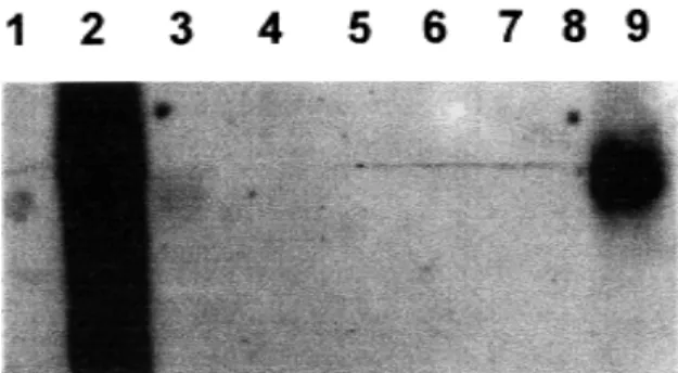
Northern blot analysis with the p27 probe, revealed that this gene was only transcribed by P. brasiliensis, with a positive signal both in the
mycelium and the yeast phases of the fungus; no signals were obtained with the mRNAs from other fungi (Figure 1).
The DNA extracted from fungal cells by means of glass beads plus the extraction buffer and phenol, showed to be of high quality and purity, as determined by the ratio A260/280 which was > 1.8.
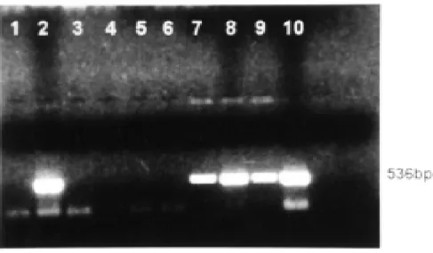
PCR amplification was obtained with all the primers used; thus the ITS1/ITS4 set which corresponds to universal specific fungal primers gave a positive band of 620 bp with all the fungi evaluated (data not shown). The gp43-1/gp43-2, the gp2 gpM (nested) and the LO/UP primers were specific for P. brasiliensis and they only amplified with material originating from this fungus but not with those from the remaining fungi tested. Figure 2 shows the specificity obtained with the LO/UP primers. The latter primers showed to be highly sensitive, allowing amplification of low amounts (3pg) of P. brasiliensis
(approximately 90 cells).
Soil samples artificially contaminated with P. brasiliensis, permitted amplification after elimination of the naturally contaminating PCR inhibitors. The addition of 5.0x10⁶ mycelial cells of P. brasiliensis to 1 g of non-sterile sediments resulted in a larger quantity of amplified DNA. The amount of P. brasiliensis DNA detected by means of PCR using the
species-specific primer (LO/UP) was 40 ng (approximately 1.2x10⁶ cells), as shown in Figure 3.
As it concerns mycological studies, primary agar cultures done with minced lymph node tissues from the first armadillo, showed growth of several colonies of a mold. These were dimorphic as demonstrated when colonies grown at room-temperature were transferred to new media and incubated at 36° C. The corresponding cultures exhibited the multiple budding cells that characterize P. brasiliensis9. The products of amplification of the lymph node and liver of this first armadillo, are shown in Figure 4.
The results of mycological studies and PCR amplifications from animal tissues are shown in the Table 2.
Fig. 1 - Northern-blot analysis of the p 27 gene. Lane 1. Negative control pBluescript without insert, 2. Positive control (pBluescript with gene of p27), 3. P. brasiliensis ATCC 32069 (mycelial), 4. S. cerevisae, ATCC 9763, 5. B. dermatitidis, 6. H. capsulatum, 7. C. albicans, ATCC 90028, 8. C. neoformans, ATCC 90112, 9. P. brasiliensis (yeast)
Fig. 2 - DNA amplification by PCR with specific primers LO/UP in agarose gel electrophoresis stained with ethidium bromide. The following DNA templates were used for PCR amplification (from line 3 on): Lanes: 1. Molecular weight markers (pUC18 Hae III digested), 2. Negative control, 3. Positive control P. brasiliensis, ATCC 32069 (mycelial phase), 4., 5. P. brasiliensis ATCC 60855 (mycelial and yeast phases), 6., 7. H. capsulatum (mycelial and yeast phases respectively), 8. B. dermatitidis, 9. S. cerevisae; ATCC 9763, 10. C. albicans; ATCC 90028, 11. C. neoformans; ATCC 90112, 12. S. schenckii, 13. A. fumigatus, 14. Negative control.
Table 2
Results of cultures and DNA amplification in animal tissues
Armadillo Isolation in Amplification by PCR
Nº culture Lungs Lymph node Liver Spleen
1* + - - +
-2 - - - -
-3 - - - -
-4 - - - -
-5 - + + +
DISCUSSION
This report describes a method for amplification of P. brasiliensis
genes through the use of PCR. This technology has previously been employed in the identification of other clinically important pathogens13,29, as well as of P. brasiliensis in an animal model13. In the later study,
authors inoculated 5.8 x 10⁶ yeast cells and obtained positive amplicons from the animals’ sera 8 weeks afterwards; the level of sensitivity was 10 pg. PCR can also be similarly applied to the search of environmental microorganisms2,4.
Fig. 3 - DNA amplification by PCR with specific primers LO/UP in agarose gel electrophoresis stained with ethidium bromide. Lanes: 1. Molecular weight markers (pUC18 Hae III digested), 2. Negative control, 3. Positive control P. brasiliensis, 4. & 5. Products of amplification from P. brasiliensis DNA extracted from soil.
Fig. 4 - DNA amplification by PCR with specific primers LO/UP in agarose gel electrophoresis stained with ethidium bromide. The following DNA templates were used for PCR amplification: Lanes: 1. Negative control, 2. Positive control, 3 & 4. Lung of first and fourth armadillos, 5 & 6. Spleen of first and five armadillos, 7. Lymph node of first armadillo, 8. Liver of first armadillo, 9. Lung of mouse inoculated with lymph node from the same armadillo, 10. Liver of mouse inoculated with armadillo’s liver.
The ability to extract DNA from fungal cells through mechanical disruption techniques and the use of SDS buffer and phenol for nucleic acid extraction, proves adequate for biochemical and/or genetic analyses and is also effective when obtaining DNA products of high quality30.
The gene codifying for the main P. brasiliensis antigenic protein of
P. brasiliensis DNA, and they were used to corroborate the positive findings in the armadillo’s studies8. Similar results have been obtained using the gene that codifies for the 27 kDa antigen protein which was cloned and characterized in our laboratory19,23. With the use of these primers it has been possible to detect low concentrations of fungal DNA (3pg, equivalent to 90 cells). We have also tested the mRNA levels of this 27kDa protein in different fungal cultures by Northern blot analysis and demonstrated that this protein mRNA is exclusively transcribed in
P. brasiliensis (Figure 1).
Isolation of P. brasiliensis from soils by means of conventional techniques such as culture and animal inoculation, has low repro-ducibility6. In order to improve these conditions, we standardized the PCR methodology in artificially P. brasiliensis contaminated soils
samples, an important step in the search of its precise ecological niche and its possible use for future epidemiological studies. The main problem in the search of fungal DNA in soils by PCR, is the presence of inhibitors contained in these samples. Soils with high organic contents have humic acids with phenolic groups, which exhibit similar solubility to that of the DNA. As a consequence, they are not totally removed during classical extraction protocols and remain as contaminants in the final DNA preparation4,20. An alternative to eliminate the presence of inhibitors is to consider that there are important differences in the size of DNA macromolecules and most common soluble PCR inhibitors; these inhibitors are effectively removed from the agarose blocks by diffusion during the washing steps, whereas genomic DNA remains trapped20. In our study, the PCR detection limit for P. brasiliensis species-specific
sequences from soils, proved to be 40 ng of DNA extracted from 1 g of soil (approximately 1.2x10⁶ cells).
The infection with P. brasiliensis has been established in the nine-banded armadillo D. novemcinctus, an animal amply distributed in Latin
America where it occupies areas that coincide, at least partly, with the paracoccidioidomycosis endemic regions. Additionally, in the armadillo’s burrows, the environmental conditions would increase the possibilities for a stable fungal microniche12.
The positive fungal amplification obtained in some organs of a positive armadillo, as well from tissues of another three culture-negative armadillos, suggest that PCR is not only a more sensitive technique than the conventional ones used with the same purpose9, but has also some advantages over culture of non sterile organs, such as lungs, usually heavily contaminated with various microorganisms. In one of the armadillos, the results obtained by PCR was confirmed by isolation of the agent in culture, the gold standard technique. The latter supports previous findings by other authors3,9,21, indicating that P.
brasiliensis, can be isolated not only from humans samples, but also
from armadillos, such as the one captured in an endemic area of Colombia9. The fact that only one armadillo from the 5 studied, was positive for culture while 3 revealed to be infected by DNA amplification, tends to suggest that PCR is indeed a more sensitive technique16.
The results reported here suggest that the PCR amplification of P. brasiliensis DNA by the use of species-specific primers, may play an
important role in the search of this fungus habitat and also for future epidemiological studies. At present analysis of soil samples obtained from different endemic areas in Colombia and Brazil is being pursued. Several questions arise from this study, such as: how do armadillos get
infected?. Once the microniche is detected, what environmental measures could be taken to avoid human infection?. Answers to these and other questions would require further explorations.
RESUMO
PCR com «primers» específicos de Paracoccidioides brasiliensis: uso potencial em estudos ecológicos
O microambiente adequado do Paracoccidioides brasiliensis não
foi ainda bem esclarecido, talvez porque os métodos utilizados não sejam suficientemente sensíveis. Aplicamos com este propósito, a reação em cadeia da polimerase (PCR) usando três jogos de primers específicos do
P. brasiliensis, correspondendo a dois dos genes do P. brasiliensis. Este
fungo, assim como outros fungos, foram cultivados e seus DNAs obtidos por ruptura mecânica e purificados com mistura de fenol-clorofórmio com álcool isoamílico. Os DNAs serviram para a reação de PCR utilizando-se primers específicos para dois dos genes do P. brasiliensis
que codificam para as proteínas antigênicas, denominadas, 27 kDa e 43 kDa. O limite mínimo de detecção para o gene 27 kDa foi de 3 pg. A amplificação para os dois genes foi positiva só com o DNA do P. brasiliensis; além disso, o mRNA para o gene de 27kDa estava presente
apenas no material do P. brasiliensis, como mostrado pela análise por
Northern-blot. A padronização da técnica do PCR permitiu a amplificação do DNA do P. brasiliensis em solos contaminados artificialmente com o
fungo e em tecidos de tatus infectados na natureza. Estes resultados indicam que a técnica do PCR podería ter um papel muito importante na pesquisa do habitat do P. brasiliensis e, além disso, podería ser utilizada
em outros estudos ecológicos.
ACKNOWLEDGEMENT
This work was supported by COLCIENCIAS, Santafé de Bogotá, Colombia, project number 2213-05-386-96. The kind cooperation offered by the personnel in the Laboratory of Mycology is greatly appreciated.
REFERENCES
1. ALBORNOZ, M.B. - Isolation of Paracoccidioides brasiliensis from rural soil in Venezuela. Sabouraudia, 2: 248-252, 1971.
2. ATLAS, R.M.; SAYLER, G.; BURLAGE, R.S. & BEJ, A.M. - Molecular approaches for environmental monitoring of microorganisms. Biotechniques, 12: 706-717, 1992. 3. BAGAGLI, E.; SANO, A.; COELHO, K.I. et al. - Isolation of Paracoccidioides brasiliensis from armadillos (Dasypus noveminctus) captured in an endemic area of paracoccidioidomycosis. Amer. J. trop. Med. Hyg., 58: 505-512, 1998. 4. BEJ, A.K.; MAHBUBANI, M.H.; DICESARE, J.L. & ATLAS, R.M. - Polymerase chain
reaction-gene probe detection of microorganisms by using filter-concentrated samples. Appl. environ. Microbiol., 57: 3529-3534, 1991.
5. BOWMAN, B.A. - A model PCR/probe system for the identification of fungal pathogens. In: PERSING, D. H.; SMITH, T. F.; TENONER, F. C. & WHITE, T. J., ed. Diagnostic molecular Microbiology. Washington, American Society for Microbiology, 1993. p. 423-430.
6. BRUMMER, E.; CASTAÑEDA, E. & RESTREPO, A. - Paracoccidioidomycosis: an update. Clin. Microbiol. Rev., 6: 89-117, 1993.
8. CISALPINO, P.S.; PUCCIA, R.; YAMAUCHI, L.M. et al. - Cloning, characterization, and epitope expression of the major diagnostic antigen of Paracoccidioides brasiliensis. J. biol. Chem., 271: 4553-4560, 1996.
9. CORREDOR, G.G.; CASTAÑO, J.H.; PERALTA, L.A. et al. - Isolation of Paracoccidioides brasiliensis from the nine-banded armadillo Dasypus novemcinctus, in an endemic area for paracoccidioidomycosis in Colombia. Rev. iberoamer. Micol., 16: 37-41, 1999.
10. ESPINEL-INGROFF, A. & KERKERING, T.M. - Spectrophotometric methods of inoculum preparation for the in vitro testing of filamentous fungi. J. clin. Microbiol., 29: 393-394, 1991.
11. FERREIRA, M.S.; FREITAS, L.H.; LACAZ, C.S. et al. - Isolation and characterization of a Paracoccidioides brasiliensis strain from a dogfood contaminated with soil in Uberlandia, Brazil. J. med. vet. Mycol., 28: 253-256, 1990.
12. FORJAZ, M.H.H. - Estudo da epidemiología da Paracoccidioidomicose. Rastreamento de áreas endêmicas e de “reserváreas” no Brasil, através do traçado do perfil migratório-residencial-profissional, de pacientes diagnosticados em São Paulo. São Paulo, 1989. (Thesis - Escola Paulista de Medicina/UNIFESP).
13. GOLDANI, L.Z. & SUGAR, A.M. - Short report: use of the polymerase chain reaction to detect Paracoccidioides brasiliensis in murine paracoccidioidomycosis. Amer. J. trop. Med. Hyg., 58: 152-153, 1998.
14. GROSE, E. & TAMSITT, J.R. - Paracoccidioides brasiliensis recovered from the intestinal tract of 3 bats (Artibeus lituratus) in Colombia. Sabouradia, 4: 124, 1965. 15. GUEZUELE, E. - Aislamiento de Paracoccidioides sp. de heces de pinguino de la
Antártida. In: ENCUENTRO INTERNACIONAL SOBRE PARACOCCIDIOIDO-MICOSIS, 4., Caracas, 1989. Proceedings. p. B2
16. INNIS, M.A.; GELFAND, D.H.; SNINSKY, J.J. & WHITE, T.J. - PCR protocols. San Diego, Academic Press, 1990.
17. JOHNSTON, C.G. & AUST, S.D. - Detection of Phynerochaetae chrysosporium in soil by PCR and restriction enzyme analysis. Appl. environ. Microbiol., 60: 2350-2354, 1994.
18. MCEWEN, J.G.; GARCIA, A.M.; ORTIZ, B.L. et al. - In search of the natural habitat of Paracoccidioides brasiliensis. Arch. med. Res., 26: 305-306, 1995.
19. MCEWEN, J.G.; ORTIZ, B.L.; GARCIA, A.M. et al. - Molecular cloning, nucleotide sequencing, and characterization of a 27-kDa antigenic protein from Paracoccidioides brasiliensis. Fungal Genet. Biol., 20: 125-131, 1996.
20. MOREIRA, D. - Efficient removal of PCR inhibitors using agarose-embedded DNA preparations. Nucleic Acids Res., 26: 3309-3310, 1998.
21. NAIFF, R.D.; FERREIRA, L.C.L.; BARRETT, T.V. et al. - Paracoccidioidomicose enzoótica em tatus (Dasypus novemcinctus) no Estado do Pará. Rev. Inst. Med. trop. S. Paulo, 28: 19-27, 1986.
22. NEGRONI, P. - El Paracoccidioides brasiliensis vive saprofíticamente en el suelo argentino. Pren. méd. argent., 53: 2381-2382, 1966.
23. ORTIZ, B.L.; DÍEZ, S.; URÁN, M.E. et al. - Use of 27-Kilodalton recombinant protein from Paracoccidioides brasiliensis in serodiagnosis of paracoccidioidomycosis. Clin. diagn. Lab. Immunol., 5: 826-830, 1998.
24. RESTREPO, A. - Ecology of Paracoccidioides brasiliensis.In: FRANCO, M.; LACAZ, C.S.; RESTREPO, A. & DEL NEGRO G., ed. Paracoccidioidomycosis. Boca Raton, CRC Press, 1994. p. 121-130.
25. RESTREPO, A. & JIMENEZ, B. - Growth of Paracoccidioides brasiliensis yeast phase in a chemically defined medium. J. clin. Microbiol., 12: 279-281, 1980. 26. SAMBROOK, J.; FRITSCH, E.F. & MANIATIS, T. - Molecular cloning: a laboratory
manual. 2. ed. New York, Cold Spring Harbor, 1989. v: 1
27. SILVA-VERGARA, M.L.; MARTINEZ, R.; CHADU, A. et al. - Isolation of a Paracoccidioides brasiliensis strain from the soil of a coffee plantation in Ibiá, State of Minas Gerais, Brazil. Med. Mycol., 36: 37-42, 1998.
28. TSAI, Y.L. & OLSON, G.H. - Rapid method for direct extraction of DNA from soil and sediment. Appl. environ. Microbiol., 57: 1070-1074, 1991.
29. VAN BURIK, J.A.H.; MYERSON, D.; SCHRECKHISE, R.W. & BOWDEN, R.A. -Panfungal PCR assay for detection of fungal infection in human blood specimens. J. clin. Microbiol., 36: 1169-1175, 1998.
30. VAN BURIK, J.A.H.; SCHRECKHISE, R.W.; WHITE, T.C.; BOWDEN, R.A. & MYERSON, D. - Comparison of six extraction techniques for isolation of DNA from filamentous fungi. Med. Mycol., 36: 299-303, 1998.
31. VESCIO, P.A. & NIERZWICKI-BAUER, S.A. - Extraction and purification of PCR amplifiable DNA from lacustrine subsurface sediments. J. microbiol. Meth., 21: 225-233, 1995.