1 This work is the partial result of the research developed for Master degree by the first author, under supervision of the second author, at the Department of Experimental Psychology of the Institute of Psychology (University of São Paulo), and it was financially supported by the Foundation for Research Support of the State of São Paulo (FAPESP), the National Council for the Scientific and Technologic Development (CNPq), and the Coordination for the Development of Superior Level Personnel (CAPES).
CLINICAL PSY CLINICAL PSY CLINICAL PSY CLINICAL PSY
CLINICAL PSYCHOPHISICS:CHOPHISICS:CHOPHISICS:CHOPHISICS:CHOPHISICS: APPLICA APPLICA APPLICA APPLICA APPLICATION OF PSYTION OF PSYCHOPHYTION OF PSYTION OF PSYTION OF PSYCHOPHYCHOPHYCHOPHYCHOPHYSICALSICALSICALSICALSICAL
METHODOL METHODOL METHODOL METHODOL
METHODOLOGY OGY OGY TOGY OGY TTTTO AID DIAO AID DIAO AID DIAO AID DIAGNOSIS.O AID DIAGNOSIS.GNOSIS.GNOSIS.GNOSIS. METHOD DESCRIPTION METHOD DESCRIPTION METHOD DESCRIPTION METHOD DESCRIPTION METHOD DESCRIPTION11111
Emilia Longhi Marcelo Fernandes da Costa
Abstr Abstr Abstr Abstr
Abstracacacacact:t:t:t: The Psychophysics applied to the clinic with humans may providet: alternative tools to assist access to objective and quantifiable internal conditions of the patient, which could only be obtained otherwise, through their stories and descriptions. An example of this partnership and implementation of Psychophysics is the commercial unit C-Quant (Oculus Optikgeräte, Germany), whose psychophysical method of access to the value of light scattering in the retina was developed by the group of Dutch researchers led by prof. Dr. Thomas van den Berg, Netherland’s Institute of Neuroscience (NIN). Access to the amount of light scattering in the retina is useful for the diagnosis of various eye diseases such as cataracts. In this article the psychophysical method in this unit (Comparison of compensation) is described.
Key Key Key Key
148
CLINICAL PSYCHOPHISICS: APPLICATION OF PSYCHOPHYSICAL... E3 MILIA LONGHIE MARCELO FERNANDESDA COSTAIn many clinical situations, access to the patient’s condition can be done directly, without interference of this, as in blood collection for analysis. However, when it comes to sensory or emotional situation, for example, such access can not be obtained directly. It is the patient who needs to tell what you feel, describe what you see, and that includes a personal bias in any analysis. The Psychophysics arises in the second half of the nineteenth century with Fechner, try to provide an alternative access to private data, commonly called “psychic”, which ultimately correspond to the way the subject sees and perceives the world and yourself. The main purpose of the psychophysical methodology, therefore, is to offer the clinical researcher or a more objective way of measuring access and internal states, stimuli from the subject offered by the researcher.
The main difference is the ability to measure these states, since, with numbers in hand with which to work, one can quantify a feeling or perception, the scientific work (both research and clinical applications) acquires greater reliability and is subject statistical analysis and possible replication by other groups.
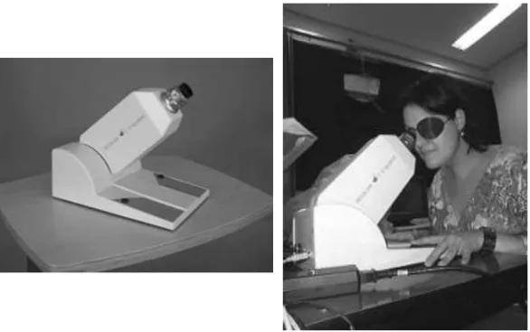
Several psychophysical methods have been developed over these 150 years, but their use over time has become limited to research groups at universities, and little was done to the psychophysical rehabilitation of the clinic. However, this situation has been changing in recent years: the use of new methodologies psychophysical apparatus in commercial use is a reflection of the relationship between the university (as a researcher) and professional practice. An example of this relationship is to develop a device to be used in ophthalmology clinics in the world, a device that uses this psychophysical method developed by the research group led by prof. Dr. Thomas van den Berg, Netherland’s Institute of Neuroscience (NIN), and marketed by a renowned German company of ophthalmic products, Oculus. This device, called the C-Quant commercially (Figure 1) allows the clinician access to the amount of scattered light within the patient’s eye, a value that can be used to assist in the diagnosis of cataracts.
Figure 1. C-Quant (Oculus Optikgeräte, Alemanha).
1. Blurring phenomena and Intraocular Light Scatter
Glare, as defined by CIE (Commission Internationale de l’Éclairage) as “disabling glare” (disability glare), consists of a momentary blindness exists when a person is exposed to a bright stimulus in the visual field, as in the case of an automobile headlight coming toward someone (Vos, 1984; Vos & van den Berg, 1999).
Sensitivity to glare has relation with the scattering of light in the eye, usually in a situation of glare, the light from the glare source forms an image on the retina, and a portion of light is also scattered by the retina, forming a veil of light that covers the retina (van den Berg & van Rijn, 2005, ch. 2, p. 53).
The amount of scattering of light on the retina is different for each subject and may be different even between the eyes of the same subject. It depends on a number of factors such as age, pigmentation of the iris and choroid, the existence of conditions such as cataracts or corneal damage, as well as being secondary to procedures such as refractive surgery.
In the normal eye, there are four major sources that contribute to the total light scattering in the retina, cornea, lens, iris and fundus. The contribution is calculated for each structure: cornea (third), lens (third) and the iris and fundus (third) (van den Berg & van Rijn, 2005).
150
CLINICAL PSYCHOPHISICS: APPLICATION OF PSYCHOPHYSICAL... E3 MILIA LONGHIE MARCELO FERNANDESDA COSTAable to adversely affect vision. This sensitivity depends on two factors: the amount of light glare and eye sensitivity to glare. The conventional method to measure sensitivity to glare was to measure visual acuity or contrast sensitivity in the presence of a source of obfuscation, this method has disadvantages, particularly the need for dark adaptation, which differs from the daily experience of the person in the presence a source of glare.
An indirect way to measure sensitivity to glare is to measure how much light is scattered in the eye, ie, the dispersion in the retina – this is appropriate since the scattering of light on the retina is the physical parameter that causes the effect of glare (van den Berg & van Rijn, 2005, ch. 2, p. 53-4). To access the value of obfuscation, it is a measure of equivalent luminance, observing which the luminance of a point source of light that causes momentary blindness in the subject, preventing to observe a target (any stimulus) present next to the source of light. When a guy says you can not see the target, according to the luminance of the light source is increased, the luminance value at this moment is equivalent to the threshold of blurring of the subject.
As the sensitivity depends on the blurring of these two factors (light source and characteristics of the eye), any change in one of these factors affect the passage of light and therefore increases the glare. In the case of the light source, when its intensity is increased, the intensity of scattered light also increases in the retina, a phenomenon known as “light curtain” (veiling light) or light cascading effect, since the scattered light appears in one eye form of veil or cascade, whereas in the case of the eye, any change in the visual pathways that impede or alter the light path can cause an increase in the dispersion. The most common case of increasing stray light and glare in cataract happens, because the natural lens becomes more opaque with time – with increasing opacity of the lens, increases the dispersion of the light falling on the retina, causing an increase in glare (van Rijn & van den Berg, 2005, Valbuena, Bandeen-Roche, Rubin, Munoz & West 1999).
2. Psychophysical Method
Direct Compensation method:
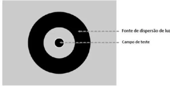
The initial instrument for measuring light scattering in the retina had a screen test as shown in Figure 2, with a central circle and two peripheral rings, and the center circle was the staging area, and the outer ring, the source of dispersion light.
Figure 2. Example of the test screen of the intraocular light dispersion quantifier based on the Direct Comparison method (adapted from Franssen & Coppens, 2007). In this procedure, the participant was to fixate on the center circle, and as the test was started, the peripheral ring (source scatter) started flashing, being perceived by participants as being intermittently black and white. When the ring was white, called phase-on, and when the ring was black, off-stage. (Figure 3-1). Thus, the phase-on, the ring was designed in the peripheral retina, but due to optical imperfections of the eye, a small portion of light that originated in the ring is dispersed to other parts of the retina including the fovea. As the participant is staring at the center circle, who was black, the fovea was the image of a black circle, but because of the scattered light from the ring, this circle appears slightly gray, although in reality is black, since all this light comes from the Ring Road. In phase-off, there was no light coming from the ring, and thus the cen-tral circle appears black to the participant because no scattered light. In the end, like the phases alternate on and off, the participant perceives the central circle flashing, alternating between black and gray in phase with the flashing of the peripheral ring.
The direct compensation method worked reasonably, but the experiments (Franssen, Coppens and van den Berg, 2006) showed participants’ difficulties concerned with determining whether the cen-tral circle was blinking when the peripheral ring flashed with intensity. Furthermore, this methodology did not allow experimenters to obtain measures of reliability of the results. So, to get a psychophysical method that would allow a simpler task for the volunteer and that was reliable was developed Compensation Comparison Method.
Compensation Comparison Method
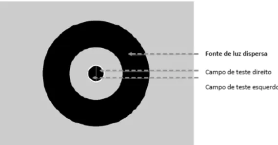
This method was developed from the direct compensation method, the big difference is in the center circle, which is split in this method (Figure 4), which in practical terms, facilitated the task of the participant, in the previous method was to eliminate the task Central and the twinkle in the current method compare which central light is flashing yellow (forced choice paradigm of two alternatives). In one half of the circle, the phase-off, the light is added compensation, but the other half remains dark, ie, one half corresponds to the starting point of the method of direct compensation, and the other half of the field test (circle center) corresponds to a compensation value in the method of direct compensation. Thus, the participant should compare the two halves of the central circle, each time comparing the different intensities of flashing light, the participant’s task is to determine which of the two halves of the field test flashes more strongly: right or left. During the test, the hemifield with light compensation is varied randomly.
154
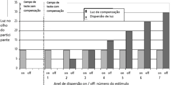
CLINICAL PSYCHOPHISICS: APPLICATION OF PSYCHOPHYSICAL... E3 MILIA LONGHIE MARCELO FERNANDESDA COSTA The hemifield without compensation is black light throughout the experiment, but due to light scattering from the Ring Road, the participant realizes that hemifield flashing once the ring starts flashing. The scattering of light also affects the other hemifield, but the presence of light compensation makes the perception of the hemifield with flash compensation is different from the perception of flashing hemifield without compensation. Depending on the amount of light existing compensation, the participant can see the flickering hemifield with compensation as stronger or weaker than the other hemifield. If the participant chooses the side with compensation as the flashing stronger, the test program makes this response a number “1”, and if he chooses the side without compensation as the flashing stronger, that reply is transformed in a number “0”. Thus, the responses of the participant during the test are binary (1 or 0), identifying when the side with the light compensation was chosen or not. Figure 5 shows an illustrated diagram of choices, depending on the amount of light existing compensation.Figure 5. Hypothetical example of the comparison of hemifields, portraying the retinal incident amount of light in phases on and off (in arbitrary values) and the amount of disperse light proceeding from the peripheral ring (adapted from Franssen & Coppens, 2007).
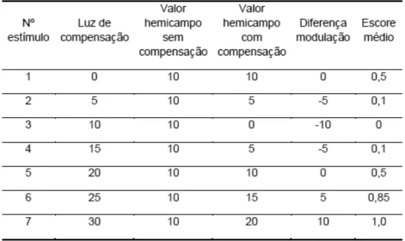
compensation is 5, then the modulation that the participant perceives the difference is (10-5 = 5), which is less than the amount of scattered light in the other hemifield, so the participant strongly tend to choose the hemifield without light compensation. In the stimulus 3, there is no variation in perceived by the participant hemifield light compensation, because the amount of compensatory light is equal to the scattered light (10-10 = 0), as well as the participant does not notice the blinking light compensation hemifield , it is easy to determine that the hemifield that flashes is the stronger without compensation.
The procedure is the same for every point, determine what the participant must realize in each hemifield, making the subtraction of the values of light compensation and dispersion in the hemifield with light compensation, and comparing with the other hemifield, no light compensation. Table 1 is a summary of the comparison, considering that only 3 stimulus there will be 100% chance that the participant chooses the hemifield without compensation, since the difference in the other stimuli is not the most, and there may be errors in the response.
Table 1. Summary of the comparisons between hemifields with and without compensation, according to the example of the text.
In the example above, only seven stimuli are presented, however, the C-Quant 25 stimuli are shown to be possible to determine the reliability of results and also to be able to determine the value of dispersion parameter.
156
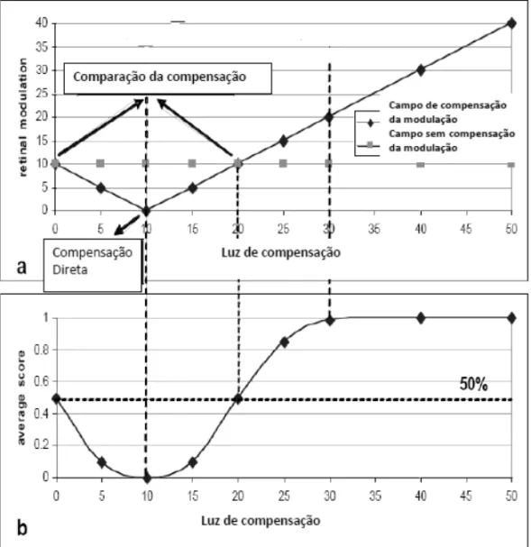
CLINICAL PSYCHOPHISICS: APPLICATION OF PSYCHOPHYSICAL... EMILIA LONGHI E MARCELO FERNANDES DA COSTA3 function (Figure 6). By characteristics of the human visual system, the psychometric function is shown on a logarithmic scale (Figure 6b) and nonlinear (Figure 6a). Through the construction of the psychometric cur-ve, you can determine the value of the dispersion parameter, which corresponds to the point of direct comparison, in which the player will always choose the hemifield without compensation, since the light compensation is equal to the scattered light.Figura 6. Data plotting of the example (Table 1) depending on the amount of light, and determination of the psychometric curve in logarithmic unities (adapted from Franssen & Coppens, 2007)
psycho-physical method used (comparison of compensation) is quite complex, the partnership between research and application became possible to use simple, contributing to the advancement, in this case, diagnostic techniques in ophthalmology. Interestingly, this device is to show the possibility of union between psychophysics and Clinic, giving this new tool.
P P P P
Psicsicsicsicofísica clínica:sicofísica clínica:ofísica clínica:ofísica clínica:ofísica clínica: aplicação de met aplicação de met aplicação de metodologia psic aplicação de met aplicação de metodologia psicodologia psicodologia psicodologia psicofísica no auxílio de diagnósticofísica no auxílio de diagnósticofísica no auxílio de diagnósticofísica no auxílio de diagnósticofísica no auxílio de diagnósticososososos... D
D D D
Descrição de métescrição de métescrição de métescrição de métescrição de métodoodoodoodoodo
Resumo: Resumo: Resumo: Resumo:
Resumo: A Psicofísica aplicada à Clínica com seres humanos pode prover ferramen-tas alternativas que auxiliem o acesso objetivo e quantificável a condições internas do paciente, que só poderiam ser obtidas, de outra forma, através de seus relatos e descrições. Um exemplo dessa parceria e aplicação da Psicofísica é o aparelho co-mercial C-Quant (Oculus Optikgeräte, Alemanha), cujo método psicofísico de acesso ao valor de dispersão de luz na retina foi desenvolvido pelo grupo de pesquisadores holandeses liderados pelo Prof. Dr. Thomas van den Berg, do Netherland Institute of Neuroscience (NIN). O acesso ao valor de dispersão de luz na retina é útil para auxili-ar no diagnóstico de várias doenças oculauxili-ares, como catauxili-arata. Neste auxili-artigo o método psicofísico presente no aparelho (Comparação da Compensação) é descrito.
P P P P
Palaalaalaalaalavrvrvrvras-chavras-chavas-chaas-chaas-chavvvve:e:e:e:e: Psicologia. Psicofísica. Distúrbios da visão. Catarata.
P P P P
Psysysysychophsychophchophchophchophysique Clinique:ysique Clinique:ysique Clinique:ysique Clinique: application de la méthodologie psyysique Clinique: application de la méthodologie psy application de la méthodologie psy application de la méthodologie psychoph application de la méthodologie psychophchophchophysique de l’aidechophysique de l’aideysique de l’aideysique de l’aideysique de l’aide au diagnostic
au diagnostic au diagnostic au diagnostic
au diagnostic... D D D Description de la méthode Description de la méthodeescription de la méthodeescription de la méthodeescription de la méthode
Résumé: Résumé: Résumé: Résumé:
158
CLINICAL PSYCHOPHISICS: APPLICATION OF PSYCHOPHYSICAL... E3 MILIA LONGHIE MARCELO FERNANDESDA COSTA cataracte. Dans cet article, la méthode psychophysique dans cette unité (comparaison de la rémunération) est décrite.M MM M
Mots-clés:ots-clés:ots-clés:ots-clés:ots-clés: Psychologie. Psychophysique. Troubles de la vision. La cataracte.
P PP P
Psicsicsicsicsicofísica Clínica:ofísica Clínica:ofísica Clínica:ofísica Clínica: aplicación de la metofísica Clínica: aplicación de la met aplicación de la met aplicación de la metodología psic aplicación de la metodología psicofísica parodología psicodología psicodología psicofísica parofísica parofísica parofísica para aa aa aa aa ayudar al diagnósti-yudar al diagnósti-yudar al diagnósti-yudar al diagnósti-yudar al diagnósti-c
cc
ccoooo... Do D D Descripción del mét Descripción del métescripción del métescripción del métescripción del métodoodoodoodoodo
Resumen: Resumen:Resumen: Resumen:
Resumen: La Psicofísica aplicada a la clínica con los seres humanos puede proporci-onar herramientas alternativas para facilitar el acceso objetivo y cuantificable a las condiciones internas del paciente, que sólo se podría obtener de otra manera, a tra-vés de sus historias y descripciones. Un ejemplo de esta asociación y la aplicación de la psicofísica es la unidad comercial de C-Quant (Oculus Optikgeräte, Alemania), cuyo método psicofísico de acceso al valor de dispersión de la luz en la retina fue desarrollado por el grupo de investigadores holandeses dirigidos por el Prof. Dr. Thomas van den Berg, del Netherland Institute of Neuroscience (NIN). El acceso al valor de dispersión de luz en la retina es útil para el diagnóstico de diversas enferme-dades oculares, como cataratas. En este artículo el método psicofísico presente en esta unidad comercial (Comparación de la compensación) es descrito.
P PP P
Palabralabralabralabralabras claas claas claas claas clavvvvve:e:e:e:e: Psicología. Psicofísica. Alteraciones de la visión. Catarata.
Ref RefRef Ref
Refererererencerencencencenceseseseses
Franssen, L., e Coppens, J. E. (2007). Straylight at the retina – scattered papers. Amsterdam: Gildeprint Drukkerijen B.V.;
Franssen, L., Coppens, J. E., van den Berg, T. J. T. P. (2006). Compensation comparison method for assessment of retinal straylight. Invest Ophtalmol Vis Sci. 47: 768-76;
Valbuena, M., Bandeen-Roche, K., Rubin, G.S., Munoz, B. &. West, S. K. (1999). Self-reported assessment of visual function in a population-based study: the SEE project. Salisbury Eye Evaluation. Invest Ophthalmol.Vis.Sci., 40, 280-288;
van den Berg, T. J. T. P. e van Rijn, L.J. (2005). Relevance of glare sensitivity and impairment of visual function among European drivers. Report EU project: SUB-B27020B-E3-GLARE-2002-S07.18091. disponível em www.glare.be;
Vos, J. J. (1984). Disability glare – a state of the art report. CIE J. 3(2): 39-53;
160
CLINICAL PSYCHOPHISICS: APPLICATION OF PSYCHOPHYSICAL... EMILIA LONGHI E MARCELO FERNANDES DA COSTA3Emilia L Emilia LEmilia L Emilia L
Emilia Longhi,onghi,onghi,onghi,onghi, Master in Psychology, Department of Experimental Psychology, Institute of Psychology, University of São Paulo. Address: Av. Prof. Mello Moraes, 1721. Zip Code: 05508-030, São Paulo, SP, Brazil. E-mail: [email protected]
M MM M
Marararararcccccelo Felo Fernandes Celo Felo Felo Fernandes Cernandes Cernandes Cernandes Costa,osta,osta,osta,osta, Professor at the Department of Experimental Psychology, Institute of Psychology of the University of São Paulo, and Professor at the Center of Neurosciences and Behavior at the University of São Paulo. Address: Av. Prof. Mello Moraes, 1721. Zip Code: 05508-030, São Paulo, SP, Brazil. E-mail: [email protected]
Received: 21/10/2010