October 2012
Escola de Engenharia
Ana Catarina Rodrigues
Study of the efficacy of combination
therapy based on bacteriophages for
the control of infectious biofilms
UMinho|20
12
Ana Catarina Rodrigues
Study of t
he ef
ficacy of combination t
herap
y based on bacteriophages for t
Dissertation thesis for the Master
degree in Biomedical Engineering
Supervisor:
Dr. Sanna Maria Sillankorva
Co-Supervisor:
Dr. Joana Azeredo
October 2012
Escola de Engenharia
Ana Catarina Rodrigues
Study of the efficacy of combination
therapy based on bacteriophages for
the control of infectious biofilms
COMPROMETE;
Universidade do Minho, ___/___/______
i
Acknowledgements
The present work was carried out in Biological Engineering Department of University of Minho. It was supervised by Dr. Sanna Sillankorva and Dr. Joana Azeredo.
To Dr. Joana I want to express my sincere appreciation for all the support, availability and guidance provided throughout the development of this project.
Especially, to Dr. Sanna, I want to express my deepest thanks for all help given me in coordinate my project, for all the encouragement, suggestions and advices that were also important and supportive for the achievement of this work.
I also want to thank all the colleagues of the Microbiology Laboratory that always help me when I ever needed. A special thanks to colleagues of Phage Biotechnology Group for being ready to help anytime. I want highlight my colleague Priscila whom I thank deeply all the knowledge and friendship provided during my stay in the laboratory.
In this space I also want to thank the most important people in my life: to my sister and my parents. To them I want to thank all the strength, support and motivation given me, which was important for the achievement of the objectives of my work.
Finally, I would like to address my thanks to all the other people, who have not been mentioned here by names, but who helped me during my thesis work and who made my stage in Biological Engineering Department a wonderful experience.
iii
Study of the efficacy of combination therapy based on
bacteriophages for the control of infectious biofilms
Abstract
Pseudomonas aeruginosa is regarded as a “phenomenon of bacterial resistance”.
This gram negative bacterium is responsible for a high percentage of mortality in the hospitals all over the world and its prevalence can be a consequence of important reasons, such as: intrinsic resistance determined by virulence factors; acquired resistance mechanisms that lead to a low susceptibility to antimicrobial agents; and the ability of P. aeruginosa to grow on any natural and artificial surfaces leading to the development of biofilms. The emergence of new strategies to control P. aeruginosa biofilms is becoming more evident due to their resistance to traditional treatments, and (bacterio)phages have been recognized as an attractive alternative for this problem. Nevertheless, despite the potential of phages as antimicrobial agents, it is well known that bacteria can quickly adapt and create new survival strategies and the emergence of phage-resistant phenotypes is inevitable. Thus, the combination of phage and antibiotic therapies could have potentially more benefits than just using phages and antibiotics alone.
This work describes the combined effect of phages and antibiotics against planktonic cultures and biofilms of P. aeruginosa. The antimicrobial susceptibility of three reference strains of P. aeruginosa towards antibiotics belonging to four different action groups was initially evaluated and for this purpose, two important parameters were determined: Minimal Inhibitory Concentration (MIC) for planktonic cultures and Minimal Biofilm Eradication Concentration (MBEC) for biofilms. After that, phage infection assays were performed against cultures and biofilms of P. aeruginosa using four different phages from the Bacteriophage Biotechnology Group of the University of Minho. The efficacies of antibiotics and phages or both combined were evaluated in biofilms by viable cell enumeration and in planktonic cultures by measuring the absorbance (OD600nm). In general, all antibiotics tested showed little efficacy against biofilms which were also very tolerant to phage infection. The presence of degradative enzymes, such as beta-lactamase, and the survival of tolerant cells (persister cells) can explain the failure of antibiotics in reducing the cell numbers present in biofilms. In the
iv
case of phage infection, the emergence of resistant phenotypes defective in LPS mutants can lead to long-term failure of these agents.
In combined treatments, phages used together with ciprofloxacin caused total biofilm eradication. Also, other combinations resulted in interesting results. For example, the combination therapy of both phage phiIBB-PAP21 and amikacin resulted in approximately 3.66 log reduction of viable cells while individually, phage and amikacin only caused a 1.3 and 1.76 log reduction, respectively. The effectiveness of combined phages-antibiotic treatments can be due to a higher burst size, as observed with phage phiIBB-PAP21, when their host cells were exposed to antibiotics. Also, the higher biomass reductions observed when biofilms are exposed to a combined treatment suggest that phages can enhance the antibiotic penetration through matrix disruption, rendering cells freely available to be killed with antibiotics.
Overall, the combination of phage and antibiotic enhances biofilm control; however the complex universe behind this synergistic interaction suggests that this is not always a linear process. Further studies should be conducted to complement and disclose this synergistic behaviour observed in the work described herein.
v
Avaliação da eficácia de terapias combinadas á base bacteriófagos
no controlo de biofilmes infeciosos
Sumário
Pseudomonas aeruginosa é considerada um "fenómeno da resistência
bacteriana". Esta bactéria gram-negativa é responsável por uma elevada percentagem de mortalidade nos hospitais de todo o mundo e a sua prevalência pode ser consequência de razões importantes como: resistência intrínseca determinada por vários fatores de virulência; mecanismos de resistência adquiridos que conduzem a uma baixa suscetibilidade aos agentes antimicrobianos e a capacidade de crescimento de P.
aeruginosa em todas as superfícies naturais e artificiais que favorece ao
desenvolvimento de biofilmes. A necessidade de novas estratégias de controlo de biofilmes de P. aeruginosa tem-se tornado mais evidente devido à sua resistência aos tratamentos tradicionais, e os bacteriófagos têm sido reconhecidos como uma alternativa atrativa para este problema. No entanto, apesar do potencial dos fagos como agentes antimicrobianos, sabe-se que as bactérias podem adaptar-se rapidamente e criar novas estratégias de sobrevivência e o aparecimento de fenótipos resistentes aos fagos é inevitável. Assim, a combinação de fagos com as terapias antibióticas pode ter potencialmente mais benefícios em relação á utilização isolada de fagos e antibióticos.
Este trabalho descreve o efeito combinado de fagos e antibióticos contra culturas planctónicas e biofilmes de P. aeruginosa. A suscetibilidade antimicrobiana de três estirpes de referência de P. aeruginosa contra antibióticos pertencentes a quatro grupos de ação diferentes foi inicialmente avaliada. Para este fim, dois parâmetros importantes foram determinados: Concentração Mínima Inibitória (CIM) para culturas planctónicas e Concentração Mínima de Erradicação do Biofilme (CMEB) para os biofilmes. Depois desta etapa realizaram-se ensaios de infeção fágica contra as culturas e biofilmes de P.
aeruginosa utilizando quatro fagos diferentes do Grupo de Biotecnologia de
Bacteriófagos da Universidade do Minho. A eficácia dos antibióticos e fagos ou dos dois agentes combinados foi avaliada nos biofilmes pela enumeração de células viáveis, e nas culturas planctónicas pela medição da absorvência (OD600nm). Em geral, todos os antibióticos testados apresentaram pouca eficácia contra os biofilmes, os quais também foram muito tolerantes à infeção fágica. A presença de enzimas de degradação,
vi
tais como a beta-lactamase, e a sobrevivência de células tolerantes (células persister) pode explicar a falha dos antibióticos sobre a redução do número de células presentes nos biofilmes. No caso da infeção fágica, o aparecimento de fenótipos deficientes na produção de LPS pode conduzir ao insucesso destes agentes a longo prazo.
Nos tratamentos combinados, os fagos utilizados juntamente com ciprofloxacina causaram a erradicação total do biofilme. Também outras combinações demonstraram resultados interessantes. Por exemplo, a combinação de ambos fago phiIBB-PAP21 e amicacina resultou numa redução de células viáveis de cerca de 3,66 log, enquanto individualmente, o fago e amicacina só provocou uma redução de 1,3log e 1,76log, respetivamente. A eficácia da combinação fago-antibiótico pode ser devido a elevados
burst size, tal como observado com o fago phiIBB-PAP21, quando o respetivo
hospedeiro foi exposto a antibióticos. Além disso, as maiores reduções da biomassa observadas quando os biofilmes são expostos a um tratamento combinado sugerem que os fagos podem melhorar a penetração de antibióticos através de rutura da matriz, permitindo uma libertação das células para a ação dos antibióticos.
Em geral, a combinação de fagos e antibióticos permite melhorar o controlo dos biofilmes; no entanto o universo complexo por trás desta interação de sinergia sugere que esta não é sempre um processo linear. Novos estudos devem ser realizados para complementar e divulgar este comportamento sinergístico observado neste trabalho.
vii
Table of Contents
Acknowledgements ... i Abstract ... iii Sumário ... v List of Figures ... ixList of Tables ... xiii
Abbreviations ... xv
Motivation and aim of the project... 1
Chapter 1: Review Literature ... 3
1.1 Pseudomonas aeruginosa characteristics ... 3
1.2 Pseudomonas aeruginosa virulence factors ... 3
1.3 Clinical impact of Pseudomonas aeruginosa ... 4
1.1.1 Antimicrobial resistance of Pseudomonas aeruginosa ... 6
1.4 Control strategies for Pseudomonas aeruginosa biofilms ... 8
1.4.1 Antibiotics ... 8
1.4.1.1 Categories of Antibiotics and mode of action... 9
1.4.2 Bacteriophages... 10
1.4.3 Antibiotics vs Bacteriophages ... 14
1.5 Reference List ... 16
Chapter 2: Antimicrobial Susceptibility of P. aeruginosa ... 21
2.1 Background ... 21
2.2 Materials and methods ... 25
2.2.1 Bacteria and growth conditions ... 25
2.2.2 Preparation of Antibiotics ... 25
2.2.3 Determination of Antimicrobial susceptibility ... 25
2.2.3.1 MIC determination ... 25
2.2.3.2 MBEC determination ... 26
2.2.4 Biomass Quantification ... 26
2.2.5 Microscopy analysis after BacLightTM staining ... 27
2.2.6 Adhesion assays - Flow cell ... 27
2.3 Results and discussion ... 28
2.3.1 Determination of the minimum inhibitory concentration (MIC) ... 28
2.3.2 Determination of the minimum biofilm eradication concentration (MBEC) . 29 2.3.3 Evaluation of ciprofloxacin against adhered cells ... 34
2.4 Reference List ... 37
Chapter 3: Bacteriophage Treatment ... 41
3.1 Background ... 41
3.2 Materials and methods ... 41
3.2.1 Bacteriophages... 41
viii
3.2.3 Phage titration ... 42
3.2.4 Infection of planktonic cells with phages phiIBB-PAP1, phiIBB-PAP21, phiIBB-PAA2, phiIBB-PAC23 ... 43
3.2.5 Infection of biofilm cells with phages phiIBB-PAP1, phiIBB-PAP21, phiIBB-PAA2, phiIBB-PAC23 ... 43
3.2.6 Statistical Analysis ... 44
3.3 Results and discussion ... 44
3.3.1 Infection of planktonic cultures... 44
3.3.2 Infection of biofilms ... 45
3.3.3 Evaluation of phages against adhered cells ... 47
3.4 Reference List ... 49
Chapter 4: Combined Treatment ... 53
4.1 Background ... 53
4.2 Materials and methods ... 54
4.2.1 P. aeruginosa control by combined therapies using phages and antibiotics .. 54
4.2.2 Zeta potential measurement of wild type strain and LPS mutants ... 54
4.2.3 One-step growth curve ... 56
4.3 Results and discussion ... 57
4.3.1 Combined therapy in planktonic cultures... 57
4.3.2 Combined therapy in biofilms ... 58
4.3.3 Antibiotic susceptibility and zeta potential of LPS mutant phenotypes... 63
4.3.4 Biofilm biomass ... 65
4.3.5 Effect of antibiotics on the phage growth cycle ... 67
4.3.6 Efficacy of combined antibiotic-endolysins treatment... 68
4.4 Reference List ... 71
Chapter 5: Main conclusions and Suggestions for forthcoming work ... 75
Chapter 6: Appendixes ... 79
ix
List of Figures
Chapter 1
Figure 1.1: LPS structure of P. aeruginosa ... 4 Figure 1.2: Schematic representation of stages of bacterial biofilm development. At stage 1, the bacterial cells attach reversibly to the surface, where weak forces prevalence. Next, at stage 2, the cells attach irreversibly, due mainly by exopolymeric substances, and as a consequence the cells lose their flagella-driven motility. At the next stages 3 and 4 the maturation of the biofilm occur, with the increment of biofilm architecture with formation of extracelular matrix. At stage 5 the dispersion of biofilm cells happen where single motile cells diffuse from the microcolonies ... 6 Figure 1.3: Resistance mechanism mediated by phenotypic/persister variants in the biofilm. In the first part, antimicrobial treatment of bacterial biofilms leads to the eradication of part of the biofilm susceptible population (1). A small portion of phenotypic/a persister variant (represented as maroon bacteria) survive to the treatment (2) and are able to establish biofilm development after antimicrobial therapy is suspended (3).. ... 8 Figure 1.4: Diagrammatic representation of a typical bacteriophage structure ... 11 Figure 1.5: The life cycle of lytic bacteriophages: 1 – phage adsorption and DNA injection; 2 – host genome degradation; 3 - phage DNA replication; 4 – appearance of morphogenesis intermediates, including empty heads (proheads); 5 – packaging of phage DNA into capsids; 6 – phage assembly; 7 – lysis and release of progeny phage………13
Chapter 2
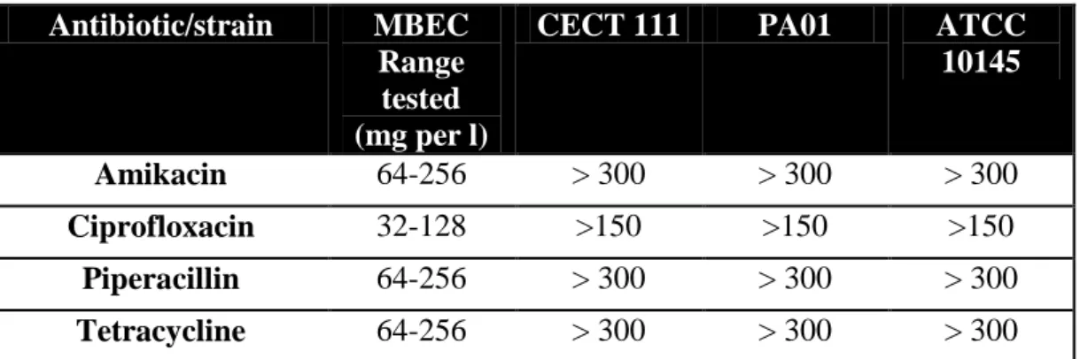
Figure 2.1: Schematic representation of the arrangement of components in the cell wall of P. aeruginosa and access of antibiotics ... 24 Figure 2.2: Assembly used in adhesion assays. ... 28 Figure 2.3: Minimum Biofilm Eradication Concentration of antibiotics against P.
aeruginosa strains: a) PA01; b) CECT 111; c) ATCC 10145. ... 30
Figure 2.4: LIVE/DEAD BacligthTM staining of P. aeruginosa CECT 111a) bacteria without antibiotic (control); b) treatment of bacteria with piperacillin; c) treatment of
x
bacteria with ciprofloxacin. (Live cells – stained in green; dead cells – stained in red)………...32 Figure 2.5: Visualization by electron microscopy of the elongation of P. aeruginosa cells in the presence of piperacillin. a) ATCC 10145, b) CECT 111, c) PA01 ... 33 Figure 2.6: Images of P. aeruginosa cells adhered over time to a glass surface after image processing by Sigma Scan Pro 5: a), b) and c) adhesion of cells; d) treatment with antibiotic ciprofloxacin. ... 35
Chapter 3
Figure 3.1: Phage infection of planktonic cells of P. aeruginosa strains PA01, CECT 111 and ATCC 10145 with different concentrations of the respective phages. ... 44 Figure 3.2: Number of viable cells present in biofilms of P. aeruginosa strains PA01, CECT 111 and ATCC 10145 after exposure to different concentrations of the respective phages ... 46 Figure 3.3: Images of P. aeruginosa cells adhered over time to a glass surface: a), b) and c) adhesion of cells; d) Infection of bacterial cells with phage phiIBB-PAP21. 1 – Bacterial cells; 2 – artefacts. ... 48
Chapter 4
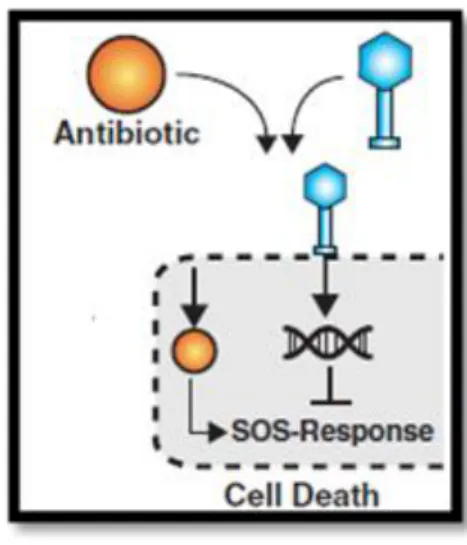
Figure 4.1: Schematic representation of a possible synergy process between phage and antibiotic ... 53 Figure 4.2: Disposable folded capillary cells used in zeta potential measurements... 55 Figure 4.3: Zetasizer Nano ZS used in zeta potential measurements ... 56 Figure 4.4: Number of cells present in biofilm after exposure to a) Combined treatment phage-antibiotic during 6h and 24 h; b) Antibiotic treatment during 24h for CECT 111 strain A - amikacin 64mg/l; C – ciprofloxacin 4mg/l; P – piperacillin and T- tetracycline 256mg/l. ... 59 Figure 4.5: Number of cells present in biofilm after exposure to a) Combined treatment phage-antibiotic during 6h and 24 h; b) Antibiotic treatment during 24h for CECT 111 strain A - amikacin 64mg/l; C – ciprofloxacin 4mg/l; P – piperacillin and T- tetracycline 256mg/l . ... 59
xi
Figure 4.6: Number of cells present in biofilm after exposure to a) Combined treatment phage-antibiotic during 6 h and 24 h; b) Antibiotic treatment during 24 h for PA01 strain A - amikacin 64mg/l; C – ciprofloxacin 4mg/l; P – piperacillin and T- tetracycline 256mg/l ... 60 Figure 4.7: Number of cells present in biofilm after exposure to a) Combined treatment phage-antibiotic during 6 h and 24 h; b) Antibiotic treatment during 24 h for ATCC 10145 strain A - amikacin 64mg/l; C – ciprofloxacin 4mg/l; P – piperacillin and T- tetracycline 256mg/l ... 60 Figure 4.8: Analysis of phage viability during synergy treatment. a) phage P1 with ciprofloxacin and amikacin; b) phage P21 with tetracycline and piperacillin; c) phage C23 with ciprofloxacin and amikacin. ... 62 Figura 4.9: Biomass quantification using Crystal Violet Staining: a) Treatment with antibiotic; b) combined treatment phage-antibiotic ... 66 Figure 4.10: One-Step-Growth curve for phage P21 with antibiotic and without antibiotic ... 68 Figure 4.11: Reduction of cells number presented in biofilms of P. aeruginosa strain PA01 after the exposure to antibiotic, endolysin and endolysin in combination with antibiotics. ... 69
Chapter 6
Figure A.1: Clinical breakpoints EUCAST for a) amikacin and b) ciprofloxacin against
P. aeruginosa ... 80
Figure A.2: Clinical breakpoints EUCAST for a) piperacillin and b) tetracycline against
xiii
List of Tables
Chapter 1
Table 1.1: Antibiotic families and mechanisms of action. ... 10 Table 1.2: Bacteriophages families. ... 11
Chapter 2
Table 2.1: Values of Minimum Inhibitory Concentration (MIC) of antibiotics for the three strains of P. aeruginosa tested ... 29 Table 2.2: Values of Minimum Biofilm Eradication Concentration (MBEC) of antibiotics for the three strains of P. aeruginosa tested ... 31
Chapter 3
Table 3.1: Characteristics of the phages ... 42
Chapter 4
Table 4.1: Lytic spectra of phages against different P. aeruginosa wild type strains and mutant phenotypes ... 55 Table 4.2: MIC values for the combined treatment phage + antibiotic on planktonic cultures ... 57 Table 4.3: Zeta potential measurements for wild-type strains and its LPS derivatives mutants ... 64 Table 4.4: Life cycle parameters determined for phages used in this project ... 67
xv
Abbreviations
ATCC - American Type Culture Collection CECT - Colleción Española de Cultivos Tipo CFU - Colony Forming Unit
DNA - Deoxyribonucleic acid h - hours
MIC - Minimum Inhibitory Concentration
MBEC - Minimal Biofilm Eradication Concentration min - minutes
MOI - Multiplicity of Infection
MTA - Molten Top-Agar OD - Optical Density
OSGC - One Step Growth Curve PEG - Polyethylene glycol PFU - Plaque Forming Unit RNA - Ribonucleic acid rpm - revolutions per minute s - seconds
TSA - Tryptic Soy Agar TSB - Tryptic Soy Broth
1
Motivation and aim of the project
Pseudomonas aeruginosa is a gram-negative bacterium that can be found in two
distinctive forms: planktonic form as unicellular organism; or sessile form, when attached to a substrate or a surface, leading to a biofilm formation. In the last years, P.
aeruginosa has received a special attention because this microorganism has been
described as a “phenomenon of bacterial resistance” and is responsible for a range of nosocomial infections or hospital–acquired infections. Actually, about 15% of the nosocomial infections worldwide, for example respiratory tract (in patients with cystic fibrosis), blood, urinary tract, ear, skin and soft tissue infections, are caused by this microorganism.
The prevalence of P. aeruginosa in the hospital environment is becoming a critical issue, and for this reason, it is important to know what are the main factors involved. The emergence of these bacteria in clinical areas is a result of many factors, such as: intrinsic resistance determined by several virulence factors, including the presence of efflux systems, restricted membrane permeability and antibiotic degrading enzymes. Also, the acquired resistance mechanisms and the ability of P. aeruginosa to grow in any natural and artificial surfaces, like medical devices, leading to the development of biofilms, play an important role in this context. P aeruginosa is known to be tolerant to a variety group of antimicrobial agents, including b-lactams,
aminoglycosides and fluoroquinolone antibiotics. Actually, infections caused by this
microorganism become even more critical since the conventional treatments with antibiotics are failing because of an increasing resistance of the bacteria to many of the agents available on the market, and its broad spectrum of virulence factors. This unpleasant reality has led to an interest in using alternative strategies to combat infections caused by P. aeruginosa, one of which is the use of (bacterio)phages. Although there are few studies on biofilms, phages have shown significant potential in controlling this protective life form of bacteria. In fact, the use of phages presents important advantages over the use of antibiotics, in particular in clinical areas, because they don’t have lethal effects on eukaryotic cells. However, the emergence of bacterial resistance to phages is inevitable leading to a rapid conversion on resistant phenotypes.
2
For these reasons, the aim of the work presented in this thesis consists on developing a strategy to control biofilms formed by P. aeruginosa based in a combination of antibiotics and phages in the same antimicrobial solution. To accomplish this goal, the following approach was performed both for P. aeruginosa planktonic cultures and biofilms:
1. Evaluation of the antimicrobial susceptibility using a set of antibiotics with distinct mechanisms of action;
2. Phage efficacy studies to compare the two therapies and highlight some differences between them;
3. Finally, study the synergy efficacy using the antibiotic-phage combined treatment.
3
Chapter 1: Review Literature
1.1 Pseudomonas aeruginosa characteristics
P. aeruginosa is a gram-negative and rod-shaped bacterium belonging to the
class of Gamma Proteobacteria. It measures 0.5 to 0.8 µm in width by 1.5 to 3.0 µm in length, and almost all strains possess a single polar flagellum that allows motility. In terms of metabolism, it is an obligatory aerobe, because it prefers oxygen as the terminal electron acceptor, but in certain situations it can grow under anaerobic conditions using nitrate as the terminal electron acceptor [1,2].
This bacterium is ubiquitous in soil and water and can be found in two distinctive forms: planktonic form, as a unicellular organism; and biofilm, when attached to abiotic surfaces or substrates. In terms of nutrition, P. aeruginosa has very simple requirements, and for this reason, growth factors and other complements are not necessary for its growth. The optimal temperature for growth is 37 degrees at a neutral pH. Nevertheless, it should be noted that P. aeruginosa is tolerant and resistant to a wide variety of physical conditions, like temperature, high concentrations of salts, disinfectants and antibiotics [1–3].
1.2 Pseudomonas aeruginosa virulence factors
Additional to the highest versatility in terms of nutrition and metabolism, P.
aeruginosa is an opportunistic pathogen in humans and animals, displaying a variety of
virulence mechanisms, derived from its intrinsic resistance which allow the establishment of the infection. These factors can be group in three main stages: adhesion, colonization and dissemination [3–5].
For the development of these processes, the role of certain components is crucial. The most important one’s refer to enzymes such as proteases, elastases, and
phenazine pigments such as pyocyanin which interferes with the tissue damage and
4
lipopolysaccharides (LPS) and extracellular polymeric substances (EPS) constitute virulence mechanisms. For example, the LPS molecules (Figure 1.1) that are present on the outer membrane of this Gram-negative bacterium contribute to their pathogenicity, because they allow bacterial adhesion mediating the entry of bacteria into eukaryotic cells. In terms of structure and composition, the LPS of P. aeruginosa is very specific being composed of a lipid A, a core oligosaccharide divided in outer and inner cores and O-antigens that involve the A and B bands [7,8–11]. Additionally, P. aeruginosa possesses several different export systems that are involved in the secretion of virulence factors, such as the injection of effector proteins directly into the cytoplasm of host cells that will interfere with the functioning of macrophages and neutrophils. Another factor that could be indirectly related to this virulence is the large and complexity genome of P.
aeruginosa, which allows this microorganism to adapt and survive to different
ecological niches. This could happen either through mutation of an existing gene product, or through the acquisition of a drug resistance plasmid [7,12].
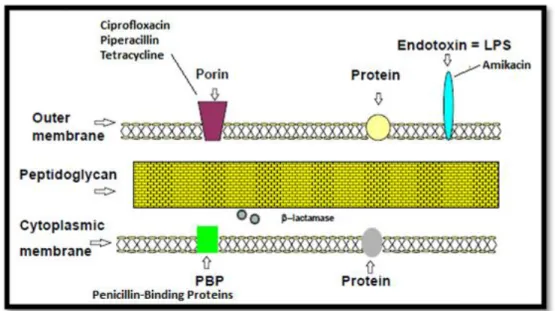
Figure 1.1: LPS structure of P. aeruginosa. Adapted from Atabek et al [7].
1.3 Clinical impact of Pseudomonas aeruginosa
Biofilms are sessile microbial communities attached to a solid surface, in which the organisms produce a hydrated matrix of extracellular polymeric substances (EPS)
5
consisting of polysaccharides, proteins, nucleic acids, and lipids [13]. Biofilms constitute an essential and protective lifestyle for bacteria in many different hostile and man-made environments, including dental plaques, water pipes, medical devices, and industrial systems [14,15]. When the biofilm formation occur on indwelling medical devices (Figure 1.2), this can lead to infection processes by detachment and dispersion of individual cells or aggregates of cells, resulting in serious damages on human tissues and organs. Additionally, the populations of bacteria within biofilms exhibit differences in the expression of surface molecules, antibiotic resistance, nutrient condition and virulence factors, and for these reasons, antimicrobial treatment that is sufficient to inactivate planktonic organisms is generally inadequate to inactivate biofilm organisms [13,15–18].
In the hospital and community environments, the development of antimicrobial resistance plays a very important role in infections caused by P. aeruginosa biofilms. This bacterium is ubiquitous in clinical settings, it can be isolated from respiratory therapy equipment (endotracheal tubes used in mechanical ventilation), sinks and physiotherapy and even hydrotherapy pools. According to surveillance data collected by the European Centre for Disease prevention and Control (ECDC) organization, P.
aeruginosa is considered the second most frequently isolated organism in cases of
intensive care unit nosocomial infections. Additionally, according to ECDC, this bacterium was responsible for 30% of pneumonias and chronic lung infection in cystic fibrosis patients, 19% of urinary tract infections, and 10% of bloodstream infections, mainly in severe burn victims. In particular, in cases of nosocomial pneumonia there is a high rate of mortality and the treatment is difficult due the long-term of antibiotic treatment [5,19–21].
Outside of the hospital environment, this microorganism can be found in swimming pools, whirlpools, hot tubs, contact lens solutions, home humidifiers, soil and vegetables [5,12,19,20].
6
Figure 1.2: Schematic representation of stages of bacterial biofilm development. At stage 1, the
bacterial cells attach reversibly to the surface, where weak forces prevalence. Next, at stage 2, the cells attach irreversibly, due mainly by exopolymeric substances, and as a consequence the cells lose their flagella-driven motility. At the next stages 3 and 4 the maturation of the biofilm occur, with the increment of biofilm architecture with formation of extracelular matrix. At stage 5 the dispersion of biofilm cells happen where single motile cells diffuse from the microcolonies. Adapted from
http://www.pasteur.fr/recherche/RAR/RAR2006/Ggb-en.html.
1.1.1 Antimicrobial resistance of Pseudomonas aeruginosa
Biofilm formation can be considered as a potential factor of antimicrobial resistance of P. aeruginosa, and there are important aspects that contribute for this situation. For instance, the mucoid exopolysaccharide matrix, that surrounds the biofilm cells, can act as an effective barrier that limits penetration of antimicrobial compounds [1,19,22]
. However, there are differences between certain classes of antibiotics in terms of biofilm penetration, because antibiotics like fluoroquinolones (such as, ciprofloxacin) have better penetration into biofilms, in opposite to aminoglycosides, which penetrate more difficultly [19,20]. This can be related to the differences observed in delivery and transport of these molecules through the outer membrane of cells and since fluoroquinolones are small hydrophilic molecules, they pass easily through the aqueous channels on the membrane provided by porin proteins. On the other hand, the aminoglycosides (cationic antibiotics) promote their own uptake by binding to the negatively charged LPS on the outer surface of the membrane [23].
7
Furthermore, the biofilm population of P. aeruginosa is heterogeneous, which means that there are fast- and slow-growing cells. This happens because of gradients of nutrients and oxygen availability into the biofilm structure [19,23]. The divergence of the cell population will create a fraction that is resistant to a range of mechanisms leading to a decrease in antimicrobial susceptibility of P. aeruginosa biofilms. This resistant subpopulation, also called persister cells, is a major source of reoccurring infections (Figure 1.3) [17,24]. Also, the architecture and complexity of biofilms provides an appropriately environment for gene transfer and cell-to-cell signaling. This mechanism is called quorum sensing, and has been shown to play an important role in the expression of virulence factors, biofilm differentiation, cell attachment and detachment, through the activation of certain genes [7,23,25].
In addition to biofilm formation, there are other factors which increase the resistance of P. aeruginosa to multiple antimicrobial agents. For example, the efflux pumps are responsible for extruding drugs, like antibiotic molecules, toxic metal ions, organic solvents and other ligands from inside the cell. In P. aeruginosa there are some important efflux pumps, such as the system MexAB-oprM that is responsible for extrusion of b-lactams, quinolones and a range of disinfectants; MexXY-oprM that extrudes aminoglycosides and MexEF-oprN that is responsible to extrudes carbapenems and quinolones [19,23].
Other important mechanism of resistance of this bacterium is the modification of the primary target for antibiotics. In fluoroquinolones, one possible situation is the mutation in the gyrA gene that encodes the A subunit of the target enzyme, DNA gyrase, of this class of antibiotics [23,25].
In terms of antibiotic uptake, the intrinsic resistance of P. aeruginosa plays also a central role in the rejection of some of these molecules. The own bacterial outer membrane, that possesses water filled channels, designated the transmembrane porins, constitutes a semi-permeable barrier to the uptake of antibiotics into the cell. For example, the OprF porin has a large exclusion limit and only allows small portions of
Beta-lactams molecules moving inside the cells [8,9,23]. Other examples include an expression of inactivating enzymes, like β-lactamases that will act on β-lactam antibiotics (for example piperacillin), degrading the β-lactamic ring present in these antibiotics and leading in this way to their inactivation [1,19,23].
On the other hand, LPS, as described above, can interfere with antimicrobial resistance of P. aeruginosa, because when bacteria present a mutation in this structure,
8
(absence of O-specific chain for example) this can affect the antibiotic uptake, in terms of acceptance or rejection. In addition, there are some studies that reveal that the oligosaccharide portion of LPS structure may play an important position in the gating mechanism of porin proteins, such as OprF [7–9,26].
Figure 1.3: Resistance mechanism mediated by phenotypic/persister variants in the biofilm. In the
first part, antimicrobial treatment of bacterial biofilms leads to the eradication of part of the biofilm susceptible population (1). A small portion of phenotypic/a persister variant (represented as maroon bacteria) survive to the treatment (2) and are able to establish biofilm development after antimicrobial therapy is suspended (3). Adapted from Drenkard et al [19].
1.4 Control strategies for Pseudomonas aeruginosa biofilms
This section will briefly review two strategies that can be used to control P.
aeruginosa biofilms: antibiotics and bacteriophages.
1.4.1 Antibiotics
Antibiotics are chemical, natural or even synthetic substances that have been used for the last 70 years to treat patients who have infectious diseases, by preventing the growth of bacteria or their destruction. One important concept is that antibiotics are not active against viruses [27,28]. Since the 1940s, the use of antimicrobial agents has been beneficial, because when prescribed and taken correctly they significantly reduce illness and death. However, often, these drugs have been used inappropriately and
1
2
9
therefore, the antibiotics that are designed to kill infectious organisms become less effective causing emergence and selection of resistant and multiresistant bacteria [27,29,30]
. Antimicrobial resistance is nowadays one of the major threats to public health, mainly in health care settings. Every year, in the European Union, it is estimated that 25.000 patients die because of serious resistant bacterial infections acquired in hospitals. Additionally, antibiotic resistance causes a direct impact to hospital and to the patient’s family: longer hospital stays increase of treatment costs; and frequently more expensive antibiotic drugs are used in treatments. There are also indirect factors that significantly affect the respective families and the society, such as: greater absenteeism at work, lower economic yield, decrease productivity by sequels and psychological and emotional changes [27,29,30].
1.4.1.1 Categories of Antibiotics and mode of action
Antibiotics can be categorised according to their action spectrum and mode of action. Regarding the first criteria, there are two types of antibiotics: narrow spectrum antibiotics and broad spectrum antibiotics. The first ones are specific for a specific bacterium and only prescribed when it is known which bacterium is causing the infection, and the second type refers to antibiotics that are used in case it is unclear which bacterium or bacteria is/are causing the infection. Contrarily to the first type, this group of antibiotics also kill harmless bacteria due to their non-specificity [31].
In terms of mode of action, antibiotics can be classified as bactericidal if they kill the susceptible bacteria; or bacteriostatic if they reversibly inhibit the growth of bacteria. In spite of bactericidal antibiotics being preferred, this does not mean that they are more effective than bacteriostatic [32]. In this context, antibiotics can be classified in five major groups (Table 1.1), according to the physiological and metabolic functions in bacterial cell [33,34] .
10
Table 1.1: Antibiotic families and mechanisms of action. Adapted from Levy et al [34]
Mechanism of action Antibiotic families Inhibition of cell wall synthesis
Penicillins; cephalosporins; carbapenems; daptomycin; monobactams; glycopeptides
Inhibition of protein synthesis
Tetracyclines; aminoglycosides; oxazolidonones; streptogramins; ketolids; macrolides; lincosamides
Inhibition of DNA synthesis Fluoroquinolones
Competitive inhibition of folic
acid synthesis Sulfonamides; trimethoprim
Inhibition of RNA synthesis Rifampin
Other Metronizadole
1.4.2 Bacteriophages
Bacteriophages, also called phages, are viruses that kill bacteria and like other viruses they are obligate parasites, so they use the bacteria host to multiply and spread. Phages are ten times more numerous in the environment than bacteria, making them the most abundant ‘life’ forms on earth, with an estimated 1032
on the planet [35–37]. Bacteriophages cannot infect mammalian cells, but only specifically target bacteria.
There are a variety of families of bacteriophage and according to the International Committee on Taxonomy of Viruses (ICTV) they are classified in terms of morphology and nucleic acid type (Table 1.2). A significant percentage of bacteriophages, about 96%, are distributed over three families that belong to the
11
Table 1.2: Bacteriophages families. Adapted from Hanlon [36].
Family Morphology Genome
Corticoviridae Icosahedral capsid with
lipid layer dsDNA
Cystoviridae Enveloped, icosahedral
capsids, lipids dsRNA
Fuselloviridae Pleomorphic, envelope,
lipids, no capsids dsDNA
Inoviridae Rod-shaped with helical
symmetry ssDNA
Leviviridae Quasi-icosahedral capsids ssRNA
Lipothrixviridae Enveloped filaments,
lipids dsDNA
Microviridae Icosahedral capsids ssDNA
Myoviridae Non-enveloped ,
Contractile tail dsDNA
Plasmaviridae Pleomorphic, envelope,
lipids, no capsids dsDNA
Podoviridae Non-enveloped , short non
contractile tail dsDNA
Rudiviridae Non enveloped, helical
rods dsDNA
Siphoviridae Non-enveloped , long non
contractile tail dsDNA
Tectiviridae Icosahedral capsid with
inner lipoprotein vesicle dsDNA
Figure 1.4: Diagrammatic representation of a typical bacteriophage structure. Adapted from Harper et al [37].
12
Bacteriophages can exhibit one of two types of life cycle: virulent (lytic phages) or temperate (lysogenic phages). Virulent phages, the only ones allowed to be used in phage therapy, cause a rapid lysis and death of the host bacterial cell leading lead to a release of a hundreds of viral particles (Figure 1.5). The infection process starts from recognition and binding of the phage to the host by a specific receptor. This binding constitutes a critical stage, and when phage are irreversible connected to the host, the injection of genetic material of the phage occurs. Then, through the metabolism of the host, the DNA replication of the phage takes place, culminating in the formation of new phage particles. Subsequently, there is the packaging of the genome and recovery of phage particles. In the case of dsDNA phages, after the assembly, the phages present into the cytoplasm of bacterial host can produce enzymes that attack the bacterial peptidoglycan, leading to the instability of cell wall. These enzymes are designed lytic enzymes or endolysins and they have a therapeutic activity, mainly against gram positive bacteria. All of this process ends in lysis of the host bacteria and release of new phages that are able to infect other bacteria [36,39] .
On the other hand, temperate phages spend part of their life cycle in a quiescent state called prophage, where their DNA is integrated into the host chromosome. This latent stage can be activated by specific stimuli, and then the phage can initiate a lytic mode infection [36,37]. Phages with a lysogenic phage cycle can transfer fragments of host bacterial DNA, such as toxin-encoding or antibiotic resistance-mediating genes into other bacterial species by a mechanism of transduction producing new virulent strains [35,36] and therefore are not to be used therapeutically.
13
Figure 1.5: The life cycle of lytic bacteriophages: 1 – phage adsorption and DNA injection; 2 – host
genome degradation; 3 - phage DNA replication; 4 – appearance of morphogenesis intermediates, including empty heads (proheads); 5 – packaging of phage DNA into capsids; 6 – phage assembly; 7 – lysis and release of progeny phage. Adapted from Kropinski [39].
In addition, there are filamentous bacteriophages that have a different life cycle in which the infection is persistent, that is, there is no death of the host bacteria, but there is a continued production of viral particles [3,36,37].
In a general context, phage therapy could be a potential approach in three different applications: 1) using phages as direct antibacterial agents, which, biotechnologically, could be fairly rapidly adapted for clinical applications; 2) isolating phage-encoded lytic enzymes that can be used as antibacterial agents alone or incorporated in the phage genome to be expressed during production of new phages by replication in their host cells. One example described in literature is the incorporation of Dispersin B (dspB) in phage T7 [14] . Furthermore, phage polysaccharide depolymerases, specific enzymes found in the tail spikes of the phage baseplate, have been isolated and used to degrade the polysaccharide matrix, helping in this way phages in getting access to the biofilm cells [14,40]. Other alternative, the most long-term application, but still very
14
promising is based in the study of determinants of phage lytic mechanisms to identify novel drug targets [41,42].
Currently, applications of phage therapy cover two main areas: clinical targeting their use to treat wounds, burns, chronic ulcers and respiratory tract infections, and food industry [43–45].
1.4.3 Antibiotics vs Bacteriophages
As any antimicrobial agents, antibiotics and bacteriophages have similarities, but also significant differences. First, phages only affect the target bacteria, while antibiotics act both on microorganisms and on the normal microflora of the patients and subsequently, at high concentrations, they may raise the risk of side effects. Phages have also a self-reproducing capability as long as their respective host is present, in opposite to antibiotics which require an administration of several doses since they are metabolized and eliminated from the body.
Many authors have emphasized phage therapy as an alternative or adjuvant approach to antibiotics mainly due to the fact that their isolation, selection and production is relatively rapid contrarily to the time-consuming development of new antibiotics which involve several critical steps. However, this doesn’t necessarily mean that it is simple to isolate a highly virulent, lytic, broad-spectrum and non-transducing phage appropriate for effective therapy [36,45–47].
A critical point that is inherent to both therapeutics is the appearance of bacterial resistance described already in section 1.3.1 for antibiotics. The development of bacterial resistance to phages happens due to a diversity of factors, such as: modification by mutation of the structure or exposure of host receptor molecules where phages adsorb; degradation of phage genome by DNAses after injection of the phage DNA into bacteria; inactivation of phages by the action of proteolytic enzymes present in biofilm matrix; entrapment of released phage particles in biofilm matrix [16,43,47,48].
The combination of phages and antibiotics is of great interest and, a few studies have shown the potential of this combined treatment for the control of bacterial biofilms [49–52]
. One the few studies reported, describes that a phage-antibiotic combination resulted in a weaker biofilm matrix, mainly through the ability of phages to create pores
15
and channels to enter into the biofilms leading to a dispersion of a great part of biofilm cells which becoming more available to the action of the antimicrobial agent [50].
On the other hand, it has also been described that combined treatments can decrease the mutation rate of the bacterial population, because a cell population hardly has, in the same genome, resistance mechanisms to survive both phage and antibiotic attack [49]. Nevertheless, it can be considered that the mechanisms behind phage-antibiotic synergistic action are still poorly explored.
16
1.5
Reference List1. Pires DPP. Avaliação da eficácia de terapias fágicas para o controlo de biofilmes mistos infecciosos. Tese de Mestrado Engenharia Biomédica, Departamento de Engenharia Biológica,Universidade do Minho. 2010.
2. Todar K. Pseudomonas aeruginosa. Available at: www.textbookofbacteriology.net.
3. Garbe von J. Isolation of Pseudomonas aeruginosa phages and their application for the analysis of lipopolysaccharides. Dissertation Thesis, University of Braunschweig – Institute of Technology. 2010.
4. Tré-Hardy M, Vanderbist F, Traore H, Devleeschouer MJ. In vitro activity of antibiotics combinations against Pseudomonas aeruginosa biofilm and planktonic cultures. International journal of antimicrobial agents. 2008;31:329-336.
5. Lister PD, Wolter DJ, Hanson ND. Antibacterial-resistant Pseudomonas aeruginosa: clinical impact and complex regulation of chromosomally encoded resistance mechanisms. Clinical microbiology reviews. 2009;22(4):582-610.
6. Kipnis E, Sawa T, Wiener-Kronish J. Targeting mechanisms of Pseudomonas aeruginosa pathogenesis. Médecine et maladies infectieuses. 2006;36(2):78-91.
7. Atabek A. Investigating Bacterial outer membrane polymers and bacterial interactions with organic molecules using Atomic Force Microscopy. Faculty of Worcester Polytechnic institute. 2006.
8. Burrows LL, Rocchetta HL, Lam JS. Assembly pathways for Biosynthesis of A-Band and B-Band Lipopolysaccharide in Pseudomonas aeruginosa. In: Glycomicrobiology.; 1997:127-143.
9. Straatsma TP, Soares TA. Characterization of the outer membrane protein OprF of Pseudomonas aeruginosa in a lipopolysaccharide membrane by computer simulation.
17
10. Soares TA, Straatsma TP, Lins RD. Influence of the B-band O-antigen Chain in the Structure and Electrostatics of the Lipopolysaccharide Membrane of Pseudomonas aeruginosa. J. Braz. Chem. Soc.,2008;19(2):312-320.
11. Ivanov IE, Kintz EN, Porter LA . Relating the physical properties of Pseudomonas aeruginosa lipopolysaccharides to virulence by atomic force microscopy. Journal of
bacteriology. 2011;193(5):1259-66.
12. Kerr KG, Snelling AM. Pseudomonas aeruginosa: a formidable and ever-present adversary. The Journal of hospital infection. 2009;73(4):338-44.
13. Hall-Stoodley L, Stoodley P. Evolving concepts in biofilm infections. Cellular
microbiology. 2009;11(7):1034-43.
14. Lu TK, Collins JJ. Dispersing biofilms with engineered enzymatic bacteriophage.
Proceedings of the National Academy of Sciences of the United States of America.
2007;104(27):11197-202.
15. Harmsen M, Yang L, Pamp SJ, Tolker-Nielsen T. An update on Pseudomonas aeruginosa biofilm formation, tolerance, and dispersal. FEMS immunology and medical
microbiology. 2010;59(3):253-68.
16. Donlan RM. Preventing biofilms of clinically relevant organisms using bacteriophage. Trends in microbiology. 2009;17(2):66-72.
17. Bedi MS, Verma V, Chhibber S. Amoxicillin and specific bacteriophage can be used together for eradication of biofilm of Klebsiella pneumoniae B5055. World
Journal of Microbiology and Biotechnology. 2009;25(7):1145-1151.
18. Soboh F, Khoury a E, Zamboni a C, Davidson D, Mittelman MW. Effects of ciprofloxacin and protamine sulfate combinations against catheter-associated Pseudomonas aeruginosa biofilms. Antimicrobial agents and chemotherapy. 1995;39(6):1281-6.
19. Drenkard E. Antimicrobial resistance of Pseudomonas aeruginosa biofilms.
18
20. Drago L, De Vecchi E, Nicola L, Tocalli L, Gismondo MR. In vitro selection of resistance in Pseudomonas aeruginosa and Acinetobacter spp. by levofloxacin and ciprofloxacin alone and in combination with beta-lactams and amikacin. The Journal of
antimicrobial chemotherapy. 2005;56(2):353-9.
21. Lyczak JB, Cannon CL, Pier GB. Establishment of Pseudomonas aeruginosa infection: lessons from a versatile opportunist. Microbes and infection / Institut Pasteur. 2000;2(9):1051-60.
22. Hoyle BD, Alcantara J, Costerton JW. Pseudomonas aeruginosa biofilm as a diffusion barrier to piperacillin. Antimicrobial agents and chemotherapy.
1992;36(9):2054-6.
23. Lambert PA. Mechanisms of antibiotic resistance in Pseudomonas aeruginosa.
Journal of the Royal Society of medicine. 2002;95(Figure 1):22-26.
24. Lewis K. Persister cells, dormancy and infectious disease. Nature reviews.
Microbiology. 2007;5(1):48-56.
25. Strateva T, Yordanov D. Pseudomonas aeruginosa - a phenomenon of bacterial resistance. Journal of medical microbiology. 2009;58(Pt 9):1133-48.
26. Lins RD, Straatsma TP. Computer simulation of the rough lipopolysaccharide membrane of Pseudomonas aeruginosa. Biophysical Journal. 2001;81:1037-1046.
27. Antibiotics. Available at: http://www.cdc.gov/drugresistance.
28. Antibiotics. Available at: www.insa.pt.
29. Antibiotic Resistance. Available at: http://www.who.int/en/.
30. Programa Nacional de Prevenção das Resistências aos Antimicrobianos. Ministério
da saúde, Departamento da Qualidade. 2009.
31.European Surveillance of antimicrobial consumption. Available at: http://app.esac.ua.ac.be/public/.
19
32. Mayer G. Antibiotics - protein synthesis, nucleic acid synthesis and metabolism. In:
Bacteriology.
33. Antibiotics Classification. Vitek technology 2, bioMérieux, Inc., Customer
Education. 2008:1-95.
34. Levy SB, Marshall B. Antibacterial resistance worldwide: causes, challenges and responses. Nature medicine. 2004;10(12 Suppl):S122-9.
35. Sulakvelidze A. Safety by Nature: Potential Bacteriophage Applications. Microbe
magazine. 2009.
36. Hanlon GW. Bacteriophages: an appraisal of their role in the treatment of bacterial infections. International journal of antimicrobial agents. 2007;30(2):118-28.
37. Harper DR, Enright MC. Bacteriophages for the treatment of Pseudomonas aeruginosa infections. Journal of applied microbiology. 2011;111(1):1-7.
38. Sillankorva SM. Utilização de Bacteriófagos no Controlo de Células Suspensas e Biofilmes de Pseudomonas fluorescens. Tese de Mestrado em Tecnologia do Ambiente, Departamento de Engenharia Biológica, Universidade do Minho. 2004.
39. Kropinski AM. Phage therapy – Everything old is new again. J Infect Dis Med
Microbiol 2006;17(5):297-306.
40. Hanlon GW, Denyer SP, Olliff CJ, Ibrahim LJ. Reduction in Exopolysaccharide Viscosity as an Aid to Bacteriophage Penetration through Pseudomonas aeruginosa Biofilms. Applied and Environmental Microbiology. 2001;67(6):2746-2753.
41. Projan S. Phage-inspired antibiotics? Nature Biotechnology. 2004;22:167-168.
42. Schoolnik GK, Summers WC, Watson JD. Phage offer a real alternative. Nature
Biotechnology. 2004;22:505-506.
43. Sillankorva SM. Use of bacteriophages to control biofilms. PhD Dissertation thesis in Chemical and Biological Engineering. University of Minho. 2008.
20
44. Abedon ST, Kuhl SJ, Blasdel BG, Kutter EM. Phage treatment of human infections.
Bacteriophage. 2011;1(2):66-85.
45. Azeredo J, I, W S. The use of phages for the removal of infectious biofilms. Current
Pharmaceutical Biotechnology. 2008;9:261-266.
46. Sulakvelidze A, Alavidze Z. MINIREVIEW Bacteriophage Therapy. Antimicrobial
agents and chemotherapy 2001;45(3):649-659.
47. Fu W, Forster T, Mayer O, et al. Bacteriophage cocktail for the prevention of biofilm formation by Pseudomonas aeruginosa on catheters in an in vitro model system.
Antimicrobial agents and chemotherapy. 2010;54(1):397-404.
48. Sulakvelidze A. Phage therapy : an attractive option for dealing with antibiotic-resistant bacterial infections. In Drug Discovery today. 2005;10(12):807-809.
49. Zhang Q-G, Buckling A. Phages limit the evolution of bacterial antibiotic resistance in experimental microcosms. Evolutionary Applications. 2012.
50. Rahman M, Kim S, Kim SM, Seol SY, Kim J. Characterization of induced Staphylococcus aureus bacteriophage SAP-26 and its anti-biofilm activity with rifampicin. Biofouling. 2011;27(10):1087-93.
51. Ryan EM, Alkawareek MY, Donnelly RF, Gilmore BF. Synergistic phage-antibiotic combinations for the control of Escherichia coli biofilms in vitro. FEMS immunology
and medical microbiology. 2012;65(2):395-8.
52. Kutateladze M, Adamia R. Bacteriophages as potential new therapeutics to replace or supplement antibiotics. Trends in biotechnology. 2010;28(12):591-5.
21
Chapter 2: Antimicrobial Susceptibility of P. aeruginosa
2.1
BackgroundThe selection of an antimicrobial agent and its dosage used in the treatment of P.
aeruginosa is very important to prevent the growth of resistant bacteria, and this choice
is crucial for the efficacy of process. Certain classes of antipseudomonal drugs that are frequently administered [1] and their mechanisms of action are described below:
Amikacin
Amikacin is an antibiotic of aminoglycosides family. In general, this antibiotic is administered to treat infections caused by gram-negative bacteria, such as
Pseudomonas, Acinetobacter, and Enterobacter. In case of P. aeruginosa infections,
due to its cationic nature, this antibiotic has the capacity to bind to LPS molecules (negative charged) and other anionic molecules such as DNA, RNA and phospholipids to access the interior of the cells. Inside the cells, amikacin acts on the 30S ribosomal subunit, preventing, in this way, the formation of an initiation complex with messenger RNA. Thus, the bacterium becomes unable to synthesize proteins that are vital to its growth. Also, amikacin can interfere with the cell membrane integrity [2–4].
22
Ciprofloxacin
Ciprofloxacin is an antibacterial agent of the fluoroquinolones class. This antibiotic has a broad spectrum activity, contrarily to amikacin, because it can act on gram-negative and gram-positive microorganisms. The main consequence of the bactericidal action of ciprofloxacin is the inhibition of the enzymes topoisomerase II (also called DNA gyrase) and topoisomerase IV. These enzymes are necessary for DNA replication, transcription, repair and recombination to bacterial cell. In P. aeruginosa, ciprofloxacin enters into the cell using porin protein OprF, and the positively charged piperazine ring at the C-7 position of the quinolone allows this molecule to interact with negatively charged phosphate groups of the phospholipid bilayer [2,5,6].
Ciprofloxacin
Piperacillin
Piperacillin is a penicillin beta-lactam antibiotic. The antibacterial action of this antibiotic result from the binding to specific penicillin-binding proteins (PBPs) located inside the bacterial cell wall. Subsequently, piperacillin inhibits the last point of bacterial cell wall synthesis. Alike ciprofloxacin, piperacillin has activity on both gram-positive and gram-negative bacteria. However, it is mainly indicated to treat pseudomonal infections. In these bacteria, the diffusion of this anionic antibiotic is achieved by OprF protein in the outer membrane of the cell [2,7].
23
Piperacillin
Tetracycline
This antibiotic has a bacteriostatic action against bacteria. Tetracycline belongs to tetracyclines family, and has a short action on the bacterial cell. In gram-negative bacteria, its mechanism of action allows the inhibition of bacterial growth, because when it diffuses through the porin channels in the bacterial outer membrane as a positively charged molecule, it reversibly binds to the 30S ribosomal subunit and prevents the binding of tRNA to the mRNA-ribosome complex. As a consequence, the protein synthesis is affected [2,8].
Tetracycline
The entry pathway (porin pathway or self-promoted pathway), the molecular characteristics of each antibiotic, and the diversity of intrinsic resistance factors of P.
aeruginosa may dictate some important differences between antibiotics molecules,
24
schematically a process for the entry of antibiotics in P. aeruginosa cells in order to realize the major differences in the mechanism of reception of these molecules.
Figure 2.1: Schematic representation of the arrangement of components in the cell wall of P. aeruginosa
and access of antibiotics. Adapted from [4]
There is a diversity of laboratory methods that can be applied in vitro to determine the susceptibility of bacteria to antimicrobial agents: broth microdilution test; disk diffusion test; antimicrobial gradient method (also called E-test) and automated instruments systems. In general, the majority of the methods available provide quantitative results. The Minimum Inhibitory Concentration (MIC), is defined as the lowest concentration of an antimicrobial that will inhibit the visible growth of a microorganism after an appropriate period of incubation [9].
This chapter focuses on the main results obtained with the antimicrobial susceptibility tests against planktonic cultures (section 2.3) and biofilms (section 2.4).
25
2.2 Materials and methods
2.2.1 Bacteria and growth conditions
The bacterial strains that were used in this work were: PA01, CECT 111 and ATCC 10145. All strains were grown in Tryptic Soy Broth (TSB) medium. The solid medium was Tryptic Soy Agar (TSA): TSB + 1.2% w/v of agar. Both media were prepared according to the manufacturer’s instructions. Then, the media were sterilized by autoclaving at 121 °C for 15 minutes.
2.2.2 Preparation of Antibiotics
The standard powders of antibiotics were obtained from Sigma - Aldrich. Stock solutions were prepared and diluted according manufacturer’s recommendations and stored at -20 °C after filtration. During manipulation of antibiotics, these were always protected from light.
2.2.3 Determination of Antimicrobial susceptibility 2.2.3.1 MIC determination
To determine the MIC of the three P. aeruginosa strains (strains ATCC 10145, PAO1 and CECT 111) against the four antibiotics (amikacin, ciprofloxacin, piperacillin and tetracycline) the micro-broth dilution method was used. The experiment was performed according to the Clinical and Laboratory Standards Institute guidelines [10,11].
Briefly, for each strain of P. aeruginosa used, two colonies were transferred from an overnight grown plate (from the first sub-culture, a second sub-culture on an appropriate agar plate was made) into 1 ml of TSB to approximate the density to 0.5 of the McFarland standard. This suspension, with an inoculum concentration of 108 colony forming units per ml (CFU/ml) was then diluted to 106 CFU/ml with the TSB medium. Serial two-fold dilutions of all the antimicrobial agents with the following concentrations (mg/l): ciprofloxacin (0.03125 - 1), amikacin (0.5 - 256), piperacillin (0.5 - 256), tetracycline (0.5 -256) were prepared with NaCl (0.9%) and then 100 µl of
26
each concentration were placed in 96-well microtiter plates. After, 100 µl of the suspension were added to the respective wells. TSB (100 μl) and 100 μl of 0.9% NaCl were used for the control experiments. Finally, the lowest concentration inhibiting visible growth after 20-24 h at 37 °C and 120 rpm was recorded as the MIC and the density (600 nm) was also measured. For these experiments three independent assays (each one in duplicate) were performed.
2.2.3.2 MBEC determination
Minimal Biofilm Eradication Concentration (MBEC) is defined as the lowest concentration of an antimicrobial agent that will inhibit the growth of a biofilm. Briefly, for each strain of P. aeruginosa, two colonies were transferred from an overnight grown plate (a second culture on an appropriate agar plate was made from a first sub-culture) into 1 ml of TSB to obtain a density of 1.0 in the McFarland standard (approx. 3×10⁸ CFU/ml) and then diluted to obtain an inoculum of 107 CFU/ml in TSB medium. After, 200 μl of P. aeruginosa cultures grown were added to 96-well microplates and incubated overnight at 37 °C and 120 rpm. After 24 h of biofilm formation, all medium was removed and the wells were washed with fresh TSB medium. Following that, 100 μl of fresh TSB and 100 μl of antimicrobial solutions with different concentrations (256-64 mg/l) were added to the wells and control experiments were performed with 100 μl of TSB and 100 μl of NaCl. The duration of MBEC assays was 24 h and after the microplates were washed twice with saline solution (0.9% NaCl) to remove all unattached bacteria. Fresh saline solution (200 μl) was added to each well, the biofilm scraped and the microplates were put in a water bath sonicator for 30 min. The number of viable cells present in biofilms before and after the treatment was determined by colony-forming unit (CFU) counts using the microdrop technique. For these experiments three independent assays (each one in duplicate) were performed.
2.2.4 Biomass Quantification
To quantify the total biomass attached to each well of 96-well microtiter plate, the crystal violet assay was used. This procedure consists of washing twice the biofilms
27
with a saline solution (0.9% NaCl), fixing with 200 µl of methanol (100%) (Merck) for 15 min and after this period, the methanol was removed and the microplate allowed to dry at room temperature. Following that, 200 µl of crystal violet (1% v/v, Merck) is added to each well and the plate is let to stand still for 5 min. After, the wells were washed with water and allowed to dry at room temperature. Finally, 200 µl of acetic acid (33% v/v, Merck) was added to dissolve the stain attached to the biofilm, and the absorbance was read at 570 nm. For these experiments three independent assays (each one in duplicate) were performed.
2.2.5 Microscopy analysis after BacLightTM staining
P. aeruginosa cells were stained LIVE/DEAD® BacLight™ (Invitrogen
Bacterial Viability Kit) [12] to access their viability according to the manufacturers’ specification. Briefly, an overnight culture was adjusted to an OD600 of 0.4 – 0.5, and after that the cultures were added to a microplate and incubated for 2 h at 37 °C and 120 rpm. After this incubation period the antibiotic solutions were added and allowed to act during 2 h at the same conditions as described above. The adhered cells were scrapped from the wells and put 20 µl in a microscope slide with 5 µl of LIVE/DEAD® BacLightTM stain. The solution was mixed thoroughly and incubated at room temperature in the dark for 15 min. The fluorescence from both live and dead bacteria were observed simultaneously using a fluorescence microscope with a longpass 485 nm filter.
2.2.6 Adhesion assays - Flow cell
The cell adhesion assays were carried out using a laminar flow cell apparatus mounted on an inverted optical microscope (Nikon, Diaphot 300) coupled with a digital camera (CCD camera - Sony, AVC-D5CE) to capture images over time (Figure 2.2) [13]. Before each assay, the entire system was washed with sterile water for 30 min and without recirculation. Then, in the same condition, TSB medium was passed through the system.
28
Cultures of P. aeruginosa were grown overnight at 37 ° C, centrifuged (7,000 ×g, 10 min, 4 ° C) and the pellet resuspended in TSB to an OD of about 0.5. After this, the suspension of P. aeruginosa was placed in the balloon of the system. During the feeding of the flow cell with bacterial cells, the system operated with recirculation of the suspension. When the whole surface was coated with cells, the feeding of cells was stopped and the washing with TSB medium was started without recirculation, to remove non adhered cells. After 30 min of washing, the antimicrobial agent was placed on the other balloon of the system, and the feeding started again. At the end of the tests, the entire system was washed. It is important to refer that the flow used was 0.124 ml/min.
Subsequently, for image processing, a treatment using the software developed by Sigma (Sigma Scan Pro 5) was applied.
Figure 2.2: Assembly used in adhesion assays. Adapted from Azeredo et al [13].
2.3 Results and discussion
2.3.1 Determination of the minimum inhibitory concentration (MIC)
The micro-broth dilution method was used to determine the susceptibility of P.
aeruginosa cells to specific antibiotics. The choice of this method is based in two main
characteristics: it is a practical and reproducible method [9]. Table 2.1 presents the MIC values obtained for each antibiotic tested and for each of the P. aeruginosa strains investigated.
![Figure 1.1: LPS structure of P. aeruginosa. Adapted from Atabek et al [7] .](https://thumb-eu.123doks.com/thumbv2/123dok_br/17803010.840966/23.892.232.665.663.918/figure-lps-structure-of-aeruginosa-adapted-from-atabek.webp)


![Figure 2.2: Assembly used in adhesion assays. Adapted from Azeredo et al [13] .](https://thumb-eu.123doks.com/thumbv2/123dok_br/17803010.840966/47.892.129.768.484.719/figure-assembly-used-adhesion-assays-adapted-azeredo-et.webp)


![Figure 4.2: Disposable folded capillary cells used in zeta potential measurements. Adapted from [ 6 ]](https://thumb-eu.123doks.com/thumbv2/123dok_br/17803010.840966/74.892.120.772.149.446/figure-disposable-folded-capillary-cells-potential-measurements-adapted.webp)
