Autonomic Function Evaluation in an Intermittent Lead Exposure Animal Model
L. Shvachiy
1, V. Geraldes
1,2, M. Carvalho
1, I. Rocha
1,21Cardiovascular Autonomic Function Lab, Cardiovascular Centre of University of Lisbon; 2Institute of Physiology, Faculty of Medicine, University of Lisbon, Av. Prof. Egas Moniz, 1649-028 Lisbon, Portugal
[email protected] [email protected] [email protected]
Abstract — Lead (Pb) is a toxic metal
,
which widespread use has resulted in environmental contamination, human exposure and significant public health problems. The autonomic nervous system, being a homeostatic controller, is impaired in acute and chronic lead exposure. In fact, sympathoexcitation associated to hypertension and tachypnea has been described together with baroreflex and chemoreflex dysfunction. However, up to date, no studies described the autonomic effects of an intermittent low-level lead exposure. In the present work, we addressed in vivo, autonomic behaviour in rats under chronic Pb exposure (control) and in rats under intermittent Pb exposure. For that, arterial blood pressure (BP) and ECG were recorded in 28 weeks old animal and low frequencies (LF) and high frequencies (HF) were determined (to estimate sympathetic and parasympathetic activities) using FisioSinal software with Wavelet module. Our preliminary results depict that intermittently lead-exposed rats show a significant decrease in systolic BP, without significant changes on LF, HF and LF/HF bands, when compared to chronically Pb- exposed rats.Our data suggests that the autonomic dysfunction induced by lead exposure is similar in a chronic and intermittent Pb exposure. Nevertheless, it seems that an intermittent exposure was no effect on systolic BP values.
The present study brings new insights on the environmental factors that influence autonomic and cardiovascular systems during development, which can help apprise public policy strategies to prevent and control the adverse effects of Pb toxicity.
Keywords: Lead toxicity, autonomic activity, Fisiosinal, Wavelet analysis, Heart Rate Variability
I. INTRODUCTION
Lead is the most commonly used heavy metal worldwide for over 8000 years in a large amount of industries like automobiles, ceramics, paint, plastics, among others, in behalf of its unique properties such as high malleability, low melting point, softness, ductility and resistance to corrosion[1], [2].
Regarding this wide usage, it is obvious that there is a rise in environmental free lead and its occurrence in biological systems concerning the non-biodegradable nature of this heavy metal.
Lead toxicity, due to its ingestion, inhalation or, less probably by direct contact, can evoke irreversible effects in a wide amount of body functions affecting mainly the cardiovascular [3], [4] (being one of the causes of hypertension and promoting atherosclerosis, thrombosis, arteriosclerosis and cardiovascular disease) , hematopoietic[1], [5], reproductive[6] (both, in woman and men, leading to misscariage, inferitility and premature delivery in women and reduction of libido, abnormal spermatogenesis and infertility in men), [7] and renal (leading to acute and chronic nephropathy, by molecular and tubular changes) [8] systems.
Being easily accumulated in bone and soft tissues, and due to the ability of lead to cross the blood brain barrier and to substitute cations like Ca2+, Mg2+, Fe2+ and Na2+, lead is also a neurotoxin. These alterations, induce changes in the neurogenesis, especially when the exposure happens in the early stages of life, neurodegeneration and changes on glial cells, that support neurons and play a crucial role in synaptic transmission and plasticity. The changes in the molecular and cellular processes lead to cognitive and behaviour alterations, particularly during developmental phases, persisting during the lifetime. [1], [9], [10]
Additionally, regarding the autonomic tone, that regulates the cardiorespiratory processes, the exposure to toxic levels of lead produce a significant increase in sympathetic activity associated to high blood pressure, decreased baroreflex function, increased chemoreceptor sensitivity and tachypnoea [11].
Two types of lead toxicity could be defined. Firstly, acute toxicity, usually in high-levels, is quite uncommon and happens by occupational exposure, while chronic low-level exposure toxicity is more common, happening in a household environment and much severe if not treated in time.
However, up to date, no studies were performed to establish the autonomic effects in an intermittent low-level lead exposure. This type of exposure is increasing due to the augmented migration and to the implementation of school exchange programs. Nevertheless, people tend to come back to their hometowns after some years abroad, returning to their familiar environment. Additionally, in workplaces where the use of lead is recurrent, rotation of workers and moving of these to other workplaces without lead exposure has been performed during the past years as a possible prevention of
adverse health effects caused by occupational permanent low-level lead exposure.
Therefore, our work is focused on studying the autonomic changes provoked by a newintermittent low-level lead exposure profile.The section II (methods) describe the design of the present study, including the development of two animal models and provide a description of the procedures that were performed.for the autonomic evaluation. The preliminary results obtained in our study are presented in the section III and then discussed, comparing our results with other published evidence in section IV. Some final, summative statements, based upon our findings are reported in section V:
II. METHODS
A. Development of the animal model of chronic and intermittent lead exposure
Seven-day pregnant Wistar rats (n = 2) were exposed to lead during pregnancy, via water containing Lead acetate (0.2% w/v) that replaced tap water. After birth and weaning at 21 days, the rats of both sexes were exposed to the same lead solution as that of their mother and were divided into 2 groups (n = 6/group; Pb1 – used as control group – and Pb2). At 12 weeks, the intake of the solution ended in both groups; and this was followed by a period without exposure that lasted up to 20 weeks. The Pb2 group underwent a second exposure which lasted 8 weeks (named intermittent lead exposure group) while Pb1 wasn’t exposed to lead for the second time (named chronic lead exposure group).
B. Surgical protocol
At 28 weeks, the acute experiment was carried out in both groups of animals (n=12). Animals of both sexes were anesthetized with sodium pentobarbital (60mg/kg, ip). The femoral artery and vein were cannulated for blood pressure monitoring and injection of saline and drugs, respectively. Rectal temperature was maintained between 36.5-39ºC. The electrocardiogram (ECG) was recorded with subcutaneous electrodes inserted in three of the four members and heart rate was also obtained through this registration method (Neurolog, Digitimer[12]).
C. Data acquisition
Signals were amplified with the alternative current pre amplifier gain of 5K and a high-pass filter of 0.1Hz, filtered by band-pass filter of 500-5000Hz and a 20Hz wave width (Neurolog, Digitimer) and acquired at 1 kHz (PowerLab, ADInstruments[13]). All signals were converted to digital form and stored for further analysis.
D. Analysis of heart rate and systolic blood pressure variability
Using FisioSinal [14], an in-house developed software with Wavelet module (Db12)[15], [16], the LF, HF and LF/HF indexes were determined in basal conditions. Low frequencies (LF; 0.15-0.6 Hz) that were obtained from systolic BP are a marker of sympathetic activity, high frequencies (HF; 0.6-2.0 Hz) attained from R-R interval represent both parasympathetic and respiratory variations, and LF/HF is the ratio between sympathetic and parasympathetic systems. In this study and to the fact that the respiratory rate was high in some animals, we proceeded to an adjustment of the high frequencies band while maintaining its lower end and extending to its upper limit to 2.4 Hz.
E. Statistical analysis
Variables are expressed as mean and SEM. Statistical data was obtained by comparisons between groups of animals – Pb1 and Pb2 – using Student’s t-test. Significance was considered to p<0.05. Data was analysed using GraphPad Prism 6 software[17] (GraphPad Software, Inc., USA).
III. RESULTS
A. Cardiovascular variables and autonomic tone
Baseline levels of all measured physiological parameters were similar between the 2 groups, except for systolic blood pressure with significantly higher values in Pb1-rats compared with Pb2-rats as shown in Table 1.
TABLE 1
BASELINE LEVELS OF ALL MEASURED PHYSIOLOGICAL PARAMETERS.
BP,BLOOD PRESSURE;HR,HEART RATE;RF,RESPIRATORY FREQUENCY
Values presented as MEAN ± SEM (n=6), ap<0.05
Group Pb1 Pb2 Mean BP (mmHg2) 125 ± 4 116 ± 3 Systolic BP (mmHg2) 144 ± 3 a 126 ± 4 Diastolic BP (mmHg2) 110 ± 5 106 ± 3 Basal HR (bpm) 371 ± 8 404 ± 17 Basal RF (cpm) 56 ± 4 58 ± 5
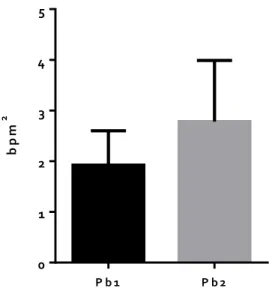
The sympathetic and parasympathetic tone evaluated through LF and HF band, respectively, as well as LF/HF band presenting the ratio between sympathetic and parasympathetic systems (1.5 ± 0.3 vs 1.7 ± 0.5 mmHg2, 1.9 ± 0.7 vs 2.8 ± 1.2 bpm2 and 1.2 ± 0.4 vs 1.1 ± 0.3 mmHg2/bpm2, respectively) didn´t change significantly between the 2 groups, as shown in figure 1, 2, and 3, respectively, for each band.
m m H g 2 P b 1 P b 2 0 1 2 3 4 5
Figure 1. Sympathetic activity presented by LF (Low Frequencies) band for each group. No significant difference in LF band values was observed in the Pb2 group – intermittent lead exposure (1.7 ± 0.5 mmHg2) when compared to the Pb1 group – chronic lead exposure (1.5 ± 0.3 mmHg2). Values presented as MEAN ± SEM (n=6). b p m 2 P b 1 P b 2 0 1 2 3 4 5
Figure 2. Parasympathetic activity presented by HF (High Frequencies) band for each group. No significant difference was detected in HF band of the Pb2 group - intermittent lead exposure (2.8 ± 1.2 bpm2) when correlated to the Pb1 group - chronic lead exposure (1.9 ± 0.7 bpm2). Values presented as MEAN ± SEM (n=6).
Figure 3. Ratio between sympathetic and parasympathetic systems presented by LF/HF band values. No significant difference between LF/HF band of the Pb2 group – intermittent lead exposure (1.1 ± 0.3 mmHg2/bpm2) and the Pb1 group - chronic lead exposure (1.2 ± 0.4 mmHg2/bpm2) was distinguished. Values presented as MEAN ± SEM (n=6).
IV. DISCUSSION
The current study is the first to address a new profile of lead exposure, the intermittent low-level lead exposure. Our reported results demonstrate that the autonomic effects of this type of exposure are similar to the chronic lead exposure, since we didn´t find differences in sympathetic and parasympathetic tones between the two types of exposure, providing an association between the autonomic function and lead intoxication, regardless of the type of lead exposure. In relation to the other physiological parameters evaluated, i.e. blood pressure, heart rate and respiratory frequency, all are similar except for systolic blood pressure that is significantly decreased in the intermittent lead exposure group (Pb2) (126 ± 4 mmHg2) when compared to the chronic lead exposure group (144 ± 3 mmHg2). The decrease on arterial compliance, which is used as an indication of arterial stiffness, provoked by a chronic lead toxicity may be the reason for the increased systolic blood pressure recorded in the chronic lead exposure group (Pb1).
Even though autonomic function is not known to be primarily affected by chronic lead exposure, the autonomic nervous system has been found compromised by Pb-poisoning, causing an overall autonomic dysfunction[11].
Additionally, it was shown that lead reduces baroreflex sensitivity, vagal parasympathetic tone, and increases sympathetic activity, by impairment of dopamine and acetylcholine transmission, as well as oxidative stress induced by lead poisoning [18]–[20].
These reported effects - baroreflex hyposensitivity, sympathetic overexcitation and decreased parasympathetic tone - are associated with several pathologies such as hypertension, acute cardiac ischemia or even heart failure. The mechanisms underlying those pathological effects haven’t been clarified but it is thought that there is an initial protective reaction which in time, turns into deleterious sympathoexcitation [11].
Here we present our preliminary results regarding the autonomic nervous system evaluation. These results indicate that the intermittent low-level lead exposure is associated with sympathetic hyperactivity, as already described for a chronic exposure, but with no effect on blood pressure values, since in the intermittent exposure the animals remain normotensive.
Regarding the fact that our results are preliminary, more evaluations should be carried on, for a full characterization of the new animal model that was developed, using other electronic equipment, machinery and software’s (such as blood pressure and ECG telemetry) or other molecular and physiological evaluations. These additional evaluations would be of extreme importance to better understand the underlying mechanisms of lead toxicity.
V. CONCLUSION
In conclusion, the present study brings new insights into how different lead poisoning profiles may influence autonomic and cardiovascular systems during developmental phases. Thus, helping to upraise public policy strategies to prevent and control the adverse effects of Pb toxicity.
REFERENCES
[1] G. Flora, D. Gupta, and A. Tiwari, “Toxicity of lead: a review with recent updates,” Interdiscip. Toxicol., vol. 5, no. 2, pp. 47–58, 2012.
[2] World Health Organization, “Exposure to Lead: A major public health concern,” World Heal. Organ., p. 6, 2010.
[3] A. Navas-Acien, E. Guallar, E. K. Silbergeld, and S. J. Rothenberg, “Lead exposure and cardiovascular disease - A systematic review,” Environ. Health Perspect., vol. 115, no. 3, pp. 472–482, 2007. [4] N. D. Vaziri, “Mechanisms of lead-induced
hypertension and cardiovascular disease.,” Am. J. Physiol. Heart Circ. Physiol., vol. 295, no. 2, pp. H454–H465, 2008.
[5] D. C. Basha, S. S. Basha, and G. R. Reddy, “Lead-induced cardiac and hematological alterations in aging Wistar male rats: Alleviating effects of nutrient metal mixture,” Biogerontology, vol. 13, no. 4, pp. 359–368, 2012.
[6] N. a. Brown, “Reproductive and developmental toxicity of styrene,” Reprod. Toxicol., vol. 5, no. JULY 1985, pp. 3–29, 1991.
[7] M. Ahamed and M. K. J. Siddiqui, “Low level lead exposure and oxidative stress: Current opinions,” Clin. Chim. Acta, vol. 383, no. 1–2, pp. 57–64, 2007. [8] M. Loghman-Adham, “Renal effects of environmental
and occupational lead exposure.,” Environ. Health Perspect., vol. 105, no. 3, pp. 103–106, 2008. [9] C. D. Toscano and T. R. Guilarte, “Lead
neurotoxicity: From exposure to molecular effects,” Brain Res. Rev., vol. 49, no. 3, pp. 529–554, 2005. [10] Y. Finkelstein, M. E. Markowitz, and J. F. Rosen,
“Low-level lead-induced neurotoxicity in children: An update on central nervous system effects,” Brain Res. Rev., vol. 27, no. 2, pp. 168–176, 1998.
[11] V. Geraldes, M. Carvalho, N. Goncalves-Rosa, C. Tavares, S. Laranjo, and I. Rocha, “Lead toxicity promotes autonomic dysfunction with increased chemoreceptor sensitivity,” Neurotoxicology, vol. 54, pp. 170–177, 2016.
[12] “NeuroLog System, Signal Processing & Conditioning Electrical Stimulation.” [Online]. Available: https://digitimer.com/products/neurolog-system/. [Accessed: 27-Nov-2017].
[13] “https://www.adinstruments.com/products/powerlab.” .
[14] C. Tavares, R. C. Martins, S. Laranjo, and I. Rocha, “Computational tools for assessing cardiovascular variability,” 1st Port. Meet. Biomed. Eng. ENBENG 2011, 2011.
[15] I. Daubechies, “Ten lectures of Wavelets,” 1992. [16] I. Daubechies, “The wavelet transform,
time-frequency localization and signal analysis,” Inf. Theory, IEEE Trans., vol. 36, no. 5, pp. 961–1005, 1990.
[17] “Home - graphpad.com.” [Online]. Available: https://www.graphpad.com/. [Accessed: 27-Nov-2017].
[18] H. Bielarczyk, X. Tian, and J. B. Suszkiw, “Cholinergic denervation-like changes in rat
hippocampus following developmental lead exposure,” Brain Res., vol. 708, no. 1, pp. 108–115, 1996.
[19] N. Bourjeily and J. B. Suszkiw, “Developmental cholinotoxicity of lead: Loss of septal cholinergic neurons and long-term changes in cholinergic innervation of the hippocampus in perinatally lead-exposed rats,” Brain Res., vol. 771, no. 2, pp. 319– 328, 1997.
[20] B. J. Brockel and D. A. Cory-slechta, “Lead-Induced Decrements in Waiting Behavior : Involvement of D 2 -Like Dopamine Receptors,” Science (80-. )., vol. 63, no. 3, pp. 423–434, 1999.