Article
Recovery of Adrenal Function in Patients with
Glucocorticoids Induced Secondary Adrenal Insufficiency
Jong Ha Baek1, Soo Kyoung Kim1, Jung Hwa Jung1,2, Jong Ryeal Hahm1,2, Jaehoon Jung1,2
1
Department of Internal Medicine, Gyeongsang National University Hospital, Gyeongsang National University School of Medicine; 2Institute of Health Sciences, Gyeongsang National University School of Medicine, Jinju, Korea
Background: The chronic use of glucocorticoids (GC) suppresses function of the hypothalamic-pituitary-adrenal axis and often results in secondary adrenal insufficiency (AI). The present study aimed to determine the recovery rate of adrenal function in pa-tients with secondary AI within 1 to 2 years and to assess the factors predictive of adrenal function recovery.
Methods: This was a retrospective observational study that enrolled patients diagnosed with GC-induced secondary AI between 2007 and 2013. AI was defined by peak serum cortisol levels <18 μg/dL during a standard-dose short synacthen test (SST). A follow-up SST was performed after 1 to 2 years, and responders were defined as those with adrenocorticotropic hormone (ACTH)-stimulated peak serum cortisol levels ≥18 μg/dL.
Results: Of the total 34 patients diagnosed with GC-induced secondary AI at first, 20 patients (58.8%) recovered normal adrenal function by the time of the follow-up SST (median follow-up period, 16.5 months). Although the baseline serum ACTH and cor-tisol levels at the first SST did not differ between responders and non-responders, the incremental corcor-tisol response during the first SST was higher in responders than that of non-responders (7.88 vs. 3.56, P<0.01). Additionally, higher cortisol increments during the first SST were an independent predictive factor of the adrenal function recovery (odds ratio, 1.58; 95% confidence in-terval, 1.02 to 2.46; P<0.05).
Conclusion: In the present study, adrenal function recovery was achieved frequently in patients with GC-induced secondary AI within 1 to 2 years. Additionally, an incremental cortisol response at the first SST may be an important predictive factor of adre-nal function recovery.
Keywords: Adrenal insufficiency; Glucocorticoids; Recovery; Predictive factor
INTRODUCTION
Glucocorticoids (GC) have been widely used since 1940s to treat inflammatory, autoimmune, and malignant disorders. The prevalence of oral GC use among the general population varies according to age, sex, and country, as well as the year of the survey was conducted; the observed incidence rates have been
reported as 0.5% to 0.9% in the United Kingdom [1,2], 0.7% in Northeast Iceland [3], and 1.2% in the United States [4]. Al-though the exact prevalence of GC use in the general Korean population has yet to be determined, it may be similar to even higher than the rates of other countries due to the high chance that patients are being administered factitious GC in regions that do not separate dispensaries from medical practices [5,6].
Received: 7 September 2015, Revised: 1 October 2015, Accepted: 27 October 2015 Corresponding author: Jaehoon Jung
Department of Internal Medicine, Gyeongsang National University Hospital, Gyeongsang National University School of Medicine, 79 Gangnam-ro, Jinju 52727, Korea
Tel: +82-55-214-3740, Fax: +82-55-750-8800, E-mail: taesikjung@gmail.com
Copyright © 2016 Korean Endocrine Society
This is an Open Access article distributed under the terms of the Creative
Com-mons Attribution Non-Commercial License (http://creativecomCom-mons.org/
Despite their beneficial effects, the chronic and/or high-dose exposure to GC is associated with a number of adverse events including osteoporosis, Cushingoid appearance with weight gain, diabetes, cardiovascular disease, and dyslipidemia, among others [7]. Furthermore, chronic exposure to GC can in-hibit the function of the hypothalamic-pituitary-adrenal (HPA) axis via negative feedback, which may result in adrenal insuffi-ciency (AI) following the cessation of GC treatment or in criti-cally ill patients [8,9]. As a result, prolonged GC treatment is considered to be the most common cause of secondary AI [10]. Therefore, it is important to detect AI during its early stages to initiate appropriate treatment, particularly for patients with a history of chronic GC treatment.
The clinical symptoms associated with AI are elusive and nonspecific, but often include nausea, vomiting, abdominal pain, shock, and even organ failures. Because these symptoms are nonspecific, it is difficult to differentiate AI from other medical conditions and it is important to perform biochemical stimulation tests to detect AI. The standard-dose (250 μg) short synacthen test (SST) has high sensitivity and specificity and is considered to be a safe and reliable method for the evaluation of adrenal function [11-13].
Many studies have reported that patients with secondary AI exhibit recovery of HPA axis function within several months (up to 1 year or more) [14-16], but no studies have investigated the predictive biochemical cut-off values associated with this
recovery. Therefore, the present study aimed to determine the recovery rate of adrenal function in patients with secondary AI within 1 to 2 years, and to assess the factors predictive of adre-nal function recovery during the follow-up.
METHODS
Study subjects
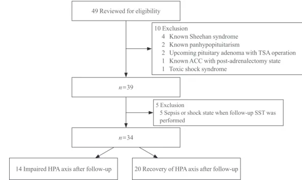
The present study conducted a retrospective screening of all patients who attended Gyeongsang National University Hospi-tal and underwent a SST between July 2007 and December 2013. Patients who were diagnosed with AI at the first SST and had a follow-up SST within 1 to 2 years were enrolled. Forty-nine patients fulfilled these criteria but 10 patients were ex-cluded due to previous history of being diagnosed with panhy-popituitarism or Sheehan syndrome (n=6), pituitary adenoma (n=2), toxic shock syndrome, and post-adrenalectomy-induced AI (n=1). Additionally, five patients were also excluded from the present study because the follow-up SST was performed under conditions of sepsis or shock (Fig. 1).
Anthropometric and laboratory measurements
The personal information of the enrolled patients including age, sex, and body measurement data, such as height, weight, and laboratory results were verified by a review of medical chart records. The body mass index (BMI) of each patient was
49 Reviewed for eligibility
n=39
n=34
14 Impaired HPA axis after follow-up 20 Recovery of HPA axis after follow-up 10 Exclusion
4 Known Sheehan syndrome 2 Known panhypopituitarism
2 Upcoming pituitary adenoma with TSA operation 1 Known ACC with post-adrenalectomy state 1 Toxic shock syndrome
5 Exclusion
5 Sepsis or shock state when follow-up SST was performed
calculated by dividing the weight in kilograms by the square of the height in meters. Patients who had any appearance of trun-cal obesity, a rounded face, supraclavicular fullness, and/or easy bruising with skin atrophy upon physical examination were regarded as having Cushingoid features. The medical chart review also revealed the reasons that each patient was ad-ministered GC.
Blood samples were obtained by venipuncture at a single central certified laboratory at Gyeongsang National University Hospital. Serum albumin levels (normal range, 3.5 to 5.2 g/dL) were measured by a bromocresol green test using a Roche modular DP analyzer (Roche Diagnostics, Basel, Switzerland), and serum cortisol (normal range, 5 to 25 μg/dL at morning, 5 to 15 μg/dL at afternoon, and 0 to 10 μg/dL at night) and adre -nocorticotropic hormone (ACTH; normal range, 5 to 60 pg/ mL) levels were measured by an electrochemiluminescence immunoassay (Roche, Manheim, Germany). The intra- and in-terassay variabilities of the cortisol and ACTH kits used in the present study were less than 3.0% and 6.0%, respectively, and the sensitivity values were 0.018 μg/dL and 0.220 pmol/L, re -spectively.
Standard-dose SST
A SST was performed to evaluate the adrenal function of the patients. The test was conducted with the patients in a supine position after the cessation of GC administration for at least 48 hours. Blood samples were collected prior to intravenous ad-ministration of tetracosactide (250 μg, Synacthen, Dalim Bio -Tech, Seoul, Korea) and then 30 and 60 minutes after adminis-tration to determine serum cortisol levels. A peak stimulated serum cortisol level ≥18 μg/dL within 60 minutes was consid -ered a normal adrenal response regardless of basal cortisol lev-el. A patient was considered a responder if their follow-up SST showed an ACTH-stimulated peak serum cortisol levels ≥18 μg/dL.
Statistical methods
With regard to the baseline characteristics of the patients, con-tinuous variables are presented as medians and interquartile ranges, and categorical variables are presented as numbers and percentages. The comparison between responders and non-re-sponders were performed using the Mann-Whitney U test. A
logistic regression analysis was conducted to estimate the pre-dictive factors associated with HPA axis recovery after the fol-low-up, and a multivariate logistic regression analysis was conducted to further evaluate the predictive factors after
adjust-ing for confoundadjust-ing factors. The first model (model 1) was justed for sex and age, and the second model (model 2) was ad-justed for age, sex, serum albumin, BMI, peak cortisol concen-tration, basal ACTH levels, and model 3 was adjusted for age, sex, serum albumin, BMI, basal cortisol concentration, basal ACTH levels. A receiver operating characteristic (ROC) curve was employed to assess the ability of cortisol increments at the first SST to predict HPA axis recovery. All statistical analyses were performed using SPSS version 18.0 (SPSS Inc., Chicago, IL, USA) and two-tailed P≤0.05 were considered to indicate
statistical significance.
RESULTS
This retrospective observational study analyzed 34 consecutive patients who were diagnosed with GC-induced secondary AI; the median age was 69.5 years, and 14 of the patients (41.2%) were male. Of these 34 patients, 20 (58.8%) recovered to nor-mal adrenal function within 1 to 2 years (the median follow-up period, 16.5 months), 16 patients (47.1%) had Cushingoid fea-tures at the initial diagnosis. GC treatment was administered for rheumatologic reasons in four patients, orthopedic reasons in 23 patients, pulmonological reasons in three patients, and malignant disorders in four patients (Table 1). All patients were prescribed physiological doses of prednisolone after being di-agnosed with secondary AI (median 5 mg/day with minimal 2.5 mg/day and maximum 10 mg/day). But unfortunately, it was not possible to evaluate the cumulative dose, duration of GC treatment, or the patients’ compliance to the GC therapy before and after AI diagnosis, because many patients (n=25) had a history of taking factitious steroids from regions without sepa-ration of dispensary from medical practice as well as this retro-spective observational study obtained data only from medical charts.
There were no significant differences between the responder and non-responder groups in terms of anthropometric and bio-chemical parameters, including basal serum ACTH and cortisol levels, and peak serum cortisol levels at the first SST. However, the degree of cortisol increments at the first SST (delta cortisol, which was defined as the peak cortisol level at either 30 or 60 minutes subtracted by the baseline) was higher in the respond-ers (on average, 7.88 vs. 3.56, P=0.004) (Table 1).
1.97; P<0.01). This association remained significant even after adjusting for age, sex, serum albumin levels, BMI, and basal se-rum cortisol and ACTH levels (in model 3: OR, 1.57; 95% CI, 1.02 to 2.46; P=0.041) (Table 2). The area under the ROC curve (AUC) was used to assess the ability of the biochemical values to predict the HPA axis recovery after the first SST. Although neither the baseline serum cortisol (AUC, 0.50; P>0.999) nor
peak cortisol (AUC, 0.65; P=0.142) levels were predictive of the recovery of adrenal function, the delta cortisol levels were predictive (AUC, 0.79; 95% CI, 0.62 to 0.96; P=0.005). The cut-off values of the serum delta cortisol levels for HPA axis re-covery were 8.15 μg/dL (a sensitivity of 50% with specificity of 79%), and 8.99 μg/dL (a sensitivity of 40% with specificity of 86%), respectively (Fig. 2).
Table 1. Patients Characteristics and Results of Standard-Dose Short Synacthen Test
Characteristic Total Non-responders Responders P valuea
No. of patients 34 14 20
Age, yr 69.5 (60.5–75.3) 70.5 (63.5–76.3) 67.0 (59.5–74.3) 0.436
Male sex 14 (41.2) 4 (28.6) 10 (50.0) 0.211b
Body mass index, kg/m2 22.4 (18.8–25.1) 22.0 (18.1–24.0) 23.0 (19.9–25.9) 0.424
Time interval from diagnosis to performing a follow-up SST, mo 16.5 (13.8–20.0) 18.5 (14.8–20.3) 15.5 (13.0–18.8) 0.217
Cushingoid feature at first SST 16 (47.1) 6 (37.5) 10 (62.5) >0.999c
Disease state 0.277
Rheumatic diseased 4 (11.8) 1 (7.1) 3 (15.0)
Orthopedic diseasee 23 (67.6) 11 (78.6) 12 (60.0)
Chronic lung diseasef
3 (8.8) 1 (7.1) 2 (10.0)
Malignancy with chemotherapyg 4 (11.8) 1 (7.1) 3 (15.0)
Serum albumin, mg/dL 3.6 (3.2–4.1) 3.6 (3.2–4.1) 3.6 (3.3–4.1) 0.691
Basal serum ACTH, pg/mL 7.5 (1.0–24.3) 13.2 (1.0–38.2) 6.3 (1.0–16.5) 0.436 Serum cortisol (basal), μg/dL 2.33 (0.72–5.83) 3.40 (0.52–7.28) 2.22 (0.81–5.42) >0.999
Serum cortisol (peak), μg/dL 11.21 (7.20–14.35) 9.11 (4.09–14.37) 11.67 (8.54–14.40) 0.148
Δ Serum cortisol, μg/dLh 6.70 (4.00–10.00) 3.56 (2.44–7.93) 7.88 (6.23–10.87) 0.004
Values are expressed as median (interquartile range) or number (%). SST, short synacthen test; ACTH, adrenocorticotropic hormone.
aMann-Whitney U test between two groups; bPearson chi-square test between two groups; cFisher exact test between two groups; dRheumatic diseases
refer to rheumatoid arthritis (n=2), gout (n=2); e
Orthopedic disease refer to spinal stenosis (n=15), osteoarthritis (n=8); f
Chronic lung disease refer to chronic obstructive pulmonary disease (n=2), asthma (n=1); g
Malignancy with chemotherapy refer to esophageal cancer (n=1), glottis cancer
(n=1), chronic myeloid leukemia (n=1), non-small cell lung cancer (n=1); h
Peak serum cortisol levels subtracted by basal serum cortisol.
Table 2. Logistic Regression of Basal Serum ACTH and Cortisol, Peak Serum Cortisol, and Serum Cortisol Increment in Relation to
Adrenal Function Recovery
Variable OR (95% CI)
Univariate analysis Model 1a Model 2b Modelc
Basal ACTH, pg/mL 0.99 (0.97–1.01) Serum cortisol (basal), μg/dL 0.94 (0.78–1.13)
Serum cortisol (peak), μg/dL 1.15 (0.97–1.35)
Δ Serum cortisol, μg/dLd
1.48 (1.10–1.97) 1.45 (1.03–2.05) 1.44 (1.01–2.05) 1.58 (1.02–2.46)
ACTH, adrenocorticotropic hormone; OR, odds ratio; CI, confidence interval.
aModel 1: adjustment with age, sex, body mass index; bModel 2: adjustment with age, sex, serum albumin, body mass index, peak cortisol
DISCUSSION
The present retrospective observational study was performed to estimate the percentage of patients who recovered from AI within 1 to 2 years, and to assess predictive baseline laboratory parameters to differentiate patients who will recover normal adrenal function during the follow-up from those who will not. In the present study, 20 secondary AI patients (58.8%) recov-ered normal adrenal function during the follow-up period (me-dian 16.5 months), and a higher incremental cortisol response to ACTH stimulation at the first SST was an independent pre-dictive factor of HPA axis recovery. To the best of our knowl-edge, the present study is the first to evaluate the recovery rate of adrenal function in GC-induced secondary AI in Korean pa-tients and to assess predictive biochemical factors associated with HPA axis recovery.
Considering the risk of incident secondary AI associated with GC treatment, neither the duration nor the dose of GC treatment is a reliable predictor of GF-induced AI [14,17]. Fur-thermore, there are no forms of administration or underlying diseases for which the risk of AI can be safely excluded [18]. Meanwhile, although it remains unknown exactly how long GC-induced suppression of the HPA axis will persist due to in-dividual variations [14], many studies have demonstrated that
it may take several months or even longer than 1 year [14-16]. It has been shown that neither the total nor the highest dose of GC or the duration of GC treatment are indicators of HPA axis recovery in patients with secondary AI [19]. A recent study demonstrated that the probability of recovering adrenal func-tion after treatment for endogenous Cushing syndrome is not correlated with the length or extent of hypercortisolism or the postoperative GC replacement doses, but rather with the under-lying etiology of the syndrome [20]. In the present study, even though the patients were heterogeneous regarding the underly-ing cause of GC treatment, and it was not possible to ascertain the previous cumulative dose and duration of GC therapy, many of the patients (n=20, 58.8%) achieved normal adrenal function within 1 to 2 years, which is similar to the findings of previous studies [14-16]. This suggests that patients diagnosed with GC-induced secondary AI due to any etiology or GC treatment may achieve normal adrenal function within 1 to 2 years. Thus, close monitoring of these patients is necessary to accurately assess and evaluate adrenal function in those under-going GC replacement treatment.
On the other hand, Yo et al. [21] suggested that the basal morning cortisol levels can be a good initial assessment tool in predicting an adrenal response and the HPA axis with deter-mining the need for dynamic test (a basal cortisol cut-off of >5 μg/dL gives a specificity of 95% and a sensitivity of 43% for predicting adrenal sufficiency). A prospective cohort study in patients with giant cell arteritis with long-term GC therapy demonstrated that 49% of patients had AI when performed by the SST after GC tapering, and recovery of adrenal function occurred in a mean time of 14 months, similar to that of our study [22]. In addition, the main predictive factors associated with an incidence of AI after GC treatment were total dose (>8.5 g), and duration of GC treatment (>19 months), as well as low basal cortisol concentration (<14 μg/dL).
The peak serum cortisol levels during the SST were well correlated with the results of an insulin tolerance test, which is the gold standard for the detection of AI [23,24]. Peak serum cortisol levels and the delta changes in cortisol levels after ACTH stimulation were once used as the diagnostic criteria for primary and secondary AI [25,26], but peak cortisol levels (>18 μg/dL) are now accepted as the most appropriate diag -nostic criteria to evaluate adrenal function due to the higher ac-curacy associated with their measurement [27], and because these levels are not affected by basal cortisol concentrations [24]. Meanwhile, in the case of critically ill patients, a delta cortisol level less than 9 μg/dL was used as the diagnostic
cut-1.0
0.8
0.6
0.4
0.2
0
Sensitivity
Delta serum cortisol (μg/dL)
1-Specificity
0 0.2 0.4 0.6 0.8 1.0
off value for critical illness-related corticosteroid insufficiency [28]. In the present study, not peak cortisol levels but delta cor-tisol levels during the first SST (defined as the peak corcor-tisol level at either 30 or 60 minutes subtracted by the baseline) were positively associated with the possibility of achieving ad-renal function recovery; this finding remained significant after adjusting for confounding factors. Using the ROC curve to pre-dict the cut-off point for HPA axis recovery revealed that a se-rum delta cortisol level of 8.99 μg/dL showed a low sensitivity but a relatively high specificity (86%). This may suggest that the biochemical marker levels during the SST may be not sole-ly predictive of HPA axis recovery, and that several confound-ing factors should be considered in clinical practice.
Total serum cortisol levels are known to be influenced by body composition, serum cortisol-binding globulin, and use of oral contraceptive pills [29]. This study reported that changes in the 30 minutes cortisol values are positively correlated with BMI in both gender. Meanwhile, 30 minutes total cortisol lev-els were independently associated with abdominal fat mass or waist circumference (but not with BMI) only in men. Addition-ally, cortisol-binding globulin levels, which are high in patients using oral contraceptive pills and low in patients with liver cir-rhosis, are positively correlated with serum total cortisol levels. In the present study, neither delta cortisol levels (r=–0.017,
P=0.939) nor 30-minute total cortisol levels (r=–0.014,
P=0.949) were significantly associated with BMI (n=24). However, any drugs that could possibly affect the concentra-tion of serum cortisol levels were not used, and patients with liver cirrhosis or nephrotic syndrome were excluded. The pres-ent study adjusted for the aforempres-entioned confounding factors to assess the correlation more accurately between delta cortisol levels during the SST and the recovery of adrenal function; this relationship remained significant.
There are several limitations inherent to the present retro-spective study that must be discussed. First, the cumulative dose and duration of GC treatment before and after the diagno-sis of secondary AI were not evaluated in the present study. Reginal characteristics in which there are regions without sepa-ration of dispensary from medical practice in rural and moun-tain villages of west Gyeongsangnam-do province, were one of barriers to evaluate the exact duration or cumulative dose of GCs. Even though impaired adrenal function may be present regardless of dose of GC [30], and the cumulative dose and du-ration of GC treatment are not closely associated with the re-covery of adrenal function [20], these factors should have been taken into consideration. Second, only a small number of
pa-tients from a single university hospital were included in the present study, and thus, selection bias could have influenced the results. Future studies should include larger general popula-tions with multi-center, prospective designs. Third, we could not evaluate duration of SAI before being performed the first SST and there is a possibility that some patients were in the course of adrenal function recovery before the first SST. De-spite these limitations, the present study demonstrated a good recovery rate of adrenal function within 1 to 2 years and is the first to demonstrate that delta cortisol levels are a predictive biochemical parameter associated with adrenal function recov-ery in patients with GC-induced secondary AI.
In summary, more than half of the patients with GC-induced secondary AI in the present study achieved normal adrenal function within 1 to 2 years of follow-up. Interestingly, serum cortisol increments during the SST may be an independent pre-dictive factor of the recovery of adrenal function. These find-ings indicate that patients with GC-induced secondary AI should be monitored, and their biochemical profiles in response to the SST, including changes in cortisol increments should be assessed to predict accurately adrenal function recovery within 1 to 2 years. Additional well-controlled, prospective, large-population studies investigating GC-induced secondary AI are required to strengthen the observed correlation between delta cortisol levels and HPA axis recovery.
CONFLICTS OF INTEREST
No potential conflict of interest relevant to this article was re-ported.
ACKNOWLEDGMENTS
We acknowledge the efforts of the data processing department in the hospital. We thanks to Dr. J.H. Byun for her help to ana-lyze biochemical results.
REFERENCES
1. Fardet L, Petersen I, Nazareth I. Description of oral gluco -corticoid prescriptions in general population. Rev Med In-terne 2011;32:594-9.
3. Gudbjornsson B, Juliusson UI, Gudjonsson FV. Prevalence
of long term steroid treatment and the frequency of decision making to prevent steroid induced osteoporosis in daily clinical practice. Ann Rheum Dis 2002;61:32-6.
4. Overman RA, Yeh JY, Deal CL. Prevalence of oral gluco -corticoid usage in the United States: a general population perspective. Arthritis Care Res (Hoboken) 2013;65:294-8. 5. Kim KM, Kim BR, Lee JS, Han OY, Park MS, Yim HW, et al.
A survey on pharmacists’ prescription behaviors for topical steroids in regions without separation of dispensary from med-ical practice in Korea. Korean J Clin Pharm 2011;21:161-9. 6. Lee C, Shin E, Park S, Kim H, Kim W. Performance evalu
-ation for ten-years of government separ-ation policy on pre-scription and dispensing of drugs: a literature review study. Health Policy Manage 2013;23:188-95.
7. Liu D, Ahmet A, Ward L, Krishnamoorthy P, Mandelcorn ED, Leigh R, et al. A practical guide to the monitoring and management of the complications of systemic corticoste-roid therapy. Allergy Asthma Clin Immunol 2013;9:30. 8. Jasani MK, Boyle JA, Greig WR, Dalakos TG, Browning
MC, Thompson A, et al. Corticosteroid-induced suppres-sion of the hypothalamo-pituitary-adrenal axis: observa-tions on patients given oral corticosteroids for rheumatoid arthritis. Q J Med 1967;36:261-76.
9. Venkatesh B, Cohen J. The utility of the corticotropin test
to diagnose adrenal insufficiency in critical illness: an up-date. Clin Endocrinol (Oxf) 2015;83:289-97.
10. Bornstein SR. Predisposing factors for adrenal
insufficien-cy. N Engl J Med 2009;360:2328-39.
11. Gleeson HK, Walker BR, Seckl JR, Padfield PL. Ten years on: safety of short synacthen tests in assessing adrenocorti-cotropin deficiency in clinical practice. J Clin Endocrinol Metab 2003;88:2106-11.
12. Arlt W. The approach to the adult with newly diagnosed
ad-renal insufficiency. J Clin Endocrinol Metab 2009;94:1059-67.
13. Agha A, Tomlinson JW, Clark PM, Holder G, Stewart PM.
The long-term predictive accuracy of the short synacthen (corticotropin) stimulation test for assessment of the hypo-thalamic-pituitary-adrenal axis. J Clin Endocrinol Metab 2006;91:43-7.
14. Livanou T, Ferriman D, James VH. Recovery of hypothala -mo-pituitary-adrenal function after corticosteroid therapy. Lancet 1967;2:856-9.
15. Tempark T, Phatarakijnirund V, Chatproedprai S,
Watchar-asindhu S, Supornsilchai V, Wananukul S. Exogenous
Cush-ing’s syndrome due to topical corticosteroid application: case report and review literature. Endocrine 2010;38:328-34.
16. Graber AL, Ney RL, Nicholson WE, Island DP, Liddle GW. Natural history of pituitary-adrenal recovery following long-term suppression with corticosteroids. J Clin Endocri-nol Metab 1965;25:11-6.
17. Henzen C, Suter A, Lerch E, Urbinelli R, Schorno XH, Bri -ner VA. Suppression and recovery of adrenal response after short-term, high-dose glucocorticoid treatment. Lancet 2000;355:542-5.
18. Broersen LH, Pereira AM, Jorgensen JO, Dekkers OM. Ad -renal insufficiency in corticosteroids use: systematic review and meta-analysis. J Clin Endocrinol Metab 2015;100:2171-80.
19. LaRochelle GE Jr, LaRochelle AG, Ratner RE, Borenstein DG. Recovery of the hypothalamic-pituitary-adrenal (HPA) axis in patients with rheumatic diseases receiving low-dose prednisone. Am J Med 1993;95:258-64.
20. Berr CM, Di Dalmazi G, Osswald A, Ritzel K, Bidlingmai-er M, GeyBidlingmai-er LL, et al. Time to recovBidlingmai-ery of adrenal function after curative surgery for Cushing’s syndrome depends on etiology. J Clin Endocrinol Metab 2015;100:1300-8. 21. Yo WS, Toh LM, Brown SJ, Howe WD, Henley DE, Lim
EM. How good is a morning cortisol in predicting an ade-quate response to intramuscular synacthen stimulation? Clin Endocrinol (Oxf) 2014;81:19-24.
22. Jamilloux Y, Liozon E, Pugnet G, Nadalon S, Heang Ly K, Dumonteil S, et al. Recovery of adrenal function after long-term glucocorticoid therapy for giant cell arteritis: a cohort study. PLoS One 2013;8:e68713.
23. Wood JB, Frankland AW, James VH, Landon J. A rapid test of adrenocortical function. Lancet 1965;1:243-5.
24. Stewart PM, Corrie J, Seckl JR, Edwards CR, Padfield PL. A rational approach for assessing the hypothalamo-pitu-itary-adrenal axis. Lancet 1988;1:1208-10.
25. Plumpton FS, Besser GM, Cole PV. Corticosteroid
treat-ment and surgery. 1. An investigation of the indications for steroid cover. Anaesthesia 1969;24:3-11.
26. Baruah MP. Sub-clinical addison’s disease. Indian J
Endo-crinol Metab 2012;16(Suppl 2):S176-7.
27. Widmer IE, Puder JJ, Konig C, Pargger H, Zerkowski HR,
Girard J, et al. Cortisol response in relation to the severity of stress and illness. J Clin Endocrinol Metab 2005;90:4579-86.
CL, Arlt W, et al. Recommendations for the diagnosis and management of corticosteroid insufficiency in critically ill adult patients: consensus statements from an international task force by the American College of Critical Care Medi-cine. Crit Care Med 2008;36:1937-49.
29. Klose M, Lange M, Rasmussen AK, Skakkebaek NE, Hil -sted L, Haug E, et al. Factors influencing the adrenocortico
-tropin test: role of contemporary cortisol assays, body com-position, and oral contraceptive agents. J Clin Endocrinol Metab 2007;92:1326-33.
30. Schlaghecke R, Kornely E, Santen RT, Ridderskamp P. The