Study of Microleakage at Class V Cavities Prepared by
Er:YAG Laser Using Rewetting Surface Treatment
R.F.Z. LIZARELLI, D.D.S., M.Sc., Ph.D.,1P.C.G. SILVA, D.D.S., M.Sc., Ph.D.,2
S.T. PORTO NETO, D.D.S., M.Sc., Ph.D.,2and V.S. BAGNATO, M.Sc., APP, Ph.D.1
ABSTRACT
Objective: This study was conducted to analyze microleakage in Class V cavity preparation, using rewetting (or not) just after burr or Er:YAG laser preparation of enamel and dentin walls in permanent teeth. Back-ground Data:Several studies reported microleakage around composite restorations when cavity preparation was done or treated by Er:YAG laser. As the hybridized laser is removed when this laser is used to cut dental hard tissue, there is a need for new materials or techniques to minimize gaps and microleakage. Results:
Primer solution showed significant effect in enamel and dentin, at the level of 5%, when Er:YAG laser was used as a cutting tool. Using primer solution after phosphoric acid in preparations with the laser, microleak-age was similar in degree to when cavities were prepared with the burr. Conclusion: Re-wetting surface just after Er:YAG irradiation and chemical treatment with phosphoric acid using HEMA aqueous solution seems to improve the quality of bioattachment between the adhesive system and enamel/dentin, showing similarities between restoration behaviors independently of the cutting tool, whether burr or laser.
51 INTRODUCTION
T
HERE IS A CONSTANT NEEDfor materials or techniques that ensure adhesion to tooth structure to minimize the poten-tial for leakage. The marginal adaptation of restorative materi-als is influenced by shrinkage from polymerization and a high coefficient of thermal expansion.1The laser is now establishedas a suitable tool for the selective and precise removal of cari-ous dental tissue.2–8If correctly used, lasers minimize the loss
of healthy tissue and promote comfort for patients during the procedure. The absence of noise and mechanical vibration re-sults in reduced pain, making the procedure tolerable for most patients.
The interaction of Er:YAG laser and dental hard tissues obeys an explosive thermo-mechanical ablation mediated by water.9Absorption of heat by water is the most likely
mecha-nism of ablation of dental hard tissue at 2940 nm. During rapid heating, the inertial confined water can create enormous sub-surface pressures that lead to the explosive removal of the sur-rounding mineral matrix. This can occur at temperatures well below the melting point of the mineral phase of enamel. Sev-eral studies of hard tissue ablation indicate that large particles
are ejected with high velocity from the irradiated tissue, which strongly supports the proposed mechanism of a water-medi-ated explosive process.
Several studies reported microleakage around composite restorations when cavity preparation was done or treated by Er:YAG laser10–14; however, there is no agreement between all
experiments or researchers. Understanding how much dental tissue is in fact modified by this laser and how the laser could be affecting the interaction between the irradiated surface and the adhesive system constitutes is an important topic for clini-cians. Because the Er:YAG laser is highly absorbed by water, the resultant irradiated surface can present dehydrated features. This may make it difficult for the adhesive agent to diffuse into the tissue.
Kataumi et al.15 showed that dentin irradiated by Er:YAG
laser does not present a smear layer; even smear plugs are ab-sent. After the application of adhesive agent over irradiated dentin (with or without phosphoric acid etching), the so-called “plastic dentin” could not be observed, and the resin tags were thinner than those found in the other group (i.e., burr). Er:YAG laser irradiation produced an acid-resistant dentin, especially the peritubular dentin, which showed increased acid resistance
1Instituto de Física de São Carlos, USP, São Carlos, Brazil. 2Faculdade de Odontologia de Araraquara, UNESP, Brazil. Volume 22, Number 1, 2004
when compared with the intertubular dentin. This study15showed
that Er:YAG laser irradiation affected the superficial dentin surface as well as the subsurface, to a depth of 20 µm.
Since there are no scientific reports about the use of rewet-ting in cavity preparation after Er:YAG irradiation, the present study considers different conditions of chemical treatments— that is, preparing enamel and dentin irradiated surfaces before composite resin restoration. We test a modified adhesive tech-nique to improve bioattachment between enamel and dentin substrate after cavity preparation by Er:YAG laser and a one-step adhesive agent.
MATERIALS AND METHODS
Teeth preparation
In this study, we used 18 intact, recently extracted human third molar teeth. The teeth were obtained from a dental clinic with a protocol approved by an institutional review board. The teeth were kept in an appropriate environment (saline solution) to avoid dryness.
Teeth were randomly classified in five different groups in terms of treatment. Each group had seven samples. In all sam-ples, cavities of class V were prepared, considering buccal and lingual surfaces. In groups I and II, we used a carbide burr; in groups III, IV, and V, the Er:YAG laser was used. The charac-teristics of each group were as follows: GI—cavity prepara-tion, 37% etching acid, adhesive agent, and composite; GII—cavity preparation, 37% etching acid, primer agent, ad-hesive agent, and composite; GIII—cavity preparation, adhe-sive agent, and composite resin; GIV—cavity preparation, 37% etching acid, adhesive agent, and composite resin; GV— cavity preparation, 37% etching acid, primer agent, adhesive agent, and composite resin restoration.
The materials used were as follows: 37% phosphoric acid (3M, USA), adhesive agent Single Bond lot 1105 (3M, USA), primer agent Scotchbond Multi-Purpose 2 lot 3008 (3M, USA), and composite resin Filtek Z250 A2 lot 2004-07 (3M, USA). Each time that etching by phosphoric acid was used, the sample was washed and softly dry using an absorbed cone paper.
We chose a primer solution composed of 10% HEMA (2-hy-droxyethyl methacrylate) aqueous solution because it is
capa-ble of re-expanding collapsed demineralized dentin effectively. Some hydrophilic monomers, such as HEMA, are extremely soluble in either water or acetone. If HEMA can replace water in the spaces around collagen fibrils, it can serve as a polymer-izable solvent for subsequently placed adhesive monomers, given sufficient diffusion time.16
Laser system
The Er:YAG laser system we used was Twin Light (Fotona Medical Lasers, Slovenia) with 2940 nm, peak energy of 500 mJ, repetition rate of 2–15 Hz, pulse width of 200–450 µsec, articulate arm, handpiece with sapphire window and non-contact, with spot size of 0.466 mm2.
Experimental irradiation
Teeth were exposed to focused Er:YAG laser with the follow-ing parameters: 300 mJ of energy per pulse, 10 Hz of repetition rate, 3.0 W of average power, during 90 sec of exposition time, resulting in a fluence of 64.38 3103J/cm2and an intensity of
64.38 3102W/cm2; using a carbide burr in a high-speed
hand-piece, cavities were prepared with an approximate volume of 1.0
3 2.0 32.0 mm3 (width, length, and depth respectively), as
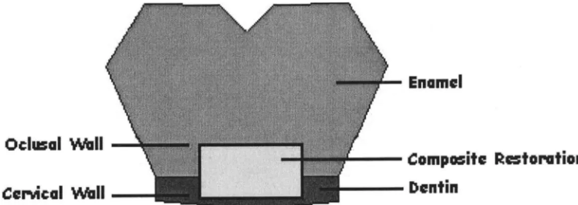
shown in Figure 1. The oclusal wall was finished at the level of enamel, and the cervical wall was finished in dentin.
After the restorations were polished with gold (no. 3195F) and silver (no. 3195FF) diamond (KG Sorensen, Brazil) 24 h later, all samples were maintained in distilled water at 37°C for 7 days. Then, the samples were polished again, using sequen-tial Sof Lex discs (3M, USA).
In order to proceed with the microleakage test, all restored teeth (except for the area of the filled cavity and an area 2.0 mm outside of the margins of the filled cavity) were com-pletely coated with two layers of epoxy resin and one layer of nail varnish, and then subjected to the thermal cycling varying temperature in 10°C and 50°C, for 500 cycles, for 15 sec in each temperature per cycle. After thermal cycling, all samples were immersed in 50% aqueous silver nitrate solution for 2 h, followed by immersion in a photorevealing solution under flu-orescent light for 16 h.
After washing with water, the samples were bisected longi-tudinally using a carborundum disc and each part was coated in
acrylic resin, individually. Each sample was sectioned, with one cut dividing the composite resin restoration in two parts. Both parts of each sample were evaluated under graduated magnification (times 40) to assess the degree of microleakage. The degree of microleakage using the dye penetration method was scored in a blinded manner based on a four-grade scale as shown in Table 1 and Figure 2.
RESULTS AND DISCUSSION
Scores for each sample were analyzed using the following statistical tests: Mann-Whitney to compare oclusal and cervi-cal walls and Kruskal-Wallis to compare the different treat-ments for each cavity preparation.
The Mann-Whitney test showed a statistically significant difference, at 1% (p= 0.01). This means that at 99% probabil-ity, oclusal and cervical walls are not similar. This result sup-ports the Kruskal-Wallis test considering different treatments and different walls.
The Kruskal-Wallis test showed a statistically significant difference at 1% (p= 0.01), when all treatments were com-pared together—that is, all treatments for enamel and all treat-ments for dentin. In pairs, at 5% (p= 0.05) as a significance level, some interesting findings were observed (Tables 2 and 3). Table 2 shows interactions of treatment for the oclusal wall, and Table 3 the same conditions for the cervical wall. Figures 3 and 4 show graphs for oclusal and cervical walls when the de-gree of microleakage is a function of the different treatments; all samples (seven in each group) are shown.
Evaluating the oclusal wall, in enamel substrate, there is a sta-tistically significant difference between GI (burr + phosphoric acid + adhesive agent + composite) and GII (burr + phosphoric acid + primer + adhesive agent + composite) in comparison with all other groups: GIII (laser + adhesive agent + composite), GIV (laser + phosphoric acid etched + adhesive agent + composite), and GV (laser + phosphoric acid etched + primer + adhesive
FIG. 2. Scheme of microleakage scores.
TABLE1. CRITERION FORDEGREE OFMICROLEAKAGE
Score Criterion
0 No dye penetration
1 Dye penetration up to a third of the length of the wall
2 Dye penetration between one and two thirds of the length of the wall 3 Dye penetration beyond of two thirds of the length of the wall
TABLE2. KRUSKAL-WALLISSTATISTICALTEST BETWEENPAIRS OFSAMPLES, CONSIDERINGOCLUSALWALL
(SIGNIFICANCELEVEL= 5%)
Comparison between average of samples Critical values (p)
Pairs of samples Difference between media 0.05 0.01 0.001 Significance
GIo 3GIIo 5,9286 7,2611 9,7786 12,9647 ns GIo 3GIIIo 18,9286 7,2611 9,7786 12,9647 5% GIo 3GIVo 17,3571 7,2611 9,7786 12,9647 5%
GIo 3GVo 9,1429 7,2611 9,7786 12,9647 5%
agent + composite). These findings show that whatever treatment is used after cavity preparation with the Er:YAG laser, it is very difficult to find a degree of microleakage lower than or similar to that when the burr is used to prepare the enamel surface.
It is possible that the different interaction mechanisms of Er:YAG laser and phosphoric acid with the enamel surface. Phosphoric acid acts by removing minerals components, such as Ca (calcium) and P (phosphorus), elements of hydroxiap-atite, and by the other side, Er:YAG laser ablates enamel re-moving water and phosphate groups, resulting in a surface rich in Ca, however dehydrated. The adhesive agent used is hydro-philic and interacts with a hydrated surface indeed with some pore to permits micro-retentions through interpenetrations of this adhesive agent into enamel tissue.
Other findings showed a statistical similarity between GII (burr + phosphoric acid etched + primer + adhesive agent + composite) and GV (laser + phosphoric acid etched + primer + adhesive agent + composite), and a difference between GIV (laser + phosphoric acid etched + adhesive agent + compos-ite) and GV (laser + phosphoric acid etched + primer +
adhe-sive agent + composite). The use of primer (10% HEMA aqueous solution) application just after 37% phosphoric acid results in a microleakage showing similar degrees with the same primer was applied before the adhesive agent in cavities done by burr, it can show that primer is very important even for enamel surfaces after Er:YAG laser irradiation to prevent microleakage.
As statistical tests showed a significant difference between GIV and GV, this result confirms the important function of primer application just after Er:YAG laser irradiation in the enamel surface.
Considering dentin surface (cervical wall), the statistical simi-larity is present just between GI (burr + phosphoric acid + adhe-sive agent + composite) and GV (laser + phosphoric acid etched + primer + adhesive agent + composite). This indicates that cavi-ties prepared with the Er:YAG laser need to be treated in advance with phosphoric acid and primer, even when using a one-step ad-hesive agent. It could be a result of the capability of Er:YAG laser in removing more water from a tissue with the use of water spray and more sensible to allow adhesive agent diffusion.
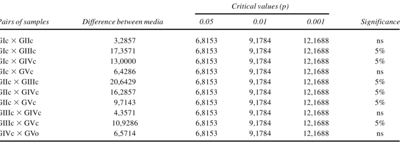
TABLE3. KRUSKAL-WALLISSTATISTICALTEST BETWEENPAIRS OFSAMPLES, CONSIDERINGCERVICALWALL
(SIGNIFICANCELEVEL= 5%)
Comparison between average of samples Critical values (p)
Pairs of samples Difference between media 0.05 0.01 0.001 Significance
GIc 3GIIc 3,2857 6,8153 9,1784 12,1688 ns GIc 3GIIIc 17,3571 6,8153 9,1784 12,1688 5% GIc 3GIVc 13,0000 6,8153 9,1784 12,1688 5% GIc 3GVc 6,4286 6,8153 9,1784 12,1688 ns GIIc 3GIIIc 20,6429 6,8153 9,1784 12,1688 5% GIIc 3GIVc 16,2857 6,8153 9,1784 12,1688 5% GIIc 3GVc 9,7143 6,8153 9,1784 12,1688 5% GIIIc 3GIVc 4,3571 6,8153 9,1784 12,1688 ns GIIIc 3GVc 10,9286 6,8153 9,1784 12,1688 5% GIVc 3GVo 6,5714 6,8153 9,1784 12,1688 ns
FIG. 3. Graph of all groups of treatment type as a function of microleakage scores, considering oclusal wall (enamel) (1 = GI; 2 = GII; 3 = GIII; 4 = GIV; and 5 = GV).
When the same conditions were repeated changing the cutting tool (burr or laser), there is no statistical similarity in the cervical wall, dentin as a substrate; GI (burr + phosphoric acid + adhe-sive agent + composite) was different from GIV (laser + phos-phoric acid + adhesive agent + composite) and GII (burr + phosphoric acid + primer + adhesive agent + composite) was different from GV (laser + phosphoric acid + primer + adhesive agent + composite), at the level of 5%. It means that using primer (10% HEMA aqueous solution) application just after 37% phosphoric acid results in a smaller microleakage, however Er:YAG laser can not replace phosphoric acid etching.
Er:YAG laser seems to affect bioattachment more in dentin surface than enamel surface, as we can see in Figures 3 and 4. Chemical composition could explain this fact; dentin has more water than enamel, so after irradiation with Er:YAG laser dentin becomes more affected than enamel, resulting in diffi-culty in promoting an adequate surface to allow diffusion of the adhesive agent.
CONCLUSION
In conclusion, we have presented a microleakage study con-sidering a new topic: re-wetting of enamel and dentin surfaces irradiated by Er:YAG laser. Our results showed some steps to-wards the improvement of marginal adaptation of composite resin Class V restorations, when Er:YAG laser was used for cavity preparations:
1. Er:YAG laser cannot replace phosphoric acid etching. 2. Er:YAG laser seems to affect bioattachment more in dentin
surface than in enamel surface.
3. The application of primer solution (10% hydroxyl ethyl methacrylate aqueous solution) as a re-wetting solution just after etching with 37% phosphoric acid results in smaller microleakage for enamel and dentin surfaces.
4. We recommend the use of this primer solution (10% HEMA) after cavity preparation with the Er:YAG laser system.
ACKNOWLEDGMENTS
We acknowledge financial support from CNPq (PRONEX) and FAPESP (CEPID program). We also acknowledge L.T. Moriyama (IFSC-USP) and 3M, of Brasil (Sr. A. Andreazi Filho, Manager), for materials used in this work.
REFERENCES
1. Barnes, D.M., McDonald, N.J., Thompson, V.P., et al. (1994). Mi-croleakage in facial and lingual class 5 composite restorations: a comparison. Oper. Dent. 19, 133–137.
2. Hibst, R., Keller, U., and Steiner, R. (1988). Die Wirkung gepul-ster Er:YAG—laserstrahlung auf zahngewebe. Laser Med. Surg. 4, 163–165.
3. Paghdiwala, A.F., Vaidyanathan, T.K., and Paghdiwala, M.F. (1993). Evaluation of erbium:YAG radiation of hard dental tissues analysis of temperature changes, depth of cuts and structural ef-fects. Scanning Microsc. 7, 989–997.
4. Pelagalli, J., Gimbel, C.B., Hansen, R.T., et al. (1997). Investiga-tion study of the use of Er:YAG laser versus dental drill for caries removal and cavity preparation—phase I. J. Clin. Laser Med. Surg. 15, 109–116.
5. Dostálová, T., Jelínková, H., Krejsa, O., et al. (1997). Dentin and pulp response to erbium:YAG laser ablation: a preliminary evaluation of human teeth. J. Clin. Laser Med. Surg. 15, 117– 122.
6. Tokonabe, H., Kouji, R., Watanabe, H., et al. (1999). Morphologi-cal changes of human teeth with Er:YAG laser irradiation. J. Clin. Laser Med. Surg. 17, 7–12.
7. Hadley, J., Young, D.A., Eversole, L.R., et al. (2000). A laser-pow-ered hydrokinetic system for caries removal and cavity prepara-tion. J. Am. Dent. Assoc. 131, 777–785.
8. Yamada, Y., Hossain, M., Nakamura, Y., et al. (2001). Removal of carious dentin by mechanical, chemomechanical and Er:YAG laser in deciduous teeth. J. Oral Laser Appl. 1, 109–114.
9. Fried, D. (2000). IR laser ablation of dental enamel. Proc. SPIE 3910, 136–148.
10. Wright, G.Z., McConnell, R.J., and Keller, U. (1993). Micro-leakage of class V composite restorations prepared conventionally and with Er:YAG laser: a pilot study. Pediatr. Dent. 15, 425–426. 11. Khan, M.F.R., Yonaga, K., Kimura, Y., et al. (1998). Study of mi-croleakage at class I prepared by Er:YAG laser using three types of restorative materials. J. Clin. Laser Med. Surg. 16, 305–308. 12. Niu, W., Eto, J.N., Kimura, Y., et al. (1998). A study on
microleak-age after resin filling of class V cavities prepared by Er:YAG laser. J. Clin. Laser Med. Surg. 16, 227–231.
13. Lizarelli, R.F.Z., Kurachi, C., Porto Neto, S.T., et al. (2000). Comparative study in vitroof microleakage in class V cavity preparation with and without Er:YAG laser. Proc. SPIE 3910, 254–260.
14. Lizarelli, R.F.Z., Silva, P.C.G., Kurachi, C., et al. (2002). Estudo-piloto comparativo da microinfiltração in vitroentre preparos cav-itários classe V, através de ponta diamantada em alta rotação ou laser de Er:YAG seguido ou não de ataque ácido. J. Brasil. Dent. 1, 33–41.
15. Kataumi, M., Nakajima, M., Yamada, T., et al. (1998). Tensile bond strength and SEM evaluation of Er:YAG laser irradiated dentin using dentin adhesive. Dent. Mater. J. 17, 125–138. 16. Nakabayashi, N., and Pashley, D.H. (1998). Hybridization of
den-tal hard tissues.Tokyo: Quintessence.
Address reprint requests to:
Rosane F.Z. Lizarelli, D.D.S., M.Sc., Ph.D. Instituto de Física de São Carlos–USP P.O. Box 369 CEP 13560-900 São Carlos, SP, Brasil