Vol-7, Special Issue-Number4-July, 2016, pp744-751 http://www.bipublication.com
Research Article
Comparison of the Quality of Pap smear Slides Taken from the Cervix Using
Two Fixation Methods
Keshavarz Najmeh*1, Abdali Khadijeh2, Akbarzadeh Mojgan3, Amooei Sedighe4 and Tabatabaei Sayyed Hamidreza5 1,2-Department of Midwifery, Fatemeh College of Nursing and Midwifery,
3-Department of Pathology, School of Medicine,
4-Department of Obstetrics and Gynecology, School of Medicine,
5-Department of Epidemiology, Health and Nutrition Faculty,
Shiraz University of Medical Sciences, Shiraz, Iran
*Corresponding Author Email: [email protected], Tel :+989179281638
ABSTRACT
Introduction: Pap smear or Pap test is a cytological method based on sampling from the cervix (or neck of the womb) in women and preparation of smear and direct observation under microscope. Pap smear is now used as a common test for screening in women. Since the Pap smear method has been used as a screening test for early detection of cervical cancer, the incidence of aggressive cervical cancer and mortality caused by it has reduced by 70%. So, conducting of experiments to improve the quality of prepared slides and reduction of red blood cells by proper fixators for better diagnosis of the disease are the purposes of this research. This study is aimed to comprise the adequacy of Pap smear taken from the cervix using conventional fixative spray and Carnoy’s fixative methods.
Methods: this study was performed on 202 blood Pap smear slides taken from 101 cervixes of women with abnormal
uterine bleeding. These patients referred to Zeinabieh and Shahid Faghihi Hospitals related to Shiraz University of Medical Sciences in 2012 and 2013. After observation of two bleedings, two Pap smear slides were prepared from each sample; both of prepared slides from cervical cells of each sample were fixed at first by conventional fixative spray and one of them was then placed in Carnoy’s solution for 22 minutes and air-dried. The slides were then stained with Papanicolaou stain and examined by two different cytologists in a double-blind fashion. The obtained data were analyzed by SPSS.
Results: the results showed that the presence of squamous cells (p=0.004), glandular cells (p=0.001), transformation zone cells (p=0.001), cellular inadequacy (p=0.002), distribution of inflammatory cells (p=0.001) and metaplastic cells (p=0.001) were more on the spray-fixed slides. The mean percentage of blood on the slides (p=0.091) and distribution of microbial agents (p=1) on the spray-fixed and Carnoy’s-fixed slides did not show statistically a significant difference.
Discussion and Conclusion: based on the results of this study, Carnoy’s solution do not help to diagnosis of endometrial cells and their malignancies in women with abnormal uterine bleeding.
Keywords:Quality of Slides, Pap smear, Cervix, Fixation
INTRODUCTION
Fixation is one of the most aspects of histology and is an essential step in preparation of histological specimens and diagnosis of them.
and easily and to track cellular changes. Proper fixation seems to be necessary to obtain this goal [1].
The purposes of fixation are preservation of autolysis steps, bacterial attack and shape and volume of cells during the fixation process, so that different sections stain easily and the cells remain unchanged [2]. Based on studies, Carnoy’s solution is recommended in case that examination of nucleic acid is desired. Carnoy’s fixative does not interfere with activity of nucleases but may wrinkle the tissue. Carnoy’s
fixative has high rate and is used during paraffin steps of emergency biopsy for 5 hours. The sections that are fixed for an hour in Carnoy’s solution can be directly transferred into absolute alcohol or into alcohol-chloroform solution in 1:1 ratio. In addition, since Carnoy’s solution contains acetic acid in its compositions so this solution has a property to lyse proteins due to the presence of the acid. This property can be used to clean blood-stained samples [2].
On the other hand, Papanicolaou test is one of the most effective tools available for early diagnosis of cancer [3]. This test has been reduced the incidence of cancer by approximately 79% and reduced the mortality from it by approximately 70%, since 1995 [4]. False-negative results take place due to errors in sampling, interpretation, preparation of glass slides, improper fixation, drying the samples in air and covering the surface of glass slides by mucus, secretions and blood [5, 6]. Presence of blood on the prepared slides from cervical cells has negative impact on quality and results of smear [7]. As stated in previous studies, 85% of glass slides have small to medium (3 to 7%) smearing to blood and cellular abnormalities of bloody slides are further, which indicates that a decline in smearing to blood can be effective in better results of the test [8]. Bleeding occurs usually after sampling process in 22% of patients with abnormal Pap smear test, so blood is present in most cases and reduction of it will increase the quality of the prepared slides [9]. According to earlier studies, blood-stained slides of specimens
prepared from Pap smear reduce the ability of diagnosis in tests. So finding a solution for removal of blood from the samples will reduce the related costs [10]. Different studies, including the study conducted by Nguyen GK et al. in 2005 on thyroid nodule specimens prepared by fine-needle aspiration (FNA) method showed that putting blood specimens in Carnoy’s solution for 3-5 minutes prior to staining lyses the red blood cells. So, the use of Carnoy’s solution may effectively improve diagnosis of uterus diseases and can reduce the costs. This study is aimed to compare two methods including fixation on bloody Pap smear slides using Carnoy’s solution and conventional fixative spray on specimens taken from women with abnormal uterine bleeding.
METHODS
This research is an experimental study performed in 2012 and 2013 in gynecological clinics of Zeinabieh and Shahid Faghihi Hospitals related to Shiraz University of Medical Sciences.
time, they did not use vaginal douching and/or any type of lubricant 48 hours prior to the sampling, they did not also use vaginal cream for a week. Written informed consent was obtained from every patient to participate in this clinical trial. A questionnaire containing demographic information, technical information related to gynecological diseases, a report related to observations of sampler and report related to cytology expert was then completed for each patient. Each patient was lying in lithotomy position and speculum was slowly inserted into vagina. No tool was used for insertion of speculum and warm water used if lubrication was needed. The cervix and vagina status was examined carefully. After observation of uterine bleeding, two specimens of Pap smear were prepared in the conventional method. Both slides prepared from cervical cells of each patient were fixed by conventional fixative spray at first. Then one of the two slides was also placed in Carnoy’s solution and dried in air. To avoid bias, the slides were fixed by conventional fixative spray and Carnoy’s solution in a crisscross manner. Determination of the percentage of blood on the slides was performed by a pathologist and based on the observation of the number of red blood cells in per microscopic field of vision [10]. According to some criteria including the percentage of blood on the slides and staining quality of the nucleus and cytoplasm, the resolution of the slides in this study was categorized as follows: the percentage of blood on the slides was in the range of fine resolution 0-40%, moderate resolution 40-70% and bad resolution 70-100%. The nucleus and cytoplasm staining was in the range of fine resolution (proper staining), moderate resolution (weak/proper
staining) and bad resolution (weak staining of both the nucleus and cytoplasm) [8, 10]. The prepared specimens were then stained by Papanicolaou method and examined by two different cytologists in a double-blind fashion and the results were recorded. The obtained data were analyzed by statistical package for social sciences (SPSS). The results of the study on spray-fixed and Carnoy-fixed slides were compared through two methods. The statistical tests used in this research include, paired t-test, McNemar’s test and Wilcoxon’s signed rank test.
RESULTS
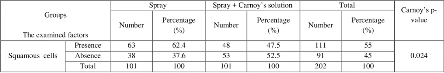
McNemar’s test was used to evaluate cellular adequacy in the conventional fixative spray group and spray + Carnoy’s solution group. Chi-square test (or Fisher’s exact test) was used for comparison of Carnoy’s solution in the studied groups. The results are listed in Table 1.
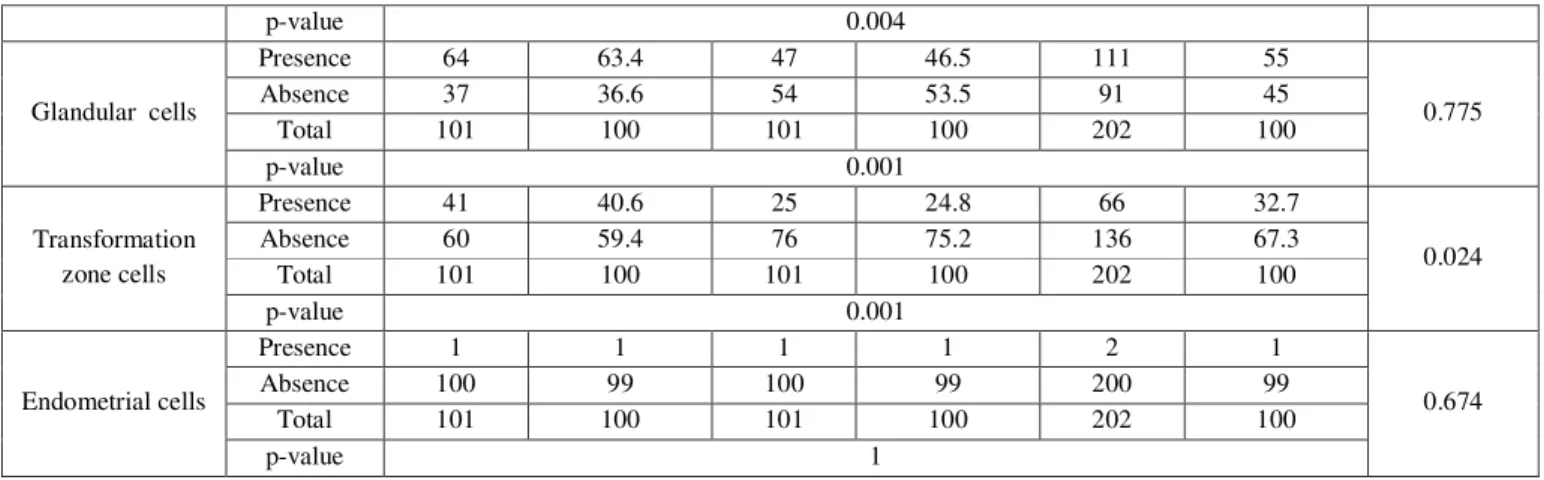
According to the recorded data in Table 1 and on the bases of McNemar’s test, there is a significant difference in comparing distribution of squamous cells on the spray-fixed and the Carnoy’s-fixed slides. The presence of squamous cells is further on the spray-fixed slide (p=0.004). Difference in comparing distribution of glandular cells on the spray-fixed and the Carnoy’s-fixed slides is statistically significant and the presence of glandular cells is further on the spray-fixed slide (p=0.001). Difference in comparing distribution of transformation zone cells on the spray-fixed and the Carnoy’s-fixed slides is statistically significant and the presence of transformation zone cells is further on the spray-fixed slide (p=0.001). The difference in distribution of endometrial cells on the spray-fixed and the Carnoy’s-fixed slides is not statistically significant (p=1).
Table 1- The results related to cellular adequacy in the two fixation methods used in this study
Groups
The examined factors
Spray Spray + Carnoy’s solution Total
Carnoy’s p-value
Number Percentage
(%) Number
Percentage
(%) Number
Percentage (%)
Squamous cells
Presence 63 62.4 48 47.5 111 55
0.024
Absence 38 37.6 53 52.5 91 45
p-value 0.004
Glandular cells
Presence 64 63.4 47 46.5 111 55
0.775
Absence 37 36.6 54 53.5 91 45
Total 101 100 101 100 202 100
p-value 0.001
Transformation zone cells
Presence 41 40.6 25 24.8 66 32.7
0.024
Absence 60 59.4 76 75.2 136 67.3
Total 101 100 101 100 202 100
p-value 0.001
Endometrial cells
Presence 1 1 1 1 2 1
0.674
Absence 100 99 100 99 200 99
Total 101 100 101 100 202 100
p-value 1
Difference in comparing distribution of cellular inadequacy on the spray-fixed and the Carnoy’s-fixed slides is statistically significant and cellular inadequacy is more on the Carnoy’s-fixed slide (p=0.002) (Table 2). There is statistically no significant difference in comparing the mean percentage of blood on the spray-fixed and the Carnoy’s-fixed slides (p=0.091) (Table 3). According to the results recorded in the above table and using the Wilcoxon signed-rank test, the difference of resolution in comparing the mean and quartile range on the spray-fixed and the Carnoy’s-fixed slides is not statistically significant (p=0.051) (Table 4). The difference in comparing distribution of inflammatory cells on the spray-fixed and the Carnoy’s-spray-fixed slides is statistically
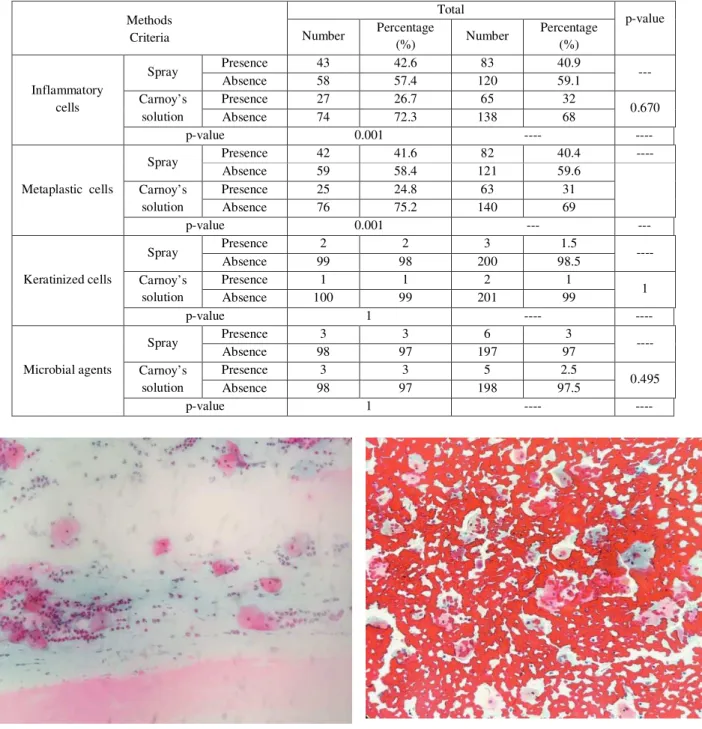
significant and distribution of these cells is further on the spray-fixed slides (p=0.001). There is statistically a significant difference in comparing the distribution of metaplastic cells on the spray-fixed and the Carnoy’s-spray-fixed slides and the distribution of these cells is further on the spray-fixed slides (p=0.001). There is statistically no significant difference in comparing the distribution of keratinized cells on the spray-fixed and the Carnoy’s-fixed slides (p=1). There is statistically no significant difference in comparing the distribution of microbial agents on the spray-fixed and the Carnoy’s-spray-fixed slides (p=1). No cases of abnormalities in the epithelial and the cylindrical cells are found in this study (Table 5).
Table 2- The results related to comparing the distribution of cellular inadequate on the spray-fixed and the Carnoy’s-fixed slides
Spray Spray + Carnoy’s solution Total
Carnoy’s p-value
Number Percentage
(%) Number
Percentage
(%) Number
Percentage (%)
Cellular inadequacy
Yes 38 37.6 55 54.5 93 46
0.003
No 63 62.4 46 45.5 109 54
Total 101 100 101 100 202 100
p-value 0.002
Table 3- The mean of blood percentage on the slides in the two fixation methods
Spray Spray + Carnoy’s solution
Carnoy’s p-value
Mean Standard
deviation Mean
Standard deviation Blood percentage on the slides
62.87 27.21 67.02 32.83
0.001
Table 4- The results related to the resolution of the slides in the two fixation methods
Middle Quartile range Carnoy’s p-value
Resolutio n of the slides
Spray 2 (2-3) ---
Carnoy’s solution 3 (2-3) 0.002
p-value 0.051 ---
* 1: good resolution; 2: medium resolution; 3: bad resolution
Table 5- The results related to the distribution of inflammatory cells, metaplastic cells and keratinized cells as well as microbial agents in the studied groups
Methods Criteria
Total
p-value
Number Percentage
(%) Number
Percentage (%)
Inflammatory cells
Spray Presence 43 42.6 83 40.9 ---
Absence 58 57.4 120 59.1
Carnoy’s solution
Presence 27 26.7 65 32
0.670
Absence 74 72.3 138 68
p-value 0.001 ---- ----
Metaplastic cells
Spray Presence 42 41.6 82 40.4 ----
Absence 59 58.4 121 59.6
Carnoy’s solution
Presence 25 24.8 63 31
Absence 76 75.2 140 69
p-value 0.001 --- ---
Keratinized cells
Spray Presence 2 2 3 1.5 ----
Absence 99 98 200 98.5
Carnoy’s solution
Presence 1 1 2 1
1
Absence 100 99 201 99
p-value 1 ---- ----
Microbial agents
Spray Presence 3 3 6 3 ----
Absence 98 97 197 97
Carnoy’s solution
Presence 3 3 5 2.5
0.495
Absence 98 97 198 97.5
p-value 1 ---- ----
Figure3: Spray+carnoys-fixed smears Figure 4: Spray-fixed smears
DISCUSSION
The results of this study formed based on findings of comparing the fixation of Pap smear blood samples taken from cervical cells of women with abnormal uterine bleeding referred to the gynecological clinics related to Shiraz University of Medical Sciences. The specimens were studied using two conventional fixative spray and spray + Carnoy’s methods. The presence of squamous cells, glandular cells and transformation zone cells were considered in this study as the criteria of cellular adequacy.
In comparing the distribution of squamous cells on the spray-fixed and the Carnoy’s-fixed slides, the presence of these cells were further on the spray-fixed slides (p=0.004). The above results revealed that the distribution of squamous cells on the Carnoy’s-fixed slides was more favorable. Underlying blood may remove from the slides by Carnoy’s solution, and create the above condition. The distribution of glandular cells on the surface of spray-fixed slides was more than on the surface of Carnoy’s-fixed slides (p=0.001). A statistically significant difference was also observed in the distribution of transformation zone cells on the surface of spray-fixed slides and spray + Carnoy’s-fixed slides; so that the presence of these cells was further on the surface of spray-fixed slides. Based on the evaluation performed on
cellular inadequacy, it became clear that a significant difference existed between cellular inadequacies. There was also no significant difference between the mean percentage of blood on the surface of spray-fixed and the Carnoy’s-fixed slides. This can be due to the used method in this study; so that the slides were fixed using fixative spray and then placed in the Carnoy’s solution. Fixation of cells with spray prevented the effectiveness of Carnoy’s solution and reduced the cleansing effect of the blood. Studies have suggested that putting blood samples in Carnoy’s solution for 3-5 minutes and prior to staining lyses the red blood cells [11] and thereby improves quality of the cells and the prepared slides. As mentioned earlier, a fixative spray was used in this study prior to the use of Carnoy’s solution. This decreased the quality of the slides.
Other studies have reported that the resolution of Carnoy’s-fixed slides was further than the spray-fixed slides, while in this study no significant difference was statistically observed between the resolution of the spray-fixed and the Carnoy’s-fixed slides [13]. This may be due to the presence of abundant blood on the slides. Note that the slides were prepared from women with abnormal uterine bleeding. The fixation process was performed one-time with spray and then the process continued again with Carnoy’s solution. The solution could not well erase the abundant volume of blood on the slides and consequently the slides resolution did not change.
The distribution of inflammatory cells on the spray-fixed and the Carnoy’s-fixed slides had statistically significant difference and the distribution of the cells was further on the spray-fixed slides (p=0.001). No significant difference was statistically found in comparing the distribution of microbial agents on the spray-fixed and the Carnoy’s-fixed slides.
This result was different from results of the study conducted by Shamsi. The distribution of microbial agents and inflammatory cells on the Carnoy’s-fixed slides was more than the spray-fixed slides in the later study. Reduced underlying blood and increased resolution of the slides were two reasons that expressed for the event and they led to better identification of these factors. Probably due to the presence of thick layer of blood smeared on the slides (due to heavy bleeding in patients with abnormal uterine bleeding to patients with contact bleeding), the possibility of diagnosis of microbial agents and inflammatory cells did not achieve even with the use of Carnoy’s solution. Kanbe and Kurosawa [15] also claimed that Carnoy’s fixative solution reduced the number of chymase-positive mast cells derived from umbilical cord blood and nasal mucosa cultures and Carnoy’s fixative solution had a negative effect on the number of chymase-positive mast cells.
There is statistically no significant difference between comparing the distribution of endometrial
cells on the spray-fixed and the Carnoy’s-fixed slides. The result is quite similar to the result obtained by Shamsi. It can be concluded from this comparison that Carnoy’s solution cannot well diagnosis the endometrial cells and their malignancies.
CONCLUSION
This research showed that a desirable result could be achieved with indirect use of Carnoy’s solution, but in some cases including cellular adequacy (the presence of squamous cells, glandular cells and transformation zone cells) the results were completely reverse; and fixation of the blood slides with conventional fixative spray showed better results than Carnoy’s fixative solution. The identification of microbial agents and inflammatory cells was not possible in this research even with the use of Carnoy’s fixative solution. This may be due to the presence of a thick layer of blood smeared on the slides. According to the results, it should be stated that Carnoy’s solution did not help in diagnosis of endometrial cells and their malignancies in women with abnormal uterine bleeding. So, conducting experiments and tests about direct use of Carnoy’s solution is recommended to enhance the quality of Pap smear slides.
REFERENCES
1. Bancroft JD,Stevens A ,Turner DR.Theory and practice of Histological techniques .chapter 2:Fixation and Fixatives.Thirded , page:21-42.Co:Churchill Livingstone.
2. Koss LG,Melamed MR.Koss Diagnostic cytology and its Histopathologic Bases 2006;15ed,vol 2.chapter 44.Laboratory techniques,section III.Techniques in Diagnostic cytology:1570-1634.
4. Kenneth GR, Ross SB. Kisner’s gynecology & womens health. 7th ed. USA: Mosby; 1999; 100-6.
5. Nuovo J, Melnikow J, Howell LP. New tests for cervical cancer screening. America Family Physician 2001; 46(5):780-6.
6. Bauer HM, Ting Y, Greer CE, Dunton CJL. Genital human papillomavirus infection in female university students as determined by a PCR-based method. JAMA 1991; 265:472-7. 7. Rahnama P, Faghihzadeh S, Ziaei S. Effect of
the sampling sequence on the ability of papanicolaou smear. Int J Gynecol Cancer 2005;(15):66-9.
8. Kadivar C , Robati M, Kadknodaci M , Ghasempour X. Challneges in pap smear screening: A qunlitication assessment of provincial health system network. International ymposiumon predictive oncology and international sterategies: 2004 February.7-10: Nice, Ferance
9. Sadri Gh H, Mahdi Zadeh M, Shahidi Sh et al. 1999. Abnormal Pap smear results in patients referred to the health centers of Isfahan Province, Iran during 1997-1998. Journal of Research in Medical Sciences, 6(2):134-135. 10.Hideshein A, Bratli M, Concepcion E, Rober
T. Collection of cervical secretions does not adversely affect pap smears. Take immediately afterward. Clinical and Diagnostic of Laboratory Immunology 1998; 5 (4): 491-3. 11.Nguyen Gk, Lee M*, Ginsberg J, Wragg T,
Bilodeau D. Fine-needle aspiration Of the thyroid:an overview. CytoJournal 2005: 2:12. 12.Weidmann J, King LC, Bibbo M. Modification
of Cytorich-Red fixative system for use on bloody pap and fine needle aspiration smears. Diagnostic Cytopatology J1999; 20(2): 95-8. 13.Leslie R, Rowe B, Jole S. A simple method to
determine the need for glacial acetic acid treatment of bloody thin prep Pap test before slide processing. Diagnostic Cytology J 2003; 5: 321-5.
14.Shamsi M, Abdali K, Montazer NR, Kumar PV, Tabatabaee HR. "Comparison of Carnoy's
solution and 96% ethyl alcohol fixation in bloody Pap smears". Acta Cytol 2008;52 (2): 187–90.