Int J Anat Res 2015, 3(4):1568-72. ISSN 2321-4287
Original Research Article
EXTRA HILAR BRANCHING OF RENAL ARTERIES: AN ANATOM ICAL
STUDY
N. Shakuntala Rao *
1, Krishna Kishore
2, Sujatha. K
3, H.R.Krishna Rao
4.
ABSTRACT
Address for Correspondence: Dr.N.Shakuntala Rao, Professor, Depart ment of Anat omy, PES Inst i-t ui-t e of M edical Sciences and Research, Kuppam-517425, Andhra Pradesh, India.
M obile No.: +91 9493502095 E-M ail: drshakunt [email protected]
Int roduction: The object ive of t his st udy w as t o observe t he pat t erns of different ar t eries t hat supply t he kidneys. The kidney has a segm ent al dist ribut ion of art eries. The kidneys are divided int o five vascular segm ent s. The art eries t hat arise from t he aor t a above or below t he m ain renal art er y and reach t he hilum ar e called accessor y renal art eries. They are persist ent em br yonic lat eral splanchnic art eries. Accessory renal art er ies m ay arise fr om t he celiac or superior m esent eric art er ies, near t he bifur cat ion or from t he com m on iliac art eries. The present st udy has att em pt ed t o f ind out accessory, and aberrant ar t eries t o kidneys w it h review of lit erat ure.
M aterials and M ethods:The st udy w as done on 52 kidneys random ly select ed from cadavers t hat w ere used for t he purpose of t eaching in t he depart m ent of Anat omy at P.E.S M edical College. The kidneys w ere rem oved from t he cadavers en-block w it h t he art er ies and veins int act . The renal art ery w as obser ved f or it s pat t ern of branching.
Observat ions and Discussion: The pre-hilar branching pat t ern w as absent only in six kidneys out of t he 52 kidneys select ed. The branches given bef ore ent ering t he hilum w ere eit her in t he f orm of a fork pat t ern or a ladder pat t ern in t he rem aining 46 kidneys. The fork pat t ern w herein t he branches arose from a single point w as found in 42 kidneys. The ladder pat t er ns w ere seen in t w o post erior segm ent art eries and t w o ant erior segm ent art eries. The ant erior division oft en show ed t he forkpat t erns w hich w ere eit her duplicat e or t riplicat e out side t he hilum m ore proxim ally, w it h furt her division int o duplicat e or t r iplicat e t erm inal branches closer t o t he hilum but signif icant ly out side.
KEY W ORDS: Polar art eries, Vascular patt er ns, Accessory Renal Art eries, Hilum , Segm ental Art eries.
INTRODUCTION
Int ernat ional Journal of Anatomy and Research, Int J Anat Res 2015, Vol 3(4):1568-72. ISSN 2321- 4287 DOI: ht t p:/ / dx.doi.org/10.16965/ ijar.2015.290
Access this Article online
Quick Response code Web site:
Received: 29 Sep 2015 Accept ed: 15 Oct 2015 Peer Review : 29 Sep 2015 Published (O): 30 Nov 2015 Revised: None Published (P): 31 Dec 2015
Int ernat ional Journal of Anat omy and Research ISSN 2321-4287
ww w.ijmhr.org/ ijar.htm
DOI: 10.16965/ ijar.2015.290
* 1 Professor, 2,3 Assistant Professor, 4 Professor & HOD.
Depart ment of Anat omy, PES Inst it ute of M edical Sciences and Research, Kuppam, Andhra Pradesh, India.
branches from t he dorsal aort a as it ascends t o t he adult posit ion in t he lum bar region. The kidneys are supplied by art eries t hat arise from t he lat eral side of aort a at t he level of lumbar vert ebrae, bet w een t he upper margin of L1 and t he low er margin of L2. They run at right angles t o aort a and in 70% of individuals a single art ery Renal art ery variat ions have been st udied by
M ATERIALS AND M ETHODS
RESULTS
supplies t he kidney on each side. The kidney has a segment al dist ribut ion of art eries. The kidneys are divided int o five vascular segment s. They are named apical, superior, middle, low er and posterior. The apical segment is formed by t he ant erior and medial region of t he superior pole. The superior segment is formed by t he superior pole and t he cent ral ant ero-superior region (ant er ior segm ent ).The w hole of low er pole forms t he inferior segment . Bet w een ant erior and inferior segment s is t he part of kidney t hat forms t he middle ant erior segment . The w hole post erior region bet w een apical and inferior segm ent s is t he post er ior segm ent . These segm ent s get t heir blood supply f r om t he segment al art eries w hich arise from the anterior and posterior divisions of t he main renal artery. These segment al art eries are end art eries. They do not anast omose freely. The renal medulla is supplied by long w ide vessels passing from t he efferent glomerular art erioles. The art eries t hat arise from t he aort a above or below t he main renal art er y and reach t he hilum are called accessory renal art eries. They are persist ent em bryonic lat eral splanchnic art eries. If t he vessels cross ant erior t o t he uret er at t he low er pole, it may cause obst ruct ion t o t he uret er and cause hydronephrosis. Accessory renal art eries may arise from t he celiac or superior mesent eric arteries, near the bifurcation or from the common iliac art eries [1].
In 30% of individuals accessory art eries have been report ed [1]. These arise from t he aort a above or below t he main renal art ery and go t o t h e hil um and ar e r egar d ed as p er sist ent em br yonic lat eral splanchnic ar t er ies. Tw o groups of renal art ery variat ions are considered. They are early division and ext ra renal art eries (ERA). The extra renal arteries are furt her divided int o hilar and polar art eries. The hilar art eries are considered accessory art eries. The art eries t h at ent er t h e p o l es o f t h e k i d ney s ar e considered as aberrant art eries [2]. The polar art eries ent er t he kidneys direct ly from t he capsule out side t he hilus. The present st udy has attempted t o find out accessory, and aberrant art eries t o kidneys w it h review of lit erat ure.
P.E.S Inst it ute of M edical Sciences & Research, India w ere used for t he st udy. 52 kidneys from various cadavers w ere removed along w ith t heir art eries from t heir locat ion. The art eries of t he kidneys w ere t hen observed for variat ion in branching before ent ering t he hilum , at t he hilum and also ent ry at ot her point s especially t he poles. The kidneys w it h variat ions w ere phot ogr aphed. The accessor y ar t er ies and aberrant arteries were not ed and photographed.
Formalin fixed cadavers w hich w ere used for t eaching purpose during rout ine dissect ion in
It w as observed t hat 12 kidneys out of t he 52 had art eries w hich ent ered t he upper pole of the kidneys. Of t he 12, right side kidneys w ere 2 and left side kidneys w ere 10. Of t he 12 kidneys t h er e w er e f iv e lef t ki dn ey s in w hi ch t he post erior division gave duplicat e and t riplicat e branches at t he hilum. One left kidney had an art ery t hat ent ered t he low er pole of t he kidney w hich w as a direct branch from t he aort a. The ar t eries t hat ent ered t he upper poles t ook origin from the main renal art ery proximal t o the origin of anterior segmental artery. The anterior segm ent al an d t h e p o st er i or segm ent al art eries divided out side t he hilum t o supply t heir vascular t errit ories. The art eries show ed fork pat t ern and ladder pat t erns. The fork pat t erns w ere more frequent t han ladder pat t erns. The branches w ere ident ifiable as apical, upper, middle, low er branches. Of t he branches t hat w ere given by t he post erior division one branch w as dir ect ed u pw ar d s t ow ar ds t h e u pp er segment at t he hilum.(FIG-3) The area of supply was not t raced in t his st udy.
Fig. 1:Show ing t he branching patt ern.
Int J Anat Res 2015, 3(4):1568-72. ISSN 2321-4287 Fig. 2: Show ing t he branching patt ern.
ASA- Ant erior Segm ent al Artery 1,2,3,4- Br anches – in ladder pat t ern.
Fig. 3: Show ing t he branching patt er n. PSA: Post er ior Segm ent al Art ery giving branch t o upper pole.
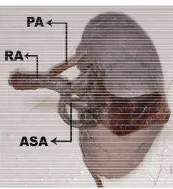
PSA- Post erior Segm ent al Art ery, PA- Polar Art ery, RA- Renal Art ery
OBSERVATIONS AND DISCUSSION
Ozkan et al [2] found in t heir angiographic st udy of 855 pat ient s t hat 24% of pat ient s had more t han one renal artery. The right side had more t han one art ery compared t o t he left side. The occurrence of more than one artery on both sides w as only 5%. They observed 71 aberrant and 69 accessor y art eries on t he r ight side and 58 aberrant and 58 accessory art eries on t he left side. In t he present st udy of t he 12 polar art eries
10 w ere seen on t he left and 2 on t he right . The polar art eries t ook origin from t he main renal art ery (FIG-1) and can be considered normal segm ent al branching based on t heir or igin. Irena Vilhova et al [3] have described r enal ar t er ies as d ou b l e, t r i p l e, accesso r y an d per forat ing art er ies. Double r enal art er ies originat ed from t he aort a, w ere ident ical in diameter, blood supply areas, and gave branches t hat ent ered t he hilum. The t riple renal art eries varied in t heir diam et ers and areas of supply but branches ent ered t he hilum. Accessory renal art ery arose from t he aort a, ent ered t he hilum and supplied only one segment eit her upper or low er pole. A perforat ed renal art ery originat ed from t he aort a, ent ered t he kidney out side t he hilum and w as comparable t o a segment al art ery supplying only one segment . The vessels w hich are aberrant are longer and narrow er and t he renal segm ent s receiving t hese vessels have low er levels of blood pressure t han t he rest of parenchyma, t hus increasing rennin secret ion. They have opined t hat an anat om ical reason cou l d b e t h e cau se f o r d i so r d er s li ke hy per t ensio n an d t hat t her e i s a n eed t o int r oduce t erm s f or classif ying plur al r enal art eries. In t he present st udy t he renal art eries t hat ent ered t he kidney out side t he hilum w ent t o t he upp er pole (FIG-1) and t hese w er e considered polar art eries because t hey arose from t he main renal art ery as a separat e branch m ore proxim al t o t he ant erior division. They cannot be considered aberrant as t hey w ere f r o m t h e m ain ar t er y and t hey cannot be addit ional or accessory as t hey did not arise from t he aort a and did not ent er t he hilum.
Saldarriaga, B et al [4]They have also observed t hat t he renal pole w as frequent ly supplied by t he ant erior division. They observed a direct branch from t he renal art ery-superior renal polar branch on t he right -hand side in 17.2% and in 13.5% on t he left hand side. The inferior polar branch w as seen in 5 specimens each on t he right and left sides. In t he present st udy t he superior polar branches were from the main renal art ery and t he inferior polar branch w as seen in only one specimen on t he left . This had taken origin from t he aorta direct ly. So t his can be considered an accessory art ery t o t he lower pole.
The frequency of more than one additional artery w as 87(22.3%) and 2 addit ional art eries w as 10 (2.6%). They found t hat addit ional art eries had great er lengt h t han main art eries. The addit ional art eries ran parallel or divergent t o t he main renal art ery. They have also observed ear ly r am if icat i on of t h e m ai n r enal ar t er y and considered it import ant in diagnost ic imaging and surgical complicat ions during t ransplant s. The first 15mm of t he renal art ery is used for anast omosis w it h t he recipient ’s iliac artery. In t he present st udy t here w as one kidney w it h an addit ional ant erior segm ent al branch w hich gave t he middle and low er branches.
Shoja et al [6] st udied t he variat ions in peri-hilar branching pat ern and morphology of t he main artery. They classified t he branching as ladder and fork pat terns. The pat tern where there w ere sequent ial branching point s w as t ermed ladder t ype. The pat t ern w it h a com mon branching point w as t ermed t he fork t ype. The fork w as eit her duplicat e or t riplicat e depending on t he n u m b er o f br an ch es. Th ey d iv i d ed t hei r observat ions int o cardinal peri-hilar morphology (more t han 5%) and infrequent m orphologies (less t han 5%). They observed t hat t he m ain art ery w as of t he fork pat t ern in 92.6%(75), duplicat ed in 80.2%(65) t riplicat ed in 12.4%(10) and ladder pat t ern w as 7.4%.(6). In t he present st u d y t h e f o r k p at t er n w as seen i n 42 k i dn ey s(FIG-1) an d 4 k i dn ey s h ad l add er pat t ern.(FIG-2) There w as no perihilar branching i n o n ly six k id n eys. Th er ef o r e i t can b e considered t hat branching out side t he hilum is a normal pat t ern w here the art eries divide t o go t o t he respect ive segment s.
Julius A.Ogeng’o et al [7] have reported single, double, t riple, and quadruple renal art eries. The double renal art eries w ere parallel, overlapped, init ially super im posed t hen div er gent and crossed t ypes. Of the double art eries there w ere superior polar and inferior polar types. The single r en al ar t er i es w ere h i l ar, p r eh i l ar, an d par enchym al br anching t ypes. The prehilar branching pat t ern show ed t erm inal branches eit her before or aft er t he hilum. The bifurcat ion p at t er n, f or k an d l ad d er p at t er n s w i t h overlapping of primary branches w as observed. Ther e w er e upt o sev en ext r apar enchy m al branches reported in t heir st udy. In t he present
st udy t he post erior division w as observed t o be having duplicat e and t riplicat e branches in fork and ladder pat t erns in 11 kidneys. They can be considered ext ra parenchymal branches as t he post erior division gives only one segm ent al branch normally.
Budhiraja V et al [8]also report ed t hat superior polar art eries t ook origin from apical segment al branch in five out of t he seven cases. In t w o cases t hey originat ed direct ly from t he aort a. The present st udy found polar art eries arising from ant erior segment al art eries. All t he polar ar t er i es ent er ed o ut si de t he h il u m an d penet rated t he capsule of t he kidney.
Neerja Rani et al [9] ident if ied variat ions in origins of segmental art eries. They observed the ant erior division gave f our branches apical, upper, middle and low er segmental arteries. In the present st udy the ant erior division gave three branches and in 12 kidneys t he polar branch as a separate branch from main renal artery.
Shinde Amol A et al [10]have report ed from a st udy of 50 kidneys, low er polar supernumerary art eries in 4% of t heir specimens. They have cit ed t he explanat ion given by Felix based on em b r yo lo gical dev el o pm ent . The ar t er ies supplying the kidneys of an 18mm fetus are from t he dorsal aort a. There are nine pairs of art eries cal l ed t h e l at er al m eso n eph r i c ar t er i es. According t o Felix t he first t w o pairs are called cranial, t he 3rd t o 5t h are called middle, and t he
6t h to 9t h are called t he caudal group. The middle
group supplies t he kidneys. If more t han one art ery persist s t hen supernumerary art eries can result . There w as only one low er polar art ery in t he present st udy.
CONCLUSION
Int J Anat Res 2015, 3(4):1568-72. ISSN 2321-4287 Conflicts of Interests: None
REFERENCES
junct ion and t he cort ex becomes reversed in t his case.
[1] . Susan Standring. Gray’s Anat om y, The Anat om ical Basis of Clinical Pract ice, 40t h ed Elsevier Churchill
Livingst one, Urogenit al syst em 2008:1231-33. [2]. Ugur Ozkan,Levent Oguzkur t ,Fahri Tercan,Osm an
Kizilkilic,Zafer Koc,Nihal Koca. Renal ar t ery origins and var iat ions: angiographic evaluat ion of 855 co n secu t i v e p at i e n t s. Di agn In t er v Rad i o l 2006;12:183-186.
[3] . Irena Vilhova,Yurij Yaroslaw ow icz Kryvko,Ryszard M aciejew ski. The r adio anat o m ical r esear ch of p l u r al r en al ar t er i es. Fo l i a M o r p h o l . 2001;60(4):337-341.
[ 4] . Sal d ar r i aga,B. ; Pi n t o ,S. A. & Bal l e st e r o s,L.E. M or phological expression of t he renal ar t er y.A dir ect anat om ical st udy in a Colom bian half-cast e populat ion. Int .J.M orphol., 2008;26(1):31-38. [5]. B.Saldarr iaga, A.F.Perez, L.E.Ballest eros. A direct
anat om ical st udy of addit ional r enal art er ies in a Colom bian m est izo populat ion. Folio M or phol. 2008;67(2):129-134.
[6]. M oham m adali M .Shoja. R, Shane Tubbs, Abolhassan Shakeri, M arios Loukas, M oham m ad R. Ardalan, Ham id T. Khosroshahi. W. Jer ry Oakes. Per i-hi lar branching pat t erns and m or phologies of t he renal art ery: a review and anat om ical st udy. Surgical and Radiologic Anat om y 30.5 (2008): 375-382. [ 7] . Juli us A.Ogeng’o, Ch ar l es O.M asaki ,Si m eon R.
Sin-keet , Johnstone M . M ut hoka, Acleus K.M urunga. Var iant anat o m y of renal ar t er ies in a Kenyan populat ion.Ann Transplant , 2010; 15(1):40-45. [ 8] . Bud hiraja V,Rast ogi R,A.K.Ast hana. Renal art er y
var i at i o n s: e m b r y o l o gi cal b asi s an d su r gi cal cor relat ion.Rom anian Journal of M orphology and Em bryology 2010;51(3):533-536.
[9]. Neer ja Rani,Seem a Singh,Pushpa Dhar and Rani Kum ar. Surgical Im por tance of Art er ial Segm ent s of Hum an Kidneys: An Angiogr aphy and Corrosion Cast St udy. J Clin Diagn Res.2014 M ar;8(3):1-3. [10]. Shinde Am ol A, Bharam be Vaishaly K. A cadaveric
st udy of low er polar supernum erary renal art eries-Em br yolo gical and clini cal consider at i on. IOSR Jo u r n al o f Den t al an d M e d i cal Sci e n ces. 2014;13(7):06-09.