Function in Ethanol Behaviour
Sonia Cavaliere, John M. Gillespie, James J. L. Hodge*
School of Physiology and Pharmacology, University of Bristol, Bristol, Avon, United Kingdom
Abstract
In humans, KCNQ2/3 channels form an M-current that regulates neuronal excitability, with mutations in these channels causing benign neonatal familial convulsions. The M-current is important in mechanisms of neural plasticity underlying associative memory and in the response to ethanol, with KCNQ controlling the release of dopamine after ethanol exposure. We show that dKCNQ is broadly expressed in the nervous system, with targeted reduction in neuronal KCNQ increasing neural excitability and KCNQ overexpression decreasing excitability and calcium signalling, consistent with KCNQ regulating the resting membrane potential and neural release as in mammalian neurons. We show that the single KCNQ channel in
Drosophila(dKCNQ) has similar electrophysiological properties to neuronal KCNQ2/3, including conserved acute sensitivity to ethanol block, with the fly channel (IC50= 19.8 mM) being more sensitive than its mammalian ortholog (IC50= 42.1 mM). This suggests that the role of KCNQ in alcohol behaviour can be determined for the first time by usingDrosophila. We present evidence that loss of KCNQ function inDrosophila increased sensitivity and tolerance to the sedative effects of ethanol. Acute activation of dopaminergic neurons by heat-activated TRP channel or KCNQ-RNAi expression produced ethanol hypersensitivity, suggesting that both act via a common mechanism involving membrane depolarisation and increased dopamine signalling leading to ethanol sedation.
Citation:Cavaliere S, Gillespie JM, Hodge JJL (2012) KCNQ Channels Show Conserved Ethanol Block and Function in Ethanol Behaviour. PLoS ONE 7(11): e50279. doi:10.1371/journal.pone.0050279
Editor:J. David Spafford, University of Waterloo, Canada
ReceivedMay 23, 2012;AcceptedOctober 23, 2012;PublishedNovember 29, 2012
Copyright:ß2012 Cavaliere et al. This is an open-access article distributed under the terms of the Creative Commons Attribution License, which permits unrestricted use, distribution, and reproduction in any medium, provided the original author and source are credited.
Funding:This work was supported by grants from the EU FP7 Marie-Curie (IRG200632), Royal Society (2008/R1), BBSRC (BB/G008973/1), and from a Wellcome PhD program (083361). The funders had no role in study design, data collection and analysis, decision to publish, or preparation of the manuscript.
Competing Interests:The authors have declared that no competing interests exist.
* E-mail: james.hodge@bristol.ac.uk
Introduction
Voltage-gated potassium (Kv) channels form a diverse gene family that, in humans, is subdivided into 12 subfamilies of 40 members [1]. Furthermore, functional Kv channels are tetramers, with multiple members of each individual subfamily able to form homo- or hetero-multimers with different properties. Such a diversity of channel types in mammals has made studying these channels in native tissue challenging; determination of the functional consequence of removal of a given channel at the whole organism level is often difficult due to genetic redundancy and compensation. Developing viable genetic models to study individual channel function is becoming increasingly important clinically, with mutations in over 60 channel genes resulting in channelopathies [2]. A potentially powerful approach is to use the genetics ofDrosophila, which typically contains a single member of each Kv channel subfamily, with nulls being adult viable; this allows exploration of the functional consequence of complete lack of a subfamily of Kv channel [3].
KCNQ (Kv7) channels mediate a range of important physio-logical functions, form a hotspot of genetic diseases and are targets for new and existing drug treatments. In human cardiac muscle,
KCNQ1 mutations cause Long and Short QT [1,2]. KCNQ1
mutations also result in adult onset type II diabetes [4,5]. In the nervous system, KCNQ2 and KCNQ3 heteromultimerise to form a channel that mediates the M-current and regulates membrane excitability in the sub-threshold range for action potential generation. Therefore, reducing neuronal KCNQ is usually
sufficient to increase excitability of most neurons, with the M-current mediating changes in excitability that occur during synaptic plasticity and memory, alcohol response and nociception [1,6,7]. KCNQ2/3 loss-of-function mutations result in a form of epilepsy. KCNQ4 loss-of-function mutations cause autosomal dominant deafness. M-current inhibitors increase excitability and have shown some promise in enhancing memory in models of dementia. Conversely, M-current openers are of great interest as anticonvulsants, analgesics and treatments of psychiatric diseases [1,8].
characterise for the first time the in vivo consequence ofdKCNQ mutations on neural activity and behaviour, showing a role for the channel in regulation of ethanol sensitivity and tolerance.
Materials and Methods
DNA reagents
Drosophila KCNQ RE26469 cDNA (Flybase FBgn0033494,
vector:pIRES2-EGFP), ratKCNQ2cDNA (GenBank AAC36722;
pcDNA3.1) and rat KCNQ3 cDNA (AC79846; pcDNA3.1) [11].
Genomic database searches were performed with Drosophila RE26469 full-length KCNQ cDNA using the WU-BLAST server at EMBL-EBI.
Cell culture
cDNAs were expressed in Human Embryonic Kidney (HEK293) cells using previously published protocols [11].
Electrophysiology and pharmacology
Whole-cell voltage-clamp recordings were made from HEK293 cells using an Axopatch 200A amplifier (Axon Instruments, Molecular Devices, Sunnyvale, California, US) as previously described [11]. To determine the effect of ethanol at a depolarised voltage, currents were elicited by a single pulse protocol in which membrane potential was held at280 mV for 100 ms, stepped to +30 mV for 1 s and stepped down to2120 mV for 250 ms. To generate I–V and G–V relations, the following multi-step protocol was used: membrane potential was held at280 mV for 100 ms, stepped in increments of 10 mV from280 mV to+30 mV then, after 1 s, stepped down to2120 mV for 250 ms. Currents were measured at the end of the sweep at maximal current for each step. I–V relations were plotted as normalised current in pA/pF (6 standard error of the mean (SEM)) against voltage. The V0.5 value was the voltage required for half the maximal activation current. Mean V0.5values are shown6SEM. Data were analysed using Graphpad Prism.
Drosophilastocks
TheKCNQ deletion mutant contains an imprecise excision of the EP2074 element (KCNQ186) that removes all the 59 and transmembrane regions of the channel and therefore is likely a null mutation [9]. The KCNQ control was a precise excision of the element (KCNQ97), leaving the gene completely intact [9]. uas-KCNQflies allowedGal4promoter-driven overexpression ofKCNQ [9], while uas-KCNQ-RNAi (Bloomington stock 27252) allowed Gal4-targeted knockdown of the channel. Wild-type flies were Canton S w- (CSw-) from a stock previously maintained in the Griffith lab. AllKCNQmutant,Gal4anduaslines were out-crossed with thisCSw-line prior to behavioural analysis. All genotypes and all other crosses were raised on corn-meal malt-molasses agar medium at 2262uC and 60610% humidity under a 12:12 h light-dark cycle.
Immunohistochemistry
Adult fly brains or third-instar larvae were dissected in HL3.1 (70 mM NaCl, 5 mM KCl, 10 mM NaHCO3, 115 mM sucrose, 4 mM MgCl2 5 mM trehalose, 1.5 mM CaCl2, and 5 mM HEPES, pH 7.3), and isolated brains from either stage were fixed in 4% paraformaldehyde in HL3.1 for 30 min before being washed in HL3.1 [14]. The preps were permeabilised in HL3.1 with 0.1% triton X (HL3.1-Tx) for 1 h, and then blocked for 1 h in HL3.1-Tx with 0.1% BSA and 2% normal donkey serum (HL3.1-Tx-BSA-NDS). To visualise the vGlut(OK371)-Gal4 pat-tern, a (1:1000) rabbit anti-DrosophilaGlut antibody was used [15]
overnight at 4uC in HL3.1-Tx-BSA-NDS. After washing three times in HL3.1-Tx for 20 min, the brains were incubated with anti-rabbit Alexa-648 conjugated secondary antibody (1:400 in HL3.1-Tx-BSA-NDS) for 2 h at room temperature. Finally, the brains were washed three times in HL3.1-Tx before being mounted in Vectashield (Vector Laboratories). Samples were stored at 4uC in the dark until examination with a Leica TCS SP5 confocal microscope. The endogenousKCNQ expression pattern was determined by visualising membrane-targeted GFP expressed using KCNQ-Gal4 reporter lines (KCNQNP3423-Gal4, uas-mCD8-GFP).
Calcium imaging
Third-instar larvae were dissected in haemolymph (HL3.1) solution containing 1.5 mM Ca2+and were subsequently imaged using previously described methodology [16,17]. Spontaneous Ca2+ signalling during peristaltic crawling was imaged at ten frames/sec in the A5 ventral ganglion motor neurons of larvae expressing GCaMP3, using the following setup: 106 water-immersion lens of a Zeiss Examiner Z1 microscope with LED, filters and AV4.8 software, with an Axiocam MRm camera system optimised for GCaMP3 imaging. A region of interest was drawn around the cell bodies of MN1-Ib, MN14-Ib, MN6/7-Ib, MN30-Ib and MNISN-Is in order to calculate the per cent increase in intensity above baseline (which was defined as the average of the first ten frames) for each time point. Images were processed using Volocity (PerkinElmer) and Image J (NIH). Data were statistically analysed and presented with Graphpad Prism software.
Ethanol behavioural assays
All experiments were performed at 25uC and 70% humidity under white light. Twenty synchronised 3–5 day old male and female flies were used. One millilitre of 40% ethanol solution was added to an absorbent pad at the bottom of a sealed bottle. Active flies initially remained on the walls or up beneath the bottle lid. During the test period, a number of flies became anesthetised and were immobilised at the bottom of the bottle. These flies were counted at 5-min intervals over the 90-min ethanol exposure to measure sensitivity [18]. To test the effect of acute activation of dopamine neurons on ethanol sensitivity, flies expressing the heat-activated TRPA1 channel in these neurons were tested at either 23uC or 30uC, with only the higher temperature sufficient to cause TRPA1 activation and depolarisation [19]. To measure tolerance, flies were given a first ethanol exposure as above and then allowed to recover for 2 h on food without ethanol. The flies’ response to the same ethanol exposure a second time was then measured [18]. Flies recovered fully after the ethanol exposures used in these experiments. Data were analysed using Graphpad Prism.
Ethanol content and preference assay
Ethanol content of flies of different genotypes was determined by exposing twenty active flies to ethanol vapour in a sealed bottle that an absorbent pad soaked in 1 ml of 40% ethanol. Flies were frozen in liquid nitrogen and stored at 280uC. Flies were homogenised with 500ml of Tris-HCl (pH 7.5) and spun at 4uC
flies. All statistical analyses for behavioural data were performed and plotted with Graphpad Prism software.
Semi-quantitative RT-PCR
Total RNA was isolated from adult fly heads using a Trizol solution (Invitrogen). An equal number of age-matched control flies were frozen in liquid nitrogen and decapitated by vortexing. The detached heads were collected and homogenised. Trizol was added directly to the homogenised heads and RNA was extracted by ethanol precipitation. RNA was DNase-treated (Ambion, Inc.) and reverse-transcribed using a first-strand complementary DNA (cDNA) synthesis kit (RevertAid, Fermentas). The first strand cDNA was obtained from a total RNA template in a single reaction, adding a reverse transcriptase enzyme and oligo (dT) primers and incubating for 1 h at 42uC [9]. PCR reactions were performed as follows: 4 min at 95uC; then 30 s at 95uC, 60 s at 60uC, and 2 minutes at 72uC for 25 cycles; followed by a final 7 min at 72uC. All primers were optimally designed and synthesised by Invitrogen.
KCNQ forward: 59-AGGAAAGCCGCTGAACTACA-39, po-sition 210–230,
KCNQ reverse: 59-CGAGGTGCCCATTCCTAATA-39, po-sition 604–584.
The PCR amplification product was analysed by electrophoresis in 1% agarose gels and visualised by ethidium bromide staining and transillumination under UV light.
Results
The level of KCNQ regulates neural excitabilityin vivo
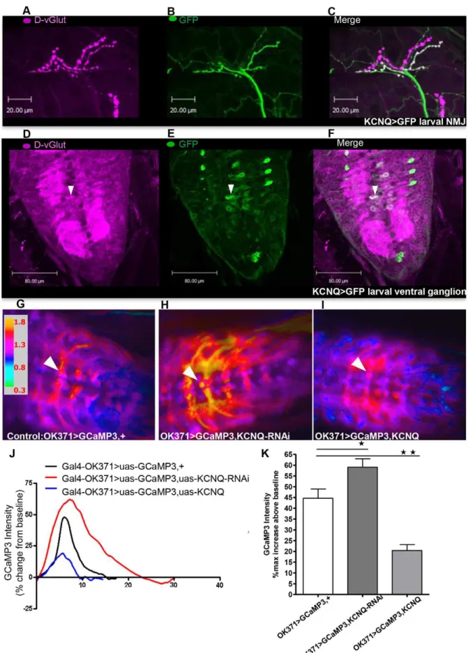
To determine how KCNQ may affect neural excitability, we first identified the neurons in which KCNQ is expressed. Consistent with previous reports [9,10], KCNQ was found to be broadly expressed in the embryonic nervous system (Figure S1). To investigate expression later in development, aGal4 enhancer trap within the KCNQ gene was used to express GFP. These experiments revealed that KCNQ continues to be broadly expressed in the nervous system, including in glutamatergic motor neurons (Figure 1B and E) that also strongly express theDrosophila vesicular glutamate transporter (D-vGlut, [15]) (Figure 1A and D). We therefore employed the vGlut(OK371)-Gal4 line that has particularly strong motor neuron expression in larvae. These neurons also expressKCNQ(Figure 1C and F). In the adult brain, KCNQ appears to be broadly expressed, including in neurons in and around the mushroom body (unpublished data). To explore how the level of KCNQ may be affecting neural excitability, we performed in vivo Ca2+ imaging in Drosophila. We used
vGlu-t(OK371)-Gal4 to express a Ca2+ reporter, GCaMP3 [16], to
examine neural activity in the KCNQ-expressing neurons. During larval peristalsis, there is a wave of Ca2+ signalling that travels through the ventral ganglion. We measured the maximum change in fluorescence in the motor neuron soma when this occurs. Expression of KCNQ-RNAi caused a significant increase in neuronal excitability (Figure 1H, J and K) compared with control (Figure 1G). Overexpression of the KCNQ channel reduced Ca2+ -induced fluorescence and hence neural excitability (Figure 1I, J, K). These bidirectional changes in KCNQ expression and neural excitability might be expected to interfere with neural release, as has been demonstrated in other systems [1,6]. Expression ofKCNQ orKCNQ-RNAiin dopa-decarboxylase (Ddc) neurons resulted in a wing-expansion phenotype (Figure S2); such a phenotype has previously been reported to occur as a result of large changes in neuronal hyper- or de-polarisation, disrupting the neuropeptide release required for wing expansion [20–22].
dKCNQ current is similar to the neuronal M-current encoded by KCNQ2/3, with both showing ethanol block
Ethanol has recently been demonstrated to inhibit the M-current in dopaminergic neurons of the ventral tegmental area (VTA, [6]); however, neither the molecular identity of the channel subunits nor the effect of ethanol on KCNQ currents was studied. Therefore, we expressed mammalian KCNQ2/3 in Human Embryonic Kidney (HEK) cells and found that application of low concentrations (10 mM) of ethanol caused acute block of the KCNQ2/3 current at+30 mV (Figure 2A and C). Ten-millimolar ethanol has little effect on the KCNQ2/3 activation curve (Figure S3A), although high concentrations of ethanol could conceivably change the channel’s gating. The IC50 for the ethanol block of KCNQ2/3 was 42.167.4 mM (Figure 2E–F). Similarly to KCNQ2/3, the dKCNQ carries a slowly activating and non-inactivating Kv current that opens at sub-threshold potentials for action potential generation (Figure 2). Under the same conditions, dKCNQ current was sensitive to ethanol block (Figure 2B and D), with little effect on the activation curve (Figure S3B) and an IC50 of 19.863.8 mM, which is significantly lower than that of mammalian KCNQ2/3 (Figure 2E–F).
Neuronal KCNQ regulates ethanol sensitivity in
Drosophila
As ethanol reduced the KCNQ current, we proceeded to determine ifKCNQloss-of-function would further increase the fly’s sensitivity to ethanol, the rationale being that they had been made more susceptible to ethanol due to their lack of functioning KCNQ channels. We employed an assay that involved exposing flies to 40% ethanol vapour and scoring sedation, whereas active flies initially remain on the walls and beneath the lid of the bottle. During the ethanol exposure, flies become sedated and are immobilised at the bottom of the bottle. These flies were counted at 5-min intervals over the ethanol exposure in order to measure sensitivity to the sedative effects of ethanol ([18], Figure 3). Wild-type flies (Figure 3B,KCNQcontrol) show an increase in sedation over 90 min. A KCNQ loss-of-function P-element mutant with greatly reducedKCNQ(Figure 3A) showed ethanol hypersensitivity (Figure 3B) compared with flies with a precise excision that leaves the gene intact and is theKCNQcontrol [9]. Pan-neural reduction inKCNQ(Figure 3A) resulted in a similar ethanol hypersensitivity to theKCNQ mutant while KCNQ overexpression in all neurons (Figure 3A) resulted in a decrease in sensitivity (Figure 3B).
Prior work has shown that aminergic (dopamine and serotonin) neurons mediate the neural response to addictive drugs such as ethanol [6,23–25]. In Drosophila, Ddc-Gal4 expresses in both serotonin and dopamine neurons; this promoter has been used to change the expression of a number of genes that alter the function of these neurons and result in modulation of the ethanol behaviour of the fly [26–28]. Therefore, to investigate the neuronal subtypes that underlie theKCNQbehavioural phenotype, we targeted the reduction ofKCNQto Ddc neurons. This caused ethanol hypersensitivity (Figure 3C); conversely, KCNQ overex-pression in Ddc neurons was sufficient to cause ethanol resistance. To further dissect the neural circuitry mediating KCNQ’s role in ethanol sensitivity, we expressed KCNQ-RNAi specifically in dopamine neurons, usingTyrosine Hydroxylase (Th)-Gal4[28]. This was sufficient to cause ethanol hypersensitivity. In contrast, changing the level of KCNQ in serotonin neurons using tryptophan hydroxylase promoter lines (Trh- andTph-Gal4, [26]) had little effect on ethanol behaviour.
potential that triggers firing action potentials without substantial inactivation of Na+
channels [19], in the same Th (dopamine) neurons (Figure 3D). This was done both to investigate the importance of Th neuronal subtypes in mediating this alcohol behaviour and to understand the mechanism of action of KCNQ-RNAiin neurons. Heat activation ofTh-Gal4,uas-TRPA1showed an ethanol hypersensitivity phenotype but did not cause any other non-specific locomotor defect (Figure S4A–B). This phenotype was also seen whenKCNQ-RNAiwas expressed in the same neurons, suggesting that both act via a common mechanism involving membrane depolarisation.
KCNQ signalling regulates development of ethanol tolerance
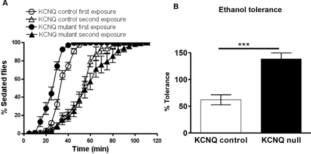
Animal models such asDrosophilacan be used to study specific aspects of human addiction, such as tolerance [12,13]. KCNQ loss of function resulted in an ethanol tolerance phenotype (Figure 4); whereas on first ethanol exposure the mutant is more sensitive than control, on second exposure the mutant becomes less sensitive (Figure 3B and 4A). This resulted in a larger shift in the ethanol behavioural response curve (Figure 4A), a measure of tolerance which was greater in the KCNQ mutant than in the control (Figure 4B). To ascertain whether these changes in ethanol behaviour resulted from alterations in KCNQ neural signalling (as opposed to purely metabolic changes), we measured the ethanol content of the flies (Figure S4C). No difference in ethanol metabolism was found between genotypes, with circulating levels of ethanol,25 mM at the end of the exposure that were reduced to 10 mM after 50 min. Furthermore, all genotypes chose equally to avoid 40% ethanol vapour in the T-maze (Figure S4D).
Drosophiladisplay age-dependent ethanol
hypersensitivity that is mimicked by KCNQ mutants KCNQ expression decreases in aged flies ([9], unpublished data), which would predict that old flies have would have increased ethanol sensitivity compared to young flies, in which KCNQ expression is higher. Therefore, we tested the effect of age on ethanol sensitivity and found that aged flies of a given genotype were more sensitive to ethanol than young flies (Figure 5), with the exception of theKCNQmutants and flies expressingKCNQ-RNAiin Elav or Ddc neurons (which were already ethanol hypersensitive as young flies).
Discussion
This study has characterised the electrophysiological properties of dKCNQ compared to mammalian neuronal KCNQ2/3 channels, showing that dKCNQ encodes a slowly activating and non-inactivating Kv current (Figure 2) similar to the M-current [10,11]. We went on to determine the effect of ethanol on KCNQ channels. Ethanol has a number of molecular targets; for instance,
,10 mM ethanol inhibits NMDA receptors and enhances GABAAreceptors [29,30], while,100 mM ethanol opens GIRK and BK K+
channels but closes Shaw K+
channels [6,30–32]. In this study, we show that theDrosophila KCNQ channel is more sensitive to ethanol than the mammalian channel, with an IC50of 19.8 mM compared to 42.1 mM for KCNQ2/3 (Figure 2E–F). This may be because flies have evolved to prefer and to live off fermenting fruit; therefore, in order to adapt to their environ-mental niche, there may have been selection for genes regulating ethanol sensitivity and behavioural response to ethanol [33,34]. The IC50 for ethanol on KCNQ2/3 is consistent with this heteromultimeric channel mediating the M-current that was also blocked by ethanol in vivo (20–120 mM; [6]). Furthermore, the KCNQ block seen here is likely to be physiologically relevant, as the blood alcohol content legally considered impairing is,20 mM [30]. Further investigation of the physiological consequence of this increased ethanol sensitivity of KCNQ in flies showed that ethanol caused sedation of wild-type flies with circulating ethanol levels of 25–30 mM (Figure S4C). This is comparable with intoxicating levels in humans and is sufficient to block the majority of KCNQ channels. KCNQ loss-of-function flies became sedated more quickly, demonstrating their increased ethanol sensitivity (Figure 3B).
Reducing KCNQ expression only in Ddc neurons caused hypersensitivity, while Ddc KCNQ overexpression resulted in resistance (Figure 3C). The KCNQ ethanol behaviour is consistent with our ethanol electrophysiology data, as exposure to compa-rable ethanol levels caused a reduction in KCNQ current. Loss-of-function alleles that further reduce the level of KCNQ in neurons made the fly even more sensitive to ethanol exposure. Conversely, neuronalKCNQoverexpression would be expected to cause a large hyperpolarisation. This might have the consequence that the depolarising effect of KCNQ ethanol blockade might have little effect, as the membrane potential would be sub-threshold for action potential firing or neurotransmitter release. ReducedKCNQ and the resultant depolarisation of Th neurons was sufficient to bring about this ethanol hypersensitivity, showing that dopami-nergic neurons mediate KCNQ’s effect on this behaviour (Figure 3C). We found that TRP activation and depolarisation of the Th neurons caused similar ethanol hypersensitivity (Figure 3D), confirming the importance of these neurons for this behaviour and suggesting that KCNQ-RNAiis likely to act via a common mechanism involving neuronal depolarisation. These results are consistent with dopamine being central for mediating the neural response to addictive drugs such as ethanol in human, mammalian and fly models [23–25,27,28,35,36]. They are also consistent with the M-current being specifically inhibited in dopamine neurons of the VTA, with ethanol likely to change the firing and release of the dopamine neurons [6]. Similarly, we found that changes in KCNQ levels in Ddc neurons resulted in a phenotype associated with impaired neural release (Figure S2F) glutamate transporter (vGlut) antibody stain (in magenta, A and D). Co-expression of vGlut and KCNQ is shown in white in the motor neuron cell bodies and neuropil in the ventral ganglion (F) and respective neuromuscular junctions (NMJs) (C) and particularly in the motor neuron cell body (white arrow head) measured in the GCaMP3 experiments. These motor neurons were labelled by thevGlut(OK371)-Gal4promoter [15] used in the GCaMP experiments. As thisGal4element is inserted in thevGlutlocus, a vGlut antibody stain (in magenta, A and D) can be used to show co-expression of a gene (such asdKCNQ) in theGal4-OK371expression pattern [15]. G–K. Spontaneous Ca2+signalling was imaged usingGal4-OK371,
uas-GCaMP3. Representative ratiometric images of larvae of control (Gal4-OK371,uas-GCaMP,+) (G.);Gal4-OK371,uas-GCaMP,uas-KCNQ-RNAi(H.); andGal4-OK371,uas-GCaMP,uas-KCNQ(I.) showing their maximum increase in fluorescent intensity above baseline, with the specific motor neuron soma used for measurements shown by the white arrowhead.J. Sample traces showing the time course of the bidirectional change in GCaMP3 signal with changes in neuronal KCNQ level.K. Histogram showing thatKCNQ-RNAi(dark grey bar in this and subsequent figures) expression in motor neurons increases (p,0.05) the amplitude of Ca2+influx, whereasKCNQoverexpression (light grey bar in this and later figures) decreases (p,0.01) the amplitude when each is compared with control (white bar in this and other figures, n.4). Data were analysed by 1-way ANOVA with a Bonferroni post-hoc test (n$8).
and that reduced KCNQ increased neural excitability and overexpression decreased excitabilityin vivo(Figure 1).
Longer-term changes in response to repeated exposure to drugs such as ethanol are required to bring about addiction [13,25]. These include functional changes in the nervous system, such as a decreased response to a given concentration of drug on repeated exposure, e.g., functional neuronal tolerance [12,25]. KCNQ
mutants also display increased ethanol tolerance compared to wildtype; therefore, functional KCNQ would decrease flies’ sensitivity to and tolerance of ethanol. All of these ethanol phenotypes seem to be caused by functional neural adaptive changes involving KCNQ, as no changes in ethanol pharmaco-kinetics were detected between genotypes. Other major regulators of neuronal excitability, such as GABAB receptors and BK Figure 2. KCNQ2/KCNQ3 and dKCNQ are acutely sensitive to ethanol inhibition.Representative traces recorded from HEK cells (A) showing that KCNQ2/3 current (black) was blocked by 10 mM ethanol (grey), as seen by the downward shift of the I–V relation (C) after ethanol application. The ethanol block was reversible (light grey traces in A). Representative traces (B) and I–V relation (D) showing acute reversible block of dKCNQ by ethanol.E.Plot showing the effect of increasing concentrations of ethanol on dKCNQ (grey) and KCNQ2/3 (black) currents. The maximum peak current amplitude evoked at 30 mV at the end of the 1350 ms pulse with or without ethanol was compared.F. Comparison of IC50s showing that dKCNQ is more sensitive (p,0.05) to ethanol inhibition than is KCNQ2/3 (n = 7). Data were analysed by Student’s unpairedt-test. For all figures: error bars are standard error of the mean and no asterisk means not significant; *p,0.05, **p,0.01 and ***p,0.001.
channels, also affect fly ethanol sensitivity and tolerance [12,37]. It is conceivable that these ethanol targets regulate the excitability of neural circuits in response to ethanol, thereby mediating part of the increase in ethanol sensitivity and tolerance seen in the KCNQ mutant. GABABreceptors couple to GIRK channels, decreasing neural excitability [38], and GABAB receptors usually promote ethanol sensitivity and tolerance in Drosophila [37], with ethanol also being able to directly bind and open GIRKs, decreasing excitability [39]. The role of BK in ethanol behaviour is more complex, with mammalian BK (Slo) channels being opened by ethanol and increasing tolerance by a number of mechanisms. In worm,slomutations decrease ethanol sensitivity, while in flies,slois transcriptionally induced by ethanol exposure, causing tolerance [12,32].
Interestingly, work from humans and mammalian models has shown that initial sensitivity to ethanol can predict future ethanol consumption and alcoholism, with the same set of genes thought to underlie both [40,41]. As KCNQ regulates both initial sensitivity to ethanol and subsequent development of tolerance, it is possible that KCNQ might be a candidate gene to predict susceptibility to alcoholism. This and recent work [42,43] suggest that KCNQ openers may offer a potential new approach to treatment of alcohol- and other drug-misuse disorders. Behavioural changes underlying addiction involve associative memory, with a role for dopamine signalling in mediating reinforcement in both flies and mammals [24,28,44].Th-Gal4expresses in dopamine neurons that innervate the mushroom body and mediate reinforcement in associative memory [28,45]. Our data are consistent with a recent Figure 3. KCNQ in dopaminergic neurons regulates ethanol sensitivity behaviour. A. Ethidium-bromide-stained agarose gel containing products of semi-quantitative PCR, showing removal ofKCNQin theKCNQmutant, reducedKCNQfrom flies expressingKCNQ-RNAiin all neurons (Elav-Gal4) and increasedKCNQfrom flies overexpressingKCNQin all neurons compared to controls (CSw-as wild-type andKCNQcontrol as the precise excision of theP-element).B.KCNQloss-of-functionP-element mutant flies (black bar in this and other figures, unless otherwise stated) or flies with pan-neural reduction inKCNQexpression (Elav-Gal4, uas-KCNQ-RNAi) exhibit a similar increase (p,0.01) in sensitivity to 40% ethanol compared with controls:CSw- wild-type (+), theKCNQ precise excision (KCNQ control),Elav-Gal4, +and uas-KCNQ-RNAi, +. Conversely pan-neuralKCNQ
overexpression (Elav-Gal4, uas-KCNQ) caused ethanol resistance (p,0.001). These changes in sensitivity were quantified as time to sedate 50% (T50%), the time taken for half of the flies of a given genotype to become sedated.C. Reduction ofKCNQin dopamine and serotonin neurons (Ddc-Gal4, uas-KCNQ-RNAi) increases sensitivity (p,0.01), while overexpression (Ddc-Gal4, uas-KCNQ) decreases sensitivity (p,0.05) with respect to controlDdc-Gal4,
+. ReducingKCNQin dopamine neurons (Th-Gal4, uas-KCNQ-RNAi) was sufficient to cause the increase (p,0.05) in ethanol sensitivity compared with control (Th-Gal4,+) while manipulatingKCNQlevels in serotonin (Tph-orTrh-Gal4) neurons alone had little effect (p.0.05). Data in B–C were analysed by 1-way ANOVA with a Bonferroni post-hoc test (n.4, 20 flies per n).D. The ethanol sensitivity ofTh-Gal4,+andTh-Gal4,uas-TRPA1flies was
measured at 23 or 30uC (n.6, 20 flies per n). Two-way ANOVA indicates significant differences in sensitivity due to interaction between temperature and genotype (p,0.0001). Post-hoc analysis showed that acute depolarisation by heat-activated (30uC) TRPA1 causes hypersensitivity (p,0.0001) when expressed in Th neurons, whereas the sensitivity ofTh-Gal4,uas-TRPA1flies at 23uC was similar to control animals at 23 or 30uC.
study showing that perturbing neurotransmission in Th neurons blocked a conditioned preference for ethanol, with pharmacolog-ical depletion of dopamine but not serotonin being sufficient to bring about this block [28]. KCNQ expression decreases in aged flies ([9], unpublished data) again correlating with the ethanol hypersensitivity phenotype (Figure 5).
Interestingly, optogenetic membrane depolarisation of dopa-mine neurons [46] recapitulated the initial increase in locomotor hyperactivity and subsequent long term sedation that is seen in Drosophila or mammals exposed to either long periods or dose-dependent increases in ethanol or cocaine [23,35]. Given the conserved role of mammalian KCNQ2/3 in the neurophysiolog-ical response to ethanol, it is likely that genetic or pharmacologneurophysiolog-ical disruption of KCNQ will result in similar ethanol behaviour phenotypes in mammals. We have, therefore, validated the use of
Drosophila to study KCNQ neuronal function and alcohol
behaviour. The fly’s compatibility with high-throughput analysis has the potential to allow both the identification of the underlying mechanism for this behaviour and screening for new therapies for alcohol related behavioural disorders and KCNQ diseases.
Supporting Information
Figure S1 KCNQ is widely expressed in the nervous system. A.Stage 17 wild-type embryos hybridised with KCNQ antisense probe showing the earliest expression ofKCNQ: that is, widespread throughout the nervous system, with little detectable expression elsewhere. B. No KCNQ expression was revealed in similarly aged KCNQ deletion mutant embryos stained with antisense probe. Wild-type embryos stained with the control sense probe (C.) andKCNQdeletion mutants hybridised with the sense probe (D.) showed little or no non-specific staining.
(PDF)
Figure S2 Changes in DDC neuron KCNQ levels leads to a wing expansion defect associated with impaired release A.Histogram showing that increasing or decreasing the level of KCNQ in Ddc neurons results in an increase (p,0.01) in the frequency of the wing expansion defect compared to control (Ddc-Gal4, CSw-). Data were analysed by 1-way ANOVA with a Bonferroni post-hoc test (n$8,,20 flies per n).
(PDF)
Figure 4. KCNQ channel levels regulate the development of ethanol tolerance. A. Flies were given two identical 90-min exposures to 40% ethanol vapour separated by a 2 h recovery; first (black circles) and second (black triangles) exposures of KCNQ mutants are separated by a greater rightward shift of the ethanol behavioural response curve (greater tolerance) than the shift between the controls’ first (white circles) and second exposures (white triangles).B. Histogram showing these changes in tolerance quantified using the following equation: T50%second exposure – T50% first exposure/T50%first exposure6100. KCNQ mutants (p,0.01) developed more tolerance than control (n = 10, 20 flies per n) as analysed by unpaired t-test.
doi:10.1371/journal.pone.0050279.g004
Figure 5. Ethanol sensitivity is similar between aged flies and
KCNQ loss-of-function mutants. The sensitivity of flies to the sedative effect of 40% ethanol was compared between young (5 days) and aged (30 days, black bars) flies of the same genotype (n$4,,100
flies per n). Two-way ANOVA indicates significant differences in memory due to interaction between age and genotype (p,0.0001). Post-hoc analysis showed thatCSw-,KCNQcontrol,Elav-Gal4/+,Ddc-Gal4/+, Ddc-Gal4/uas-KCNQ(p,0.05) andElav-Gal4/uas-KCNQ(p,0.001) flies were more sensitive to ethanol when old than young. TheKCNQmutant,
Elav-Gal4/uas-KCNQ-RNAi and Ddc-Gal4/uas-KCNQ-RNAi showed no effect of age, being equally hypersensitive to ethanol when young or old (p.0.05).
Figure S3 Ethanol does not cause a change in voltage-dependent activation of mammalian or Drosophila
KCNQ channels. The G–V relation for KCNQ2/KCNQ3 (A.) shows no apparent shift (p.0.05) by 10 mM ethanol (grey, V0.5=210.661.6 mV; slope factor = 14.261.5 mV), compared to control (black, V0.5=27.463.1 mV; slope fac-tor = 17.263.1 mV). B. The dKCNQ control current (black, V0.5=22.264.6 mV; slope factor = 20.562.9 mV) and the cur-rent in the presence of 10 mM ethanol (grey,V0.5= 11.561.0 mV; slope factor = 28.265.3 mV) show that the blocker does not cause a significant (p.0.05) shift in whole cell current activation. Activation relations were calculated from tail currents. Data were analysed by Student’s pairedt-test. n$4. For all figures: error bars are standard error of the mean and no asterisk means not significant; *p,0.05, **p,0.01 and ***p,0.001.
(PDF)
Figure S4 KCNQ signalling does not affect ethanol metabolism or avoidance. A. The sedation assay was performed in the absence of ethanol in order to control for any possible non-specific locomotor effect caused by heating flies. In the absence of ethanol, flies do not become sedated, so the per cent sedated or T50%sedated is not possible to calculate. Instead, the number of flies at the bottom of the bottle at a given time was counted.Th-Gal4,+(23uC, black square),Th-Gal4,+(30uC, white square),Th-Gal4, uas-TRPA1(23uC, black circle) andTh-Gal4, uas-TRPA1(30uC, white circle) were compared (n.9, 20 flies per n).
B.Heat activation of TRPA1 in Th neurons did not predispose flies to sedation or cause a non-specific locomotor deficit. The number ofTh-Gal4,+andTh-Gal4, uas-TRPA1flies at the bottom
of the bottle was counted at 23 or 30uC (n.9, 20 flies per n) and 2-way ANOVA indicates that genotype and temperature did not affect this number (p.0.05). C. The ethanol content of experimental and control (CSw- wild-type and KCNQ control) genotypes were quantified using an alcohol-dehydrogenase-based assay, in which the absorption levels of all genotypes were similar (p.0.05) at the end of the 90 min exposure to 40% ethanol vapour. Likewise, the rate of catabolism as reflected by the ethanol content after 50 min recovery from the exposure was the same between genotypes.D.Experimental and control (CSw-wild-type, KCNQ control and Elav-Gal4,+) flies similarly (p.0.05) avoided the arm of the T-maze containing 40% ethanol. All data were analysed by 1-way ANOVA with Bonferroni post-hoc test. (PDF)
Acknowledgments
We thank Dr Rolf Bodmer, Leslie Griffith and Ed Kravitz for flies. Additional stocks were from the Bloomington, Kyoto and Vienna stock centres. We are grateful to Dr Hermann Aberle for rabbit anti-Drosophila Glut antibody. Confocal microscopy was performed in the Bristol University Wolfson bioimaging facility. We acknowledge Dr Jon Brown, Jules Hancox, Leslie Griffith, Neil Marrion, and Ralf Stanewsky for providing valuable feedback on this manuscript.
Author Contributions
Conceived and designed the experiments: SC JMG JJLH. Performed the experiments: SC JMG JJLH. Analyzed the data: SC JMG JJLH. Contributed reagents/materials/analysis tools: SC JMG JJLH. Wrote the paper: SC JMG JJLH.
References
1. Wulff H, Castle NA, Pardo LA (2009) Voltage-gated potassium channels as therapeutic targets. Nat Rev Drug Disc 8: 982–1001.
2. Ashcroft FM (2006) From molecule to malady. Nature 440: 440–7. 3. Littleton JT, Ganetzky B (2000) Ion channels and synaptic organization: analysis
of theDrosophilagenome. Neuron 26: 35–43.
4. Unoki H, Takahasi A, Kawaguchi T, Hara K, Horikoshi M, et al. (2008) SNPs in KCNQ1 are associated with susceptibility to type 2 diabetes in East Asian and European populations. Nat Genet 40: 1098–102.
5. Yasuda K, Miyake K, Horikawa Y, Hara H, Osawa H, et al. (2008) Variants in KCNQ1are associated with susceptibility to type 2 diabetes mellitus. Nat Genet 40: 1092–1097.
6. Koyama S, Brodie MS, Appel SB (2007) Ethanol inhibition of M-current and ethanol induced direct excitation of Ventral Tegmental Area dopamine neurons. J Neurophysiol 97: 1977–85.
7. Peters HC, Hu H, Pongs O, Storm JF, Isbrandt D (2005) Conditional transgenic suppression of M channel in mouse brain reveals functions in neuronal excitability, resonance and behaviour. Nat Neurosci 8: 51–60.
8. Soldovieri MV, Miceli F, Taglialatatela M (2011) Driving with no brakes: molecular pathophysiology Kv7 potassium channels. Physiology 26: 365–76. 9. Ocorr K, Reeves NL, Wessells RJ, Fink M, Chen HS, et al (2007) KCNQ
potassium channel mutations cause cardiac arrhythmias inDrosophilathat mimic the effects of aging. Proc Natl Acad Sci USA 104: 3943–3948.
10. Wen H, Weiger TM, Ferguson TS, Shahidullah M, Scott SS, et al. (2005) A DrosophilaKCNQ channel essential for early embryonic development. J Neurosci 25: 10147–10156.
11. Cavaliere S, Hodge JJ (2011)DrosophilaKCNQ channel displays evolutionarily conserved electrophysiology and pharmacology with mammalian KCNQ channels. PLoSONE 6: e23898.
12. Atkinson NS (2009) Tolerance inDrosophila. J Neurogen 23: 293–302. 13. Kaun KR, Devineni AV, Heberlein U (2012)Drosophila melanogasteras a model to
study drug addiction. Hum Genet 131: 959–75.
14. Hodge JJ, Mullasseril P, Griffith LC (2006) Activity-dependent gating of CaMKII autonomous activity byDrosophilaCASK. Neuron 51: 327–337. 15. Mahr A, Aberle H (2006) The expression pattern of theDrosophilavesicular
glutamate transporter: a marker protein for motorneurons and glutamatergic centers in the brain. Gene Expr Patterns 6: 299–309.
16. Tian L, Hires SA, Mao T, Huber D, Chiappe ME, et al. (2009) Imaging neural activity in worms, flies and mice with improved GCaMP calcium indicators. Nat Methods 6: 875–881.
17. Cheng LE, Song W, Looger LL, Jan LY, Jan YN (2010) The role of the TRP channel NompC inDrosophilalarval and adult locomotion. Neuron 67: 373–380.
18. Wen T, Parrish CA, Xu D, Wu Q, Shen P (2005)Drosophilaneuropeptide F and its receptor, NPFR1, define a signaling pathway that acutely modulates alcohol sensitivity. Proc Natl Acad Sci USA 102: 2141–2146.
19. Pulver S, Pashkovski SL, Hornstein NJ, Garrity PA, Griffith LC (2009) Temporal dynamics of neuronal activation by channelrhodopsin-2 and TRPA1 determine behavioural output inDrosophilalarvae. J Neurophysiol 101: 3075–88. 20. Hodge JJ, Choi JC, O’Kane CJ, Griffith LC (2005)Shawpotassium channel
genes inDrosophila. J Neurobiol 63: 235–254.
21. Peabody NC, Pohl JB, Diao F, Vreede AP, Sandstrom DJ, et al. (2009) Characterization of the decision network for wing expansion inDrosophilausing targeted expression of the TRPM8 channel. J Neurosci 28: 14379–14391. 22. Hodge JJ (2009) Ion channels to inactivate neurons inDrosophila. Front Mol
Neurosci 2:13.
23. Nicola SM, Surmeier J, Malenka RC (2000) Dopaminergic modulation of neuronal excitability in the striatum and nucleus accumbens. Annu Rev Neurosci 23: 185–215.
24. Spanagel R (2009) Alcoholism: A systems approach from molecular physiology to addictive behaviour. Physiol Rev 89: 649–705.
25. Sulzer D (2011) How addictive drugs disrupt presynaptic dopamine neurotrans-mission. Neuron 69: 628–649.
26. Alekseyenko OV, Lee C, Kravitz EA (2010) Targeted manipulation of serotonergic neurotransmission affects the escalation of aggression in adult male Drosophila melanogaster. PLoS ONE 5: e10806.
27. Kong EC, Woo K, Li H, Lebestky T, Mayer N, et al. (2010) A pair of dopamine neurons target the D1-like dopamine receptor DopR in the central complex to promote ethanol stimulated locomotion inDrosophila. PLoS ONE 5: e9954. 28. Kaun KR, Azanchi R, Maung Z, Hirsh J, Heberlein U (2011) ADrosophilamodel
of alcohol reward. Nat Neuro 14: 612–621.
29. Koob GF (2004) A role for GABA mechanisms in the motivational effects of alcohol. Biochem Pharmacol 68: 1515–25.
30. Harris RA, Trudell JR, Mihic SJ (2008) Ethanol’s molecular targets. Sci Signal 1: re7.
31. Covarrubias M, Rubin E (1993) Ethanol selectively blocks a non-inactivating K+ current expressed inXenopusoocytes. Proc Natl Acad Sci USA 90: 19408–19416. 32. Treistman SN, Martin GE (2009) BK channels: mediators and models for
alcohol tolerance. TINS 32: 629–637.
33. Morozova TV, Anholt RRH, Mackay TFC (2007) Phenotypic and transcrip-tional response to selection for alcohol sensitivity in Drosophila melanogaster. Genome Biology 8: R231.
35. Bainton RJ, Tsai LT, Singh CM, Moore MS, Neckameyer WS, et al. (2000) Dopamine modulates acute responses to cocaine, nicotine and ethanol in Drosophila. Curr Biol 10: 187–194.
36. Li H, Chaney S, Forte M, Hirsh J (2000) Ectopic G-protein expression in dopamine and serotonin blocks cocaine sensitization inDrosophila melanogaster. Curr Biol 10: 211–214.
37. Dzitoyeva S, Dimitrijevic N, Manev H (2003)c-Aminobutyric acid B receptor 1 mediates behaviour-impairing actions of alcohol in Drosophila: adult RNA interference and pharmacological evidence. Proc Natl Acad Sci USA 100: 5485– 90.
38. Mezler M, Muller T, Raming K (2001) Cloning and functional expression of GABABreceptors fromDrosophila. Eur J Neurosci 13: 477–486.
39. Lewohl JM, Wilson WR, Mayfield RD, Brozowski SJ, Morrisett RA, et al (1999) G-protein-coupled inwardly rectifying K+
channels. Nat Neurosci 2: 1084–90. 40. Hodge CW, Mehmert KK, Kelley SP, McMahon T, Haywood A, et al. (1999)
Supersensitivity to allosteric GABA(A) receptor modulators and alcohol in mice lacking PKCepsilon. Nat Neurosci 2: 997–1002.
41. Mayfield RD, Harris RA, Schuckit MA (2008) Genetic factors influencing alcohol dependence. Br J Pharmacol 154: 275–87.
42. Hansen HH, Andreasen JT, Weikop P, Mirza N, Scheel-Kru¨ger J, et al. (2007) The neuronal KCNQ channel opener retigabine inhibits locomotor activity and reduces forebrain excitatory responses to the psychostimulants cocaine, methylphenidate and phencyclidine. Eur J Pharm 570: 77–88.
43. Kapfhamer D, Berger KH, Hopf FW, Seif T, Kharazia V, et al. (2010) Protein phosphatase 2a and glycogen synthase kinase 3 signaling modulate prepulse inhibition of the acoustic startle response by altering cortical M-type potassium channel activity. J Neurosci 30: 8830–8840.
44. Hyman SE, Malenka RC, Nestler EJ (2006) Neural mechanisms of addiction: the role of reward-related learning and memory. Annu Rev Neurosci 29: 565– 598.
45. Keene AC, Waddell S (2007) Drosophila olfactory memory: single genes to complex neural circuits. Nature Rev Neurosci 8: 341–354.