O R I G I N A L R E S E A R C H
Open Access
Tomographic index as auxiliary criteria for surgery
indication in fracture dislocation of acetabulum
posterior wall
Edison N Fujiki
1,4*, Eduardo N Yamaguchi
1, Edison Miachiro
1, Takechi Chikude
1, Roberto Y Ikemoto
1,
Luiz Carlos de Abreu
2, Vitor E Valenti
2,3, Luciano M R Rodrigues
1, Carlos B Monteiro
3and Carlo Milani
1Abstract
There are situations which the tomographic exam is done on the affected hip or situations where the contralateral hip presents abnormalities that make it impossible to compare. In this study we aimed to evaluate a tomographic index that does not require comparison between the both hips. Twenty two patients with unilateral acetabular fracture dislocation with fracture of posterior wall were studied. We established the relationship between the remaining posterior wall and the femoral head diameter (head/wall index-H/W index). We evaluated 45 two-dimensional computed tomography scan in normal hips and established the H/W index. In 45 normal hips we simulated a posterior wall fracture with involvement of 25% and 30% of the posterior wall and calculated the H/W index. We divided into five groups with five different H/W index (fractured group with non surgical treatment; fractured group; normal group; normal group with simulated fracture of 25% and; 30% of the posterior wall). 2.4 was the lowest limit of confidence interval of the group with 25% of the posterior wall fracture. When we analyzed the confidence interval of the 30% fracture group the upper limit of the confidence interval was 2.7, close to the lower limit of the surgical group that was 2.9. Thus, we suggest the 2.4 the H/W index limit as an auxiliary criteria to indicate whether or not to operate. H/W index is helpful to decide whether or not surgery indication in the fracture dislocation of the posterior wall of the acetabulum.
Background
Bone fractures may lead to many impaired functions [1-3]. Acetabular posterior wall fracture is the most common type of acetabular fracture comprising around 18% to 33% of all cases [4-7]. It is becoming relatively frequent in orthopedists’ daily routine because trauma-tisms are increasing. Posterior wall fractures were classi-fied by Judet-Letournel as one of the elementary types of acetabular fracture [4]. It appears to be simple on radiog-raphy; however, even after reduction it may present sig-nificant percentage of necrosis or it develops to hip arthrosis [6,8].
In our country, usually the first aid of these cases is done by orthopedists [7], who are often worried to solve
emergency cases. Clinical examination to test the articu-lar stability after reduction is the“gold” standard [9-15], however, in some cases these maneuvers are inconclu-sive or forgetfulness. Since the emergency was solved, the patient is evaluated by a hip surgeon specialist with the help of three standard plain radiographs (one antero-posterior and two Judet 45 degrees oblique pelvic radio-graphs) and a two-dimensional computed tomography (CT) scan. Nevertheless, if the fragment size is misesti-mate or it is difficult to evaluate even with a CT scan, and if there is no information regarding the hip stability criteria after reduction, certainly the decision to indicate or not indicate surgery will be difficult.
In the literature there are tomographic indexes that try to predict hip joint stability [9,16]; but all of these meth-ods compare the fractured hip with the contralateral normal hip images. On the other hand, in medical prac-tice, there are situations which the tomographic exam is done only on the affected hip, or even situations where the contralateral hip presents abnormalities that make it * Correspondence:[email protected]
1
Departamento de Cirurgia Ortopédica, Faculdade de Medicina do ABC, Santo André, SP, Brazil
4Disciplina de Cirurgia Ortopédica, Faculdade de Medicina do ABC, Av. Príncipe de Gales, 821., 09060-650 Santo André, SP, Brazil
Full list of author information is available at the end of the article
impossible to compare. Therefore, this investigation was undertaken to demonstrate the relationship between the tomographic index and instability.
Methods
A retrospective study was done in 22 patients aged between 21 to 45 years old with unilateral posterior frac-ture dislocation of the acetabulum. The inclusion criteria were: joint instability, fractured fragment size, femoral head fracture, bone fragment interposition, residual sub-luxation and wall fracture impact displaced more than 2 mm. All patients were evaluated by a senior orthopedic hip surgeon, who analyzed the clinical history, three standard plain radiographs (one anteroposterior and two Judet 45 oblique pelvic radiographs), two-dimensional computed tomography scan and joint instability under anesthesia. All experimental protocols were approved by the ethics committee in research of our Institute.
After clinical examination and images evaluation it was decided whether or not to operate. Fourteen (63.64%) patients were operated and eight (36.36%) were not operated. Twelve of the 14 operated patients showed instability in the dynamic fluoroscopy examination under anesthesia (patient placed at supine position with neutral rotation and full extension of the hip; then the hip was flexed until 90 and adduction of 20 degrees). If the joint remained congruent in the anteroposterior and oblique in fluoroscopy projection it was considered stable. The pressure used on the maneuver was not con-sidered due to the difficulty to evaluate it. The remaining two patients in spite of instability were operated because they presented large fractured size fragment on the pos-terior wall after image evaluation. The eight patients who were not operated presented stability in the exam-ination under anesthesia and were considered by the senior surgeons as small size fractured fragment of
posterior wall. Using CT (computed tomography) scan images we calculated the head wall index (H/W). The H/W index was calculated in 22 fractured hips and in 45 hips (26 patients were considered normal), we used 45 CT scans from hip images - from 26 patients - aged between 18 to 50 years old, which were considered nor-mal (without fracture, osteoarthritis, dysplasia or other hip deformity). All images were obtained using a General Electric CT (two-dimensional computed tomography scan with 5 mm slice thickness).
In the control group we calculated the H/W index in the normal hip after we divided the posterior wall in per-centages and the 25% and 30% of the distal posterior wall, respectively.
The tomographic images were digitized using a 7.2 mega pixels camera and transferred to the computer, studied and measured. We calculated the H/W index using the M2000 software for measuring angles and dis-tances [17-20]. Two senior surgeons calculated the acet-abular index for each image in all groups studied and average was calculated for each case.
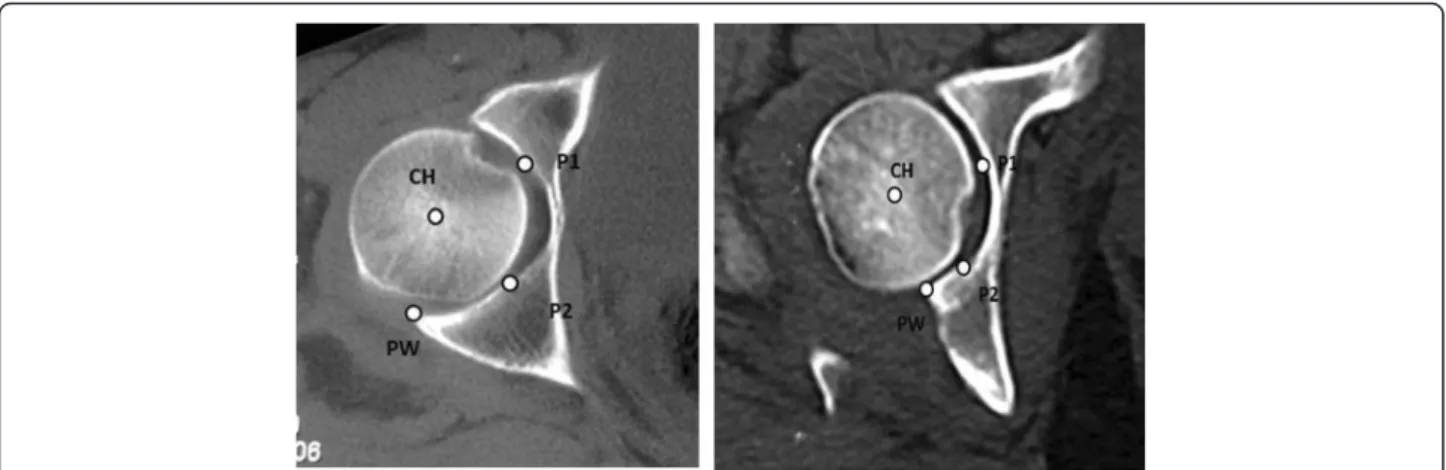
From the axial CT slices through the acetabulum, in both normal and fractured hips, we had chosen the image that showed the largest anteroposterior femoral head diameter. We defined 4 points at this tomographic slice: point 1 (P1) corresponds to the anterior transition between the articular surface and the acetabular fossa; point 2 (P2) corresponds to the posterior transition between the articular surface and the acetabular fossa; the third point (PW) corresponds to the posterior articu-lar surface edge, both in normal hips as in the remaining fractured wall; the fourth point (CH) corresponds to the femoral head center (Figure 1).
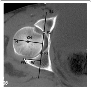
A straight baseline (BL) was drawn throw P1 and P2 points, and 2 other lines were drawn perpendicular to the BL: W line, from the PW to BL; and H line, from
Figure 1In a non fractured acetabulum, four points were defined: P1!anterior transition between the articular surface and the
acetabular fossa.P2!posterior transition between the articular surface and the acetabular fossa. PW!posterior articular surface edge.
the lateral edge of the femoral head, passing through CH to BL (Figure 2).
The relationship between H and W lines (H/W index) was studied in normal hips and fractured cases. In frac-tured wall, the W line begins just in the fracfrac-tured articu-lar surface. After calculation of the H/W index in normal CT scan group, we divided the W line in percen-tages; we took the 25% distal of the wall and calculated the H/W index in the remaining 75% of the posterior wall, simulating a 25% (Figure 3A) of the posterior wall fracture. The same procedure was done taking 30%
(Figure 3B) of distal W line and it was calculated the H/W index in the remaining 70%.
The tomographic acetabular index (H/W) of normal
CT scan group was considered the normal group. The
H/W index from the images after removing 25% and 30% of the posterior wall was namedminus 25%(−25%) and minus 30% (−30%) groups, respectively. We called
the group which underwent surgery thesurgical fracture group while the group which the patients were not oper-ated was namedconservative fracture group.
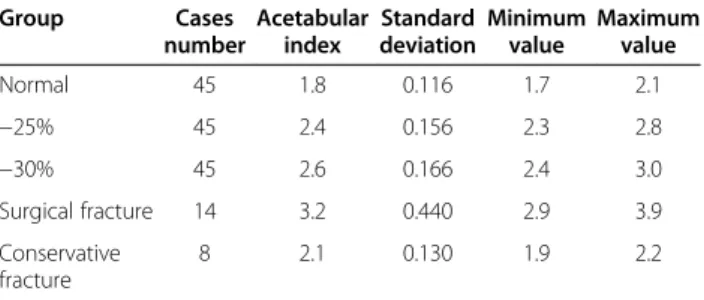
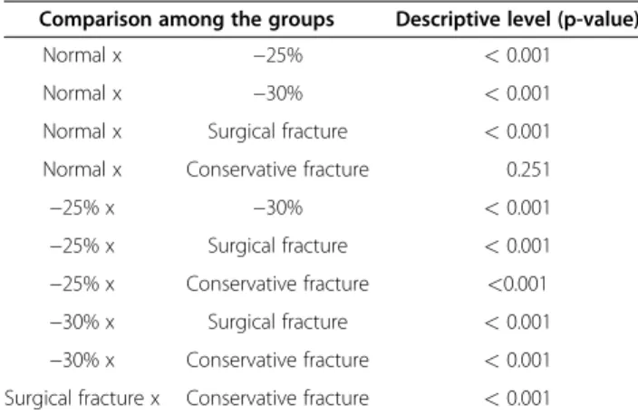
For all groups (fractured not operated group; fractured group, normal group, normal group with simulated frac-ture of 25% and 30% of the posterior wall) tomographic acetabular index and standard deviation were calculated (Table 1 and Figure 4). The 95% confidence interval for each group was calculated (Table 2). The means of the indexes of all groups were compared by applying two-way ANOVA test followed by the post hoc Tukey mul-tiple comparisons test. Differences were considered sig-nificant when the probability of a type I error was lower than 5% (p<0.05).
Results
The H/W index presented a mean of 1.83 in the normal group while the conservative fracture group presented 2.08. The mean was 3.19 in the surgical fracture group and in the −25% and −30% groups the mean were 2.44 and 2.62, respectively. There were significant differences of mean H/W index among all groups, except in normal group vs. conservative fracture group (Table 3). When we analyzed the confidence interval of the H/W index we found that the upper limit of the conservative group was similar to the lower limit in the −25% group (H/W index: 2.4). The upper limit of the confidence interval of the −25% group was near the lower limit of the −30% group (H/W index: 2.49 and 2.57, respectively). Finally, Figure 2A straight base line (BL) was drawn throw the P1 and
P2 points, and 2 other lines were drawn perpendicular to the BL line: Line W, from the PW point to BL line; and another line H, from the lateral edge of the femoral head, passing through CH point to BL line.
the lower limit of the confidence interval of the surgical fracture group was close to the upper limit for the−30% group (Figure 5).
Discussion
Evaluations of hip instability in cases of fracture disloca-tion of the posterior wall are usually made by clinical examination after reduction under anesthesia, with hip flexion of 90° and slight adduction to test the stability in association to radiographic and tomographic analysis [16,21,22]. On the other hand, clinical evaluation some-times is inconclusive, because in some cases it is asso-ciated with lesions, thereby losing this important parameter. Nonetheless, radiographic images in antero-posterior and oblique views may falsify the images of fragment sizes, depending on how they are situated in relation to the x-rays. Even tomographic images, which are more precise, may give rise to doubts regarding the sizes of fracture fragments, since the parameter of the size may be subjective, depending on each personal’s experience [16,21,22].
The method proposed by Calkins et al. [16] and Keith et al. [23] for tomographic measurements of instability
classifies it into three groups: stable, indeterminate and unstable. Other study performed on cadavers [24] reported that osteotomy of the posterior wall lower than 25% did not affect the joint stability, while osteotomy higher than 50% of the posterior wall presented signifi-cant effect on joint stability. In our opinion the indeter-minate group from 20–25% to 40–50% is the problem to make decision. Recently, Moed et al. [9] described a modi-fied method, (alternative method) to calculate the instabil-ity and demonstrated that the new alternative method is more accurate than other methods used in the literature. However, all of these measurements may be impaired if the contralateral hip presents abnormalities such as ante-version, hip dysplasia, fractures or if the tomographic image was not digitized. Conversely, the H/W index takes into account the relative variation between the femoral head and the posterior wall in the affected hip, which is the real location of the problem and depends on the pro-portion head-wall where the instability was occurred. Moreover, the method proposed in the literature makes measurements comparing different hips and consequently the mistake rate may increase [21,22].
Olson et al. [21] reported that when the posterior wall was decreased by one third of size the remainder of the
Table 1 Means and standard deviations of the indexes observed of each group
Group Cases
number
Acetabular index
Standard deviation
Minimum value
Maximum value
Normal 45 1.8 0.116 1.7 2.1
−25% 45 2.4 0.156 2.3 2.8
−30% 45 2.6 0.166 2.4 3.0
Surgical fracture 14 3.2 0.440 2.9 3.9
Conservative fracture
8 2.1 0.130 1.9 2.2
Figure 4Means and standard deviations of the acetabular indexes observed in each group.
Table 2 Confidence intervals (95%) of the mean acetabular index in each group
Group Confidence interval (95%)
Lower limit Upper limit
Normal 1.8 1.9
−25% 2.4 2.5
−30% 2.6 2.7
Surgical fracture 2.9 3.8
acetabulum was significantly overloaded when supported on one foot. In other words, even if the posterior wall loss did not cause joint instability it may be significant overloaded by the joint and it consequently cause early arthrosis. In our study there were two cases which the patients were operated without instability but the frag-ment was considered large by the senior surgeon.
We established a limit index of 2.4 for surgery or not surgery indication, stability or instability parameter, because the mean H/W index in the normal group and conservative group was similar. We did not observe stat-istical difference between these groups, however, when we analyzed the confidence interval, the upper limit of the conservative group was similar to the lower limit of the −25% group (2.4 H/W index) and there was statis-tical difference between conservative group and −25% group regarding the mean index. If surgery was not per-formed in subjects from 2.4 H/W index it was possible to mislead any case from this group. The end point of
the upper limit of conservative group in our study was 2.4 H/W index, for this reason we believe that 2.4 H/W is a safe limit to indicate surgical treatment. When we analyzed the confidence interval of the−30% group the upper limit (2.7) was near to the lower limit of the sur-gical group (2.9). We believe that 2.4 is an index that form a shield from over-indication or sub-indication to repair posterior wall fracture dislocation of the acetabu-lum, and concerning to the group called indeterminate [5] this 2.4 index contemplate it. If we accepted an index of 2.5 we would be going outside of the confidence interval for conservative treatment and increasing the risk to not operate in cases that really required surgery. Furthermore, an index of 2.5 would fall within the inter-val corresponding to remointer-val of 30% of the posterior wall. There was significant difference between removing 25% and 30% of the posterior wall and, therefore, this index cannot be 2.5. The index also cannot be lower than 2.4 because we would institute surgical treat-ment for cases that should be conservatively treated. Nevertheless, these findings are based on small conser-vative group (eight hips) and all cases (twenty two hips) presented fracture dislocation and the conservative treatment was previously determined by the senior surgeon; unfortunately, these findings may be a bias in our study.
The concept that loss of one third of the posterior wall may not be an instability factor but might give rise to future joint overload [17] is an important issue that rein-forces the choice of an index of 2.4. Our findings, which presented significant difference between−25% and−30% groups are also important. On the other hand, bad results not only depend on the joint overload, but depend on the many others circumstance such as: frac-ture types, vascular femoral head injury, acetabular wall
Table 3 Descriptive levels of two way comparisons of the indices among five groups
Comparison among the groups Descriptive level (p-value)
Normal x −25% <0.001
Normal x −30% <0.001
Normal x Surgical fracture <0.001
Normal x Conservative fracture 0.251
−25% x −30% <0.001
−25% x Surgical fracture <0.001
−25% x Conservative fracture <0.001
−30% x Surgical fracture <0.001
−30% x Conservative fracture <0.001
Surgical fracture x Conservative fracture <0.001
impact, femoral head impact, residual instability, etc. [16,21,22].
We believe that clinical criteria under anesthesia (flexion of 90 and slight adduction after performing reduction) are the gold standard [9]; however, in some cases our H/W index may be an important tool to help the surgeon to indicate or not indicate surgery. Another advantage of this method is that it is possible to make the H/W index direct from the CT scan, because the relationship between femoral head and acetabular pos-terior wall measurement is proportional and this ratio may be done in millimeters or centimeters.
Our study presents some points that should be addressed: our main issue is a possibly remaining instability of the hip joint, however, patients with fem-oral head fracture, intra-articular fragments and mar-ginal impactions were also included. In these patients the indication for operation is given even in case of a stable joint. The two patients operated were indicated to surgery prior to the method because the size of the frag-ments was considered large by the surgeon. They were submitted to the index and proved to be within the pro-posed limits. We did not perform a prospective study and we did not perform correlation between the tomo-graphic index calculated and hip instability. Further studies are worth to investigate this issue. Furthermore, it is important to make a clinical evaluation of the patients and consider overall the pathologic changes that occur with the fractures in order to indicate or not indi-cate surgery.
Our study presents a tool which is able to assist the analysis of images, especially with clinical examination, in order to provide an indication of operation, it should not be considered as a single factor to be evaluated. The advantage of this index is that it is only performed on one hip, the affected hip, unlike other different methods [9-15]. We suggest future studies to apply the new index in prospective studied. As a first study its validation could be evaluated among others patients. The validated index could also be applied in prospective and transver-sal studies.
Conclusion
We indicated a tomographic acetabular index of 2.4 for the relationship of the head with the fractured posterior wall. This index is useful to assess the presence of unstable posterior wall fracture of the acetabulum.
Competing interests
The authors declare that they have no competing interest.
Authors’contributions
All authors participated in the acquisition of data and revision of the manuscript. ENF, ENY, EM, TC, RYI, LMRR, CBM and CM conceived of the study, determined the design, performed the statistical analysis, interpreted the data and drafted the manuscript. VEV and LCA determined the design
and drafted the manuscript. All authors read and gave final approval for the version submitted for publication.
Acknowledgements
This study received financial support from Núcleo de Estudos, Pesquisas e Assessoria à Saúde da Faculdade de Medicina do ABC (NEPAS-FMABC) and FAPESP.
Author details
1
Departamento de Cirurgia Ortopédica, Faculdade de Medicina do ABC, Santo André, SP, Brazil.2Laboratório de Escrita Científica, Departamento de Morfologia e Fisiologia, Faculdade de Medicina do ABC, Santo André, SP, Brazil.3Departamento de Fonoaudiologia, Faculdade de Filosofia e Ciências, Universidade Estadual Paulista, Marília, SP, Brazil.4Disciplina de Cirurgia Ortopédica, Faculdade de Medicina do ABC, Av. Príncipe de Gales, 821., 09060-650 Santo André, SP, Brazil.
Received: 2 January 2012 Accepted: 20 June 2012 Published: 20 June 2012
References
1. Sayed D, Abd Elwanis ME, Abd Elhameed SY, Galal H:Does occupational exposure to low-dose ionizing radiation affect bone marrow thrombopoiesis?Int Arch Med2011,4:8.
2. Ichim TE, Solano F, Lara F, Paris E, Ugalde F, Rodriguez JP, Minev B, Bogin V, Ramos F, Woods EJ, Murphy MP, Patel AN, Harman RJ, Riordan NH:
Feasibility of combination allogeneic stem cell therapy for spinal cord injury: a case report.Int Arch Med2010,3:30.
3. Alvarez P, Navascués CA, Ordieres C, Pipa M, Vega IF, Granero P, Alvarez JA, Rodríguez M:Granulocytic sarcoma of the small bowel, greater omentum and peritoneum associated with a CBFβ/MYH11 fusion and inv(16) (p13q22): a case report.Int Arch Med2011,4:3.
4. Judet R, Judet J, Letournel E:Fractures of the acetabulum: classification and surgical approaches for open reduction. Preliminary report.J Bone Joint Surg1964,46:1615–1646.
5. Matta JM, Anderson LM, Epstein HC, Hendricks P:Fractures of the acetabulum. A retrospective analysis.Clin Orthop1986,205:230–240. 6. Alonso JE, Volgas DA, Giordano V, Stannard JP:A Review of the treatment
of hip dislocations associated with acetabular fractures.Clin Orthop2000,
1:32–43.
7. Guimarães JM:Fratura do acetábulo–Fratura do rebordo posterior com
impacção articular.Rev Ortop Traumat2004,8:6–7.
8. Moed BR, WillsonCarr SE, Gruson KI, Watson JT, Craig JG:Computed Tomographic Assessment of Fractures of The Posterior Wall of The Acetabulum After Operative Treatment.J Bone Joint Surg Am2003,
85:512–522.
9. Moed BR, Ajibade DA, David A, Israel H:Computed tomography as a predictor of hip stability status in poterior wall fractures of the Acetabulum.J Orthop Trauma2009,23:7–15.
10. Machotka Z, Scarborough I, Duncan W, Kumar S, Perraton L:Anterior Cruciate Ligament repair with LARS (Ligament Advanced Reinforcement System): a systematic review.Sports Med Arthrosc Rehabil Ther Technol
2010,2:29.
11. Yamamoto Y, Ishibashi Y, Tsuda E, Tsukada H, Maeda S, Toh S:Comparison between clinical grading and navigation data of knee laxity in ACL-deficient knees.Sports Med Arthrosc Rehabil Ther Technol2010,2:27. 12. Haviv B, O'Donnell J:The incidence of total hip arthroplasty after hip
arthroscopy in osteoarthritic patients.Sports Med Arthrosc Rehabil Ther Technol2010,2:18.
13. Lam MH, Fong DT, Yung PSh, Ho EP, Chan WY, Chan KM:Knee stability assessment on anterior cruciate ligament injury: Clinical and
biomechanical approaches.Sports Med Arthrosc Rehabil Ther Technol2009,
1:20.
14. Ageberg E, Flenhagen J, Ljung J:Test-retest reliability of knee kinesthesia in healthy adults.BMC Musculoskelet Disord2007,8:57.
15. Sharma H, Ayer R, Taylor GR:Complex pediatric elbow injury: an uncommon case.BMC Musculoskelet Disord2005,6:13.
18. Hirschmann MT, Adler T, Rasch H, Hügli RW, Friederich NF, Arnold MP:
Painful knee joint after ACL reconstruction using biodegradable interference screws- SPECT/CT a valuable diagnostic tool? A case report. Sports Med Arthrosc Rehabil Ther Technol2010,2:24.
19. Chen SH, Tai CL, Lin CY, Hsieh PH, Chen WP:Biomechanical comparison of a new stand-alone anterior lumbar interbody fusion cage with established fixation techniques - a three-dimensional finite element analysis.BMC Musculoskelet Disord2008,9:88.
20. Sniekers YH, Intema F, Lafeber FP, van Osch GJ, van Leeuwen JP, Weinans H, Mastbergen SC:A role for subchondral bone changes in the process of osteoarthritis; a micro-CT study of two canine models.BMC Musculoskelet Disord2008,9:20.
21. Olson SA, Bay BK, Pollak AN:The effect of variable size posterior wall acetabular fractures on contact characteristics of the hip joint.J Orthop Trauma1996,10:02–395.
22. Brooks RA, Ribbans WJ:Diagnosis and imaging studies of traumatic hip dislocations in the adult.Clin Orthop2000,1:15–23.
23. Keith JE, Brashear HR, Guilford WB:Stability of posterior fracture-dislocations of the hip. Quantitative assessment using computed tomography.J Bone Joint Surg Am1988,70:711–714.
24. Vailas JC, Hurwitz S, Wiesel SW:Posterior acetabular fracture-dislocations: fragment size, joint capsule, and stability.J Trauma1989,291:494–496.
doi:10.1186/1755-7682-5-18
Cite this article as:Fujikiet al.:Tomographic index as auxiliary criteria for surgery indication in fracture dislocation of acetabulum posterior wall.International Archives of Medicine20125:18.
Submit your next manuscript to BioMed Central and take full advantage of:
• Convenient online submission
• Thorough peer review
• No space constraints or color figure charges
• Immediate publication on acceptance
• Inclusion in PubMed, CAS, Scopus and Google Scholar
• Research which is freely available for redistribution