online | memorias.ioc.fiocruz.br
Absence of Fas-L aggravates renal injury in acute
Trypanosoma cruzi infection
Gabriel Melo de Oliveira1/+, Masako Oya Masuda2, Nazaré N Rocha3, Nestor Schor4, Cléber S Hooper5, Tânia C de Araújo-Jorge1, Andréa Henriques-Pons1
5Departamento de Controle de Qualidade Animal, Centro de Criação de Animais de Laboratório 1Laboratório de Biologia Celular,
Instituto Oswaldo Cruz-Fiocruz, Av. Brasil 4365, 21045-900, Rio de Janeiro, RJ, Brasil 2Instituto de Biofísica Carlos Chagas Filho,
Univer-sidade Federal do Rio de Janeiro, Rio de Janeiro, RJ, Brasil 3Departamento de Fisiologia e Farmacologia, Instituto Biomédico,
Universi-dade Federal Fluminense, Rio de Janeiro, RJ, Brasil 4Disciplina de Nefrologia, Escola Paulista de Medicina, Universidade Federal
de São Paulo, São Paulo, SP, Brasil
Trypanosoma cruzi infection induces diverse alterations in immunocompetent cells and organs, myocarditis and congestive heart failure. However, the physiological network of disturbances imposed by the infection has not been addressed thoroughly. Regarding myocarditis induced by the infection, we observed in our previous work that Fas-L-/- mice (gld/gld) have very mild inflammatory infiltration when compared to BALB/c mice. However, all mice
from both lineages die in the early acute phase. Therefore, in this work we studied the physiological connection re-lating arterial pressure, renal function/damage and cardiac insufficiency as causes of death. Our results show that a broader set of dysfunctions that could be classified as a cardio/anaemic/renal syndrome is more likely responsible for cardiac failure and death in both lineages. However,gld/gld mice had very early glomerular deposition of IgM and a more intense renal inflammatory response with reduced renal filtration, which is probably responsible for the premature death in the absence of significant myocarditis in gld/gld.
Key words: Trypanosoma cruzi - Fas-L - myocarditis - acute kidney injury
Financial support: Fiocruz, CNPq
+Corresponding author: gmoliveira@ioc.fiocruz.br Received 11 March 2009
Accepted 13 October 2009
Chagas disease is caused by theprotozoan parasite
Trypanosoma cruzi, which has a widespread distribu-tionin Latin America. It is estimated that 15-16 million people are infected on this continent and 75-90 million are exposed to infection (Coura & Dias 2009). Trans-mission to humans occurs primarily through blood-suckingreduviid bugs, but it may also occur through blood transfusion organ transplant, transplacental trans-mission and oral infection (Moncayo 2003, Dias et al. 2008, WHO 2004, Yoshida 2009). The disease is char-acterised by an initial acute phase and it is generally ac-cepted that patients with a more severe acute infection may develop a more aggressive chronic phase(Higushi et al. 2003, Coura 2007). Acute diffuse myocarditis is associated with parasite nests and cellular inflamma-tory foci that are composed of macrophages (Andrade 1991), CD4+ and mainly CD8+ T cells (Henriques-Pons et al.2002), differentiated as activated/memory T cells (CD62LLow, LFA-1High and VLA-4High) (dos Santos et al. 2001).The chronic phase starts with a usually long-last-ing indeterminate period, followed by a symptomatic dilated myocardiopathy in 25-30% of patients. The cy-totoxic pathway(s) that destroy cardiomyocytes and the precise correlation between inflammatory response and
heart failure are not known. It is important to evaluate which mechanisms regulate cardiac inflammatory pro-cesses and their interplay with possible causes of death from this disease. In our previous work, we observed that Fas-L-/- mice (gld/gld) have a very modest cardi-ac inflammatory response when compared to infected BALB/c. Accordingly, we observed less cardiomyo-cyte death in gld/gld, yet high mortality rates caused by unknown reasons (de Oliveira et al.2007).
Fas-L, a type II membrane homotrimeric protein, be-longs to the tumour necrosis factor family and triggers apoptosis through Fas engagement (Suda et al. 1995). However, Fas activation is also involved in the secretion of cytokines and chemokines, chemotaxis, genomic tran-scription, cellular activation and other responses (Lam-bert et al. 2003). Concerning the infection, it has been shown that Fas/Fas-L controls CD4+ T cell population through AICD (activation induced cell death) (Lopes et al.1999), parasite replication in vitro(Freire de Lima et al. 2000), NO production (Martins et al. 2001) and host cytokine response to T. cruzi infection in vivo, prevent-ing an exacerbated Th2-biased immune response (Lopes et al.1999, Guilhermo et al.2007). In other pathologies, such as Coxsackievirus B3, Fas/Fas-L plays a central role in the control of myocarditis, leading cardiac CD4+Th2 (IL-4+) protective cells to death through the cytotoxic
illness, this ischemic injury promotes intense necro-sis of many cell types (Bohle et al.1996, Schelling & Cleveland 1999). However, it is well known that human patients with chronic renal failure normally present with anaemia and congestive cardiac insufficiency. This triad named “cardio/anaemic/renal syndrome” (Silver-berg et al.2003) creates a vicious circle, where various substances, such as angiotensin II, endothelin, reactive oxygen species (ROS), epinephrine, erythropoietin and tumour necrosis factor, produced by each checkpoint, can contribute to renal and cardiac failure (Silverberg et al.2003, Palazzuoli et al.2008).
Based on our previously observed participation of Fas-L in the regulation of acute myocarditis promoted by T. cruzi, the main goal of the present study was to evaluate the influence of this molecule in the death of mice (gld/gld and BALB/c), targeting more systemic possible dysfunctions. We observed similar disturban- ces in both mouse lineages and concluded that Fas-L is not a central molecule in the regulation of a cardio/anae-mic/renal syndrome. However, the lack of Fas-L pro-motes more severe renal lesions, worsening the course of the infection and probably leading glg/gld to death despite moderate myocarditis.
MATERIALS AND METHODS
Mice - Seven-week old specific pathogen-free male
gld/gld mice and their counterpart isogenic BALB/c were obtained from the Fiocruz animal facility. Mice were housed for at least one week before parasite infec-tion at the Animal Experimentainfec-tion Division of the Cel-lular Biology Laboratory-Fiocruz under environmental factors and sanitation conforming to the Guidefor the Care and Use of Laboratory Animals [DHEW Publica-tion(NIH) 80-23, revised 1985]. This project has pro-tocol 0099/01 at the Fiocruz Committee of Ethical in Research, according to resolution 196/96 of the National Health Council of the Brazilian Ministry of Health. The number of animals used in each experiment is depicted in figure legends.
Parasites and infection - T. cruzi Y strain was maintained by in vivo passages in non-syngeneic Swiss Webster mice and blood trypomastigote forms were isolated as previously described (Araújo-Jorge 1989). Parasites were then diluted in saline and counted in a haemocytometer to adjust the inocula to 1 x 10³ para-sites/200 µ L for intraperitoneal (i.p.) injection. Some dosages and analysis were done on alternate days post infection (dpi), as indicated in graphs, to avoid exces-sive manipulation of mice.
Histopathological analysis - Mice were euthanised on dpi zero, 8 and 15 to collect hepatic and renal tissues to be processed as described elsewhere (Andrade 1990). Briefly, fragments were fixed using Millonnig-Rosman solution (Araújo-Jorge 2000) and 5 µm-thick slices of paraffin embedded samples were further processed and stained with H&E. Qualitative analysis of tissues was based on general appearance and tissue architecture, pa-renchyma and endothelial cell integrity, glomerular and
tubular preservation, edema, cellular inflammatory in-filtration and parasite nests.
Anti-IgM labelling was carried out using renal tissue slices of cryopreserved TissueTec-embedded (Sakura, CA, USA) kidneys. Endogenous peroxidase activity was blocked with a solution of 3% hydrogen peroxide in phosphate buffered saline (PBS - Sigma) for 5 min. Sec-tions were then rinsed for 20 min in PBS and incubated for 20 min with inactivated normal sheep serum (10% in
PBS) to block Fcγ receptors. To identify glomerular IgM
deposition, we used a peroxidase-conjugated rat IgG anti mouse IgM mAb (Southern, Birmingham, AL) incubat-ed for 3 h at RT in a moist chamber. Slides were then rinsed with PBS for 15 min and enzyme activity was detected with 3,3’-diaminobenzidine in chromogen so-lution (Dako Cytomation, Denmark) and counterstained with Mayer’s haematoxylin for 1 min.
Non invasive parameters
Urinalysis - Before the collection of urine samples, mice were adapted for one week (2 h/day) to individual metabolic boxes for sample collection. The urine from control and infected mice was collected on dpi zero, 6, 15 and 20 and immediately analyzed for bilirubin, uro-bilinogen, ketones, glucose, total protein, blood, nitrite, specific gravity (density), pH and leukocytes (Kit for reacting band system for semi quantitative determina-tion - Uriquest® - Lab Test Diagnóstica, Rio de Janeiro - Brazil). In addition, the time lag for the first urination and urine volume were individually registered.
Blood analysis - Serum levels of urea (URE) and CREA (CREA) - We evaluated serum levels of URE and CREA as indicators of renal function in blood samples collected from tail snips on dpi zero, 6, 15 and 20. Ten microliters of serum were collected and diluted in 1 mL of bovine serum albu-min 7% to evaluate each parameter, in accordance with the manufacturer, using VITROS® 750XRC Chemistry Ana-lyzer (Ortho-Clinical Diagnostics - Rochester, NY).
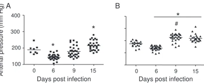
Blood pressure - Before evaluation of blood pressure, mice were adapted daily for seven days and a tail sphyg-momanometer was fitted for three consecutive readings until stabilisation. Blood pressure was individually re-corded on dpi zero, 6, 9 and 15 using an LE 5001 Pressure meter® (PanLab Instruments, Barcelona - Spain), evalu-ating caudal artery pressure in non-sedated animals. Val-ues of systolic, diastolic (DP) and the mean pressure were calculated as indicated by the manufacturer.
and B-mode. Isovolumetric relaxation time, peak early (E) and late DP transmitral flow velocities, E decelera-tion time, ejecdecelera-tion time (ET), maximal aortic velocity and flow velocity integral of aortic flow (FVI) were obtained using Echo Doppler. Cardiac output (CO) was obtained from the product of the FVI by AVA.
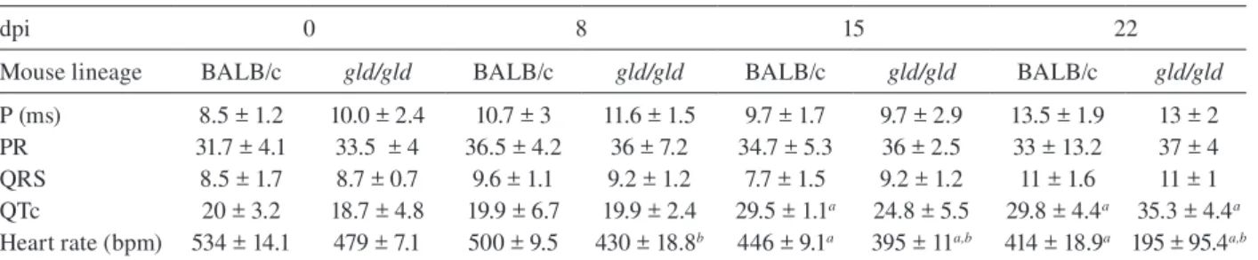
Electrocardiographic studies - All mice were i.p. tranquilised with diazepan (20 mg/kg) and transducers were carefully placed under the skin in accordance with chosen preferential derivation (DII). Traces were record-ed using a digital system (Power Lab 2/20) connectrecord-ed to a bio-amplifier in 2 mV for 1s (PanLab Instruments). Filters were standardised between 0,1 and 100 Hz and traces were analyzed using the Scope software for Win-dows V3.6.10 (PanLab Instruments). We measured heart rate (beats/mbpm), duration of the PR, QRS, QT in-tervals and P wave in ms (millisecond) on dpi zero, 8, 15 and 22. The relationship between the QT interval and RR interval was individually assessed. To obtain physi-ologically relevant values for the heart rate-corrected QT interval (QTc), in units of time rather than time to a power not equal to 1, the observed RR interval (RR0) was first expressed as a unitless multiple of 100 ms, giving a normalised RR interval (RR100 = RR0/100 ms). Next, the value of the exponent (y) in the formula QT0 = QTc x RRy
100 was assessed, where QT0 is the observed QT and both QT and QTc are in milliseconds. Taking the natural logarithm of each side of the formula (QT0) = In (QTc) +
yln(RR100), the slope of the linear relationship between the log-transformed QT and RR100 thus defined the ex-ponent to which the RR interval ratio should be raised to correct QT for heart rate (Mitchell et al.1998).
Haematology - Red blood series were evaluated on dpi zero, 6 and 13 by directly counting the number of erythrocytes per mL (red blood count - RBC) using a Neubauer chamber with 20 µL of blood diluted in 4 mL of PBS. For haematocrit determination (globular vol-ume - %), we collected 50 µL of blood using heparinised micro capillaries and centrifuged for 5 min at RT. Data are expressed as percentages and were measured using a specific ruler. Haemoglobin was determined based on free haemoglobin levels using a colorimetric assay as follows. Briefly, 250 µL of the working reagent (Hemo-globina®, Lab Test Diagnóstica, Rio de Janeiro - Brazil) were added to 10 µL of freshly collected blood, then the solution was incubated for 5 min at 37oCand thereafter the optical density (absorbance) was determined using a spectrophotometer (540 nm) (VersaMax®, Molecular Devices, Sunnyvale - CA) as recommended by the man-ufacturer. The concentration of haemoglobin globular average (CHGM - %) was calculated according to the formula: haemoglobin dosage/globular volume. There-fore, whenever observed, anaemia was classified as hy-pochromic or normochromic.
Captopril and erythropoietin treatment
BALB/c and gld/gld mice received captopril (5 mg/L, Ranbaxy Pharmaceuticals, Rio de Janeiro - Brazil) in their drinking water ad libitum from day -1 of infection until death. The captopril solution was changed three
times a week and freshly prepared from powder every time. For erythropoietin treatment, both mouse lineages received human recombinant erythropoietin (Dragon Pharmaceuticals, Vancouver, Canada) diluted in saline
(0,9%) at a dose of 1.500 U/Kg (200 μL of i.p. injections
3 times/week). Parasitaemia, expressed as parasites/mL, was evaluated in 5 µL of blood collected from tail snips at the indicated time points (Araújo-Jorge 2000).
Statistical analysis - The Mann-Whitney non para-metric test was used to compare two sets of data (Soft-ware SPSS version 8.0) and p values are indicated in figure legends.
RESULTS
Renal filtration and damage - As cardiac function may also be affected by liquid retention due to decreased renal filtration, we initially measured the urine volume in acutely infected mice (Fig. 1A, B). Our results showed that control BALB/c mice urinated a greater volume (280 µL) (Fig. 1A) than gld/gld mice (130 µL) before infection and on dpi 6 (Fig. 1B). Regarding urine urobilinogen (Fig. 1C, D), we observed a significant increase in in-fected BALB/c only on dpi 15, but in gld/gld mice it was already increased on dpi 6. Urine leukocyte evaluation (Fig. 1E, F) suggested renal damage in both lineages, but it was also detected earlier in gld/gld (dpi 6), when compared to BALB/c (dpi 15). Other parameters such as bilirubin, ketones, glucose, protein, blood, nitrite, spe-cific gravity (density) and pH showed no significant dif-ferences between both lineages (data not shown).
Renal function - Renal function was evaluated by measuring URE, CREA, Na+ and K+ in blood and both lineages showed similar levels of all parameters before infection (Fig. 2), indicating no relevant natural renal dysfunction in gld/gld. However, BALB/c and gld/gld
showed significantly higher levels of URE from dpi 6 on, although gld/gld was more affected (Fig. 2A, B). As with URE, serum levels of CREA increased after dpi 6, but only at this time point were the levels higher in gld/ gld (Fig. 2C, D). Regarding blood electrolytes (Na+ and K+), no significant differences were observed between both lineages (data nor shown).
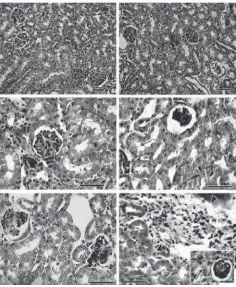
Renal damage - Histopathological analysis of control mice from both lineages showed preserved renal tissue (Fig. 3A, B). However, BALB/c mice on dpi 6 showed discrete damage of proximal tubules and glomerular atrophy (Fig. 3C). Gld/gld mice showed similar altera-tions, but with a more pronounced outcome associated with a modest inflammatory infiltration (Fig. 3D). On dpi 15, BALB/c mice presented with glomerular atrophy and apparent necrosis, with discrete glomerular haemor-rhage and cellular degeneration near proximal tubules (Fig. 3E). Interestingly, gld/gld mice presented with an intense cellular inflammatory infiltration, necrotic de-generation (Fig. 3F), glomerular atrophy and haemor-rhage (Fig. 3F, detail). We observed no parasite nests in either group (data not shown).
but on dpi 6 BALB/c (Fig. 4A) and gld/gld mice (Fig. 4B) showed a significant decrease in blood pressure (hy-potension peak). On the other hand, on dpi 15 the arte-rial pressure increased to above basal levels in BALB/c, whereas this happened from dpi 9 on in gld/gld, with a hypertension peak on dpi 9 (Fig. 4B).
Among all parameters evaluated by echocardiography (Table I) (Materials and Methods), no alterations were ob-served in control mice from either lineage, indicating no relevant natural cardiac dysfunction in gld/gld. However, on dpi 7 infected BALB/c showed abnormal cardiac function, as ascertained by altered values of LV(s), septum (s), ejection fraction, SF and ejection time. In addition, on dpi 14 these parameters had worsened and there were alterations in LV (d), left atrium and Ao/AE (relation between left atrium and aortic base), indicating the decline of cardiac function (Ta-ble I). Gld/gld showed basically the same pattern of cardiac alterations, although apparently more severe and established earlier when compared to BALB/c, especially regarding LV (d) and (s) and ET parameters (Table I).
Cardiac electric conduction system - Once more we found comparable and normal data regarding cardiac
0 50 100 150
U
R
E
(
m
g
/d
L
)
* *#
C
R
E
A
(
m
g
/d
L
)
Days post infection Days post infection 0
0.2 0.4 0.6 0.8
*
*
#
A B
C D
0 6 15 20 0 6 15 20
0 6 15 20 0 6 15 20
0 250 500
*
Urine volume (µ
L)
A
#
*
B
0 50 100
0 6 15 20
Days post infection Days post infection
Urobilinogen
(mg/dL
)
L
e
u
k
o
c
y
te
n
/µ
L
*#
C
E
D
F
* #
0 6 15 20
0 6 15 20
0 6 15 20
0 6 15 20
0 2.5 5.0
0 6 15 20
Fig. 1:urine evaluation. BALB/c (left panels, ■) and Fas-L-/- mice
(gld/gld) (right panels, ▲) were individually contained for urine
collection and we evaluated: urine volume (A, B), urobilinogen (C, D) and leukocyte number (E, F) at indicated time-points. Dashed line indicates maximal time of experiment (E, F). Asterisks in-dicate statistical difference when infected mice are compared to control mice of the same lineage (p < 0.05). Data are representative of three experiments with 5-10 mice each. #: significant difference when BALB/c and gld/gld mice are compared at the same time-point (p < 0.05).
Fig. 2:acute renal insufficiency. Serum levels of urea (URE) and creati-nine (CREA) were evaluated as indicators of renal failure. Blood samples
were collected at indicated time-points from BALB/c (left panels, ■) and
Fas-L-/- mice (gld/gld)(right panels, ▲) mice. Asterisks indicates statisti
-cal difference when infected mice are compared to control mice of the same lineage (p < 0.05). Data are representative of three experiments with 10 mice each. #: significant difference when BALB/c and gld/gld mice are compared at the same time-point (p < 0.05).
Fig. 3: renal histopathological analysis. BALB/c (left panels) and Fas-L-/- mice (gld/gld) (right panels) were euthanized on zero (A, B), 8
function in both lineages before infection, now based on ECG analysis (Table II). Besides this, a greater differ-ence was observed in the heart rate, which was reduced until dpi 22 in both lineages, but again the alteration was earlier and more pronounced in gld/gld (Table II). Only the QTc interval showed a significant increase on dpi 15 in BALB/c mice and on dpi 22 in both lineages and in
gld/gld we detected a sinusal bradycardia and discrete sinusal arrhythmia (data not shown).
Red cell evaluation - There was a significant de-crease in RBC, globular volume and haemoglobin levels on dpi 13 in both groups (Fig. 5A-F) (murine reference values: RBC = 8.7 x 10⁶/mL, Hct = 44%, Haemoglobin = 12.2 gm/dL) (27). Besides this, CHGM was around 23% in both lineages on dpi 13 (standard value 25.3%) (data not shown) (Fox et al.1984). These haematological dis-turbances, hypochromic and normocitic anaemia, could also be playing a role in the cardiac insufficiency and
maybe worsening the multiple and systemic alterations that were observed, especially in gld/gld. Moreover, hy-pochromic and normocitic anaemia are usually associ-ated with renal deficit (Silverberg et al.2005).
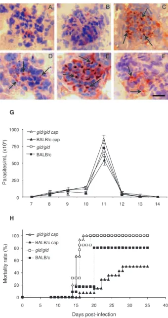
Renal lesions and cardiac failure - We evaluated whether IgM deposits could play a role in renal damage and, therefore, in renal dysfunction. We observed no relevant IgM labelling in control (Fig. 6A) or infected BALB/c mice on dpi 6 (Fig. 6B). However, on dpi 15 we observed tissue labelling apparently in the mesan-gial area (Fig. 6C). Before infection, gld/gld mice pre-sented moderate glomerular IgM deposits (Fig. 6D) and this is possibly a result of many pre-existing immune alterations observed in these mice, such as high levels of auto-immune Igs (Boes et al.2000). This precipita-tion increased and a higher glomerular labelling of IgM was observed very early in infected gld/gld mice (dpi 6) (Fig. 6E). However, there was a decrease in the labelling on dpi 15 (Fig. 6F).
To evaluate the relevance of angiotensin convert-ing enzyme (ACE) and cardiac function on mortality, BALB/c and gld/gld mice were treated daily with cap-topril, starting before infection (day-1) as a potential preventive treatment for cardiac and renal dysfunction. Captopril binds to the ACE and inhibitsACE’s catalytic production of angiotensin II (Leon et al.2003). Capto-pril had no effect on parasitaemia of BALB/c or gld/ gld mice (Fig. 6G). However, this treatment postponed and reduced BALB/c mortality and had no influence on
gld/gld death (Fig. 6H).
DISCUSSION
Previous results from our group showed that in the absence of the perforin-dependent cytotoxic pathways, the severity of acute myocarditis is increased after T. cruzi infection (Henriques-Pons et al.2002). We there-fore decided to examine whether the Fas-L-dependent pathway would also influence the evolution of myo-100
200 300 400
*
*
0 6 9 15
Days post infection
A
rt
e
ri
a
l
p
re
s
s
u
re
(m
m
H
g
)
Days post infection
0 6 9 15
* #
A B
Fig. 4: arterial pressure. Blood arterial pressure was evaluated with a tail sphygmomanometer in non-tranquilized mice. BALB/c (A) and Fas-L-/- mice (gld/gld) (B) were previously adapted and stabilized
read-ings were recorded at indicated time-points. Asterisks indicate statisti-cal difference when infected mice are compared to control mice of the same lineage (p < 0.05). Data represent three experiments with at least eight mice. #: significant difference when BALB/c and gld/gld mice are compared at the same time-point (p < 0.05).
TABLE I
Echocardiographic studies
dpi 0 7 14
Lineage
BALB/c n = 10
gld/gld n = 10
BALB/c n = 10
gld/gld n = 10
BALB/c n = 7
gld/gld n = 5
LVd (mm) 0.27 ± 0.03 0.27 ± 0.03 0.28 ± 0.02 0.4 ± 0.08a,b 0.36 ± 0.05a 0.36 ± 0.06a
LV (s) 0.11 ± 0.01 0.13 ± 0.04 0.21 ± 0.02a 0.33 ± 0.1a,b 0.28 ± 0.05a 0.32 ± 0.0a
Septum (d) 0.07 ± 0.01 0.07 ± 0.01 0.06 ± 0.0 0.06 ± 0.0 0.06 ± 0.0 0.06 ± 0.0
Septum (s) 0.13 ± 0.03 0.12 ± 0.01 0.07 ± 0.01a 0.09 ± 0.02a 0.08 ± 0.01a 0.07 ± 0.01a
Left atrium 0.12 ± 0.01 0.13 ± 0.02 0.13 ± 0.01 0.14 ± 0.03 0.16 ± 0.01a 0.22 ± 0.0a
Ao/AE 1 ± 0.1 1 ± 0.1 0.8 ± 0.1 0.8 ± 0.1 0.7 ± 0.1a 0.6 ± 0.1a
Ejec. Fraction (%) 91.3 ± 3.2 86.4 ± 8.5 58 ± 10.1a 58 ±1 0.1a 42.9 ± 20.4a 47.9 ± 11.0a 30.6 ± 5.7a,b
Short. Fraction (%) 57.6 ± 4.9 51.9 ± 11.8 26 ± 5.3a 18.6 ± 12.0 20.7 ± 5.0a 12 ± 3.1a,b
Ejec. Time (ms) 67.4 ± 5.1 71 ± 9.2 54.7 ± 5.9a 63.4 ± 3.2a,b 47.6 ± 10.4a 54 ± 1.0a
values are means ± SD. AE: left atrium; Ao: aortic base; d: diastole; gld/gld: Fas-L-/- mice; LV: left ventricle; s: systole. a: when comparing
carditis after infection and observed that in gld/gld mice there is a very mild cardiac inflammation, but a high mortality rate (de Oliveira et al.2007). There are few studies addressing the Fas-L relevance in T. cruzi infec-tion, such as in: (i) atrophy of mesenteric lymph nodes (de Meis et al. 2006) and thymus (Henriques-Pons et al. 2004), (ii) NO production and apoptosis in splenocytes (Martins et al.1999) and (iii) control of Th1/Th2 bal-ance (Lopes et al.1999). Recent publications report that a Th2-biased immune response with higher levels of IL-10 and IL-4 in gld/gld is associated with increased sus-ceptibility to experimental infection (Guilhermo et al. 2007), although it might be different in human patients (Araújo et al.2007). It is possible that T. cruzi-infected
gld/gld mice are more susceptible to the infection due to a complex array of pre-existing and infection-imposed systemic alterations, including components of the im-mune system and organ dysfunction.
In agreement with our results regarding myocardi-tis in infected gld/gld mice, it was published that cox-sackievirus B3-induced myocarditis is also decreased in the absence of Fas/Fas-L interaction (Seko et al. 2002). However, after T. cruzi infection all gld/gld died in the early acute phase, suggesting that other disturb- ances can occur and lead gld/gld to death. Thus, the goal of the present study was to investigate the partici-pation of FasL in the possible systemic alterations, oth-er than myocarditis, that could be related to the death
of T. cruzi infected mice. To date, human patients with anaemia and chronic renal failure have more severe cardiac insufficiency (Silverberg et al. 2003). More-over, anaemia is found in about one third of all cases of congestive heart failure (CHF) and is the most likely common cause of chronic renal insufficiency, which is present in about half of all CHF cases (Silverberg et al. 2004). The authors considered and discussed the exist- ence of a complex feedback known as cardio/anaemic/ renal syndrome (Silverberg et al.2003). In the present 0 2 4 6 8 10
0 6 13
E ry th ro c y te s (x 1 0 6/mL)
Days post infection Days post infection
0 5 10 15 20
0 6 13
0 10 20 30 40 50 60
0 6 13
A B C D E F * 0 2 4 6 8 10
0 6 13
* 0 20 30 40 50 60
0 6 13
G lo b u la r v o lu m e ( % ) 10 * * 0 5 10 15 20
0 6 13
H e m o g lo b in e c o n c e n tr a ti o n * *
Fig. 5:blood red series evaluation. Blood was collected from tail tips
from BALB/c (left panels, ■) and Fas-L-/- mice (gld/gld)(right panels,
♦) mice at indicated time-points to evaluate erythrocyte number (A,
B), globular volume (C, D) and hemoglobin concentration (E, F). Data are representative of three experiments with 10 mice each. Asterisks indicate statistical difference when infected mice are compared to control mice of the same lineage (p < 0.05).
Fig. 6:mechanisms leading to renal damage/dysfunction and cardiac failure. We collected kidneys from BALB/c (A-C) and Fas-L-/- mice (gld/ gld) (D-F) before infection (A-D), on 6 (B, E) and 15 (C, F) days post infection (dpi). Tissue slices were labeled with anti-IgM mAb and glo- merular deposits were identified with HPR activity. Parasitemia (G) and mortality rate (H) are shown in captopril-treated BALB/c (BALB/c cap
▲, solid line), captopril-treated gld/gld (gld/gld cap Δ, solid line), both
from dpi -1 of infection. ■, dashed line: untreated infected BALB/c; □,
dashed line: untreated gld/gld. Bar = 20 μm.
0 20 40 60 80 100
0 5 10 15 20 25 30 35 40
Days post-infection M o rt a lit y ra te ( % ) G H BALB/c cap gld/gld BALB/c gld/gld cap gld/gld cap BALB/c cap gld/gld BALB/c 0 250 500 750 1000
7 8 9 10 11 12 13 14
paper we observed a decrease in haemoglobin concen-tration on dpi 6 and this anaemia could therefore ag-gravate renal damage and cardiac insufficiency during infection in both lineages, although worsened in gld/ gld. Therefore, we treated in vivo with recombinant erythropoietin, since it has been used in the treatment of anaemia associated with renal failure, leading to the improvement of cardiac function (Silverberg et al. 2005). However, we observed no reversion in the mor-tality rate of either mouse lineage after treatment (un-published observations), thus suggesting that anaemia does not directly lead mice to death, but it may play a role when combined with other dysfunctions.
Our results showed an increase in URE and CREA, oliguria and even anuria in mice from both lineages, clearly indicating renal dysfunction (Nelson & Couto 1992). Moreover, histopathological analysis showed glomerular IgM deposits (we found no IgG precipitates) (data not shown), possibly leading to complement ac-tivation, renal damage and acute kidney injury (AKI) (Thadhani et al.1996). However, additional pathophysi-ological alterations could be taking place, such as com-pensatory neuronal-endocrine mechanisms, leading to tissue damage. We do not know why infected gld/gld
have earlier renal IgM deposits, but these deposits may be induced by a combination of pre-existing alterations of the innate immune system of these mice and the in-fection, favouring the precipitation of polyreactive IgM molecules on dpi 6. Moreover, we observed degenera-tion of proximal tubules, perivascular edema and inter-stitial congestion in both groups of mice. On dpi 15 we observed the aggravation of tubular damage in gld/gld
and intense inflammatory infiltration.
AKI may have several causes: inflammatory infil-tration, endothelial damage, ROS increase, production of nitrogen species and other mediators of tubular cells’ sub-lethal or lethal damage (Boneventre & Zuka 2004, Friedewald & Rabb 2004). After necrotic death, many pro-inflammatory mediators are released, culminating in the recruitment of blood leukocytes and cellular ac-tivation/function (Cines et al.1988). Fas/Fas-L interac-tion plays an important role in these steps (Kataoka et al. 2000, Thone & Tschopp 2001) and also in apoptosis.
TABLE II
Eletrocardiographic studies
dpi 0 8 15 22
Mouse lineage BALB/c gld/gld BALB/c gld/gld BALB/c gld/gld BALB/c gld/gld
P (ms) 8.5 ± 1.2 10.0 ± 2.4 10.7 ± 3 11.6 ± 1.5 9.7 ± 1.7 9.7 ± 2.9 13.5 ± 1.9 13 ± 2
PR 31.7 ± 4.1 33.5 ± 4 36.5 ± 4.2 36 ± 7.2 34.7 ± 5.3 36 ± 2.5 33 ± 13.2 37 ± 4
QRS 8.5 ± 1.7 8.7 ± 0.7 9.6 ± 1.1 9.2 ± 1.2 7.7 ± 1.5 9.2 ± 1.2 11 ± 1.6 11 ± 1
QTc 20 ± 3.2 18.7 ± 4.8 19.9 ± 6.7 19.9 ± 2.4 29.5 ± 1.1a 24.8 ± 5.5 29.8 ± 4.4a 35.3 ± 4.4a
Heart rate (bpm) 534 ± 14.1 479 ± 7.1 500 ± 9.5 430 ± 18.8b 446 ± 9.1a 395 ± 11a,b 414 ± 18.9a 195 ± 95.4a,b
values are means ± SD. a: when comparing infected mice to control mice of the same lineage (p < 0,05); b: when comparing BALB/c and Fas-L-/- mice (gld/gld) on the same time-point (p < 0,05); dpi: days post infection.
We do not know yet why gld/gld mice have a higher renal inflammatory response but more moderate myo-carditis, when compared to BALB/c (de Oliveira et al. 2007). Fewer cells migrate to the cardiac tissue of gld/ gld, but in both lineages we found a cardiac CD4/CD8 double negative T cell population. However, only these cells collected from gld/gld had a phenotype that sug-gested attenuation of cellular effector functions (de Oli- veira et al.2007). These findings illustrate the complex-ity of the roles played by Fas/Fas-L in the regulation of the inflammatory response in different organs during infection. On the other hand, our data regarding a cardio/ anaemic/renal syndrome indicate that both lineages have similar physiological and general alterations, but with an earlier and worse outcome in gld/gld. For example, arte-rial hypotension is observed on dpi 6 in both infected lineages, with high levels of CREA. Hypotension and shock are common events in critically ill patients, lead-ing to AKI and renal failure that may promote accumula-tion of toxic substances, vasoplegia and finally hypoten-sion. Moreover, moderate hyperchloraemic acidosis may promote vasodilatation and thus lower blood pressure, impairment of CO and decreased perfusion in both liver and kidneys, probably through down-regulation of Beta-2 receptors (Hoste & Kellun Beta-2006).
not by AT2 antagonists (Linjnem & Petrov 2003). More-over, in kidneys, ACE2 protein levels were significantly decreased in hypertensive rats, suggesting a negative regulatory role of ACE2 in blood pressure control (Da-nylczyk & Penninger 2006). We observed in the pres-ent paper that captopril treatmpres-ent postponed BALB/c mortality but had no effect on gld/gld death. This may be interpreted by at least two not mutually exclusive al-ternatives: (i) pre-existing alterations predisposed gld/ gld mice to very early and important renal damage that was not sufficiently reverted by captopril and/or (ii) al-though BALB/c and gld/gld apparently present the same sequence of central events, certain checkpoints may be more directly related to death in either lineage. Beyond the renal target of ACE activity, captopril acts to a certain extent reducing systemic arterial pressure, peripheral vascular resistance, cardiac filling pressure and increas-ing CO. Although captopril is also an anti-inflammatory agent, no alteration in T-cell proliferative response to T. cruzi or in the levels of T. cruzi and myosin-specific IgG was observed (Leon et al. 2003). Moreover, captopril upregulates bradykinin, leadingto nitric oxide synthesis (Anning et al. 1997, Gallagher et al. 1998), which may be important in the resistanceto acute T cruzi-induced myocarditis. It is also important to observe that captopril reduces cardiac necrosis and fibrosis in T. cruzi infected mice (Danylczyk & Penninger 2006).
In summary, mice infected with T. cruzi develop a cardio/anaemic/renal syndrome in the acute phase and our observations indicate antagonistic Fas-L-based im-munoregulatory functions in heart and kidney. In the ab-sence of Fas-L, there is minor myocarditis but a higher renal inflammatory response and damage (de Oliveira et al.2007), possibly due to earlier renal IgM deposits. This renal dysfunction would, in turn, increase negative effects induced by the infection in the cardiovascular system, af-fecting blood pressure and cardiac dysfunction, for ex-ample. Considering the anaemia, these alterations would lead to a cycle that could be responsible for the death of the mice. However, although BALB/c mice experience similar cardiac/renal alterations (damage/insufficiency), these events occur later in the infection, are less intense and can be partially reverted by captopril treatment.
ACkNOwLEDGEMENTS
To Mr. Luis Lopes Carvalho, for invaluable assistance in the pathological analysis, and to Dr. Alejandro Marcel Has-slocher Moreno, for important suggestions.
REFERENCES
Andrade SG 1990. Influence of Trypanosoma cruzi strain on the pathogenesis of chronic myocardiopathy in mice. Mem Inst Oswaldo Cruz85: 17-27.
Andrade ZA 1991. Pathogenesis of Chagas’ disease.Res Immunol 142: 126-129.
Anning PB, Grocott-Mason RM, Lewis MJ 1997. Effects of sulphy-dryl and non-sulphysulphy-dryl-containing ACE inhibitors on left ven-tricular relaxation in the isolated guinea pig heart. Endothelium 5: 265-275.
Araujo FF, Gomes JA, Rocha MO, Williams-Blangero S, Pinheiro VM, Morato MJ, Correa-Oliveira R 2007. Potential role of
CD4+CD25HIGH regulatory T cells in morbidity in Chagas
dis-ease. Front Biosci 1: 2797-2806.
Araújo-Jorge TC 1989. The biology of Trypanosoma cruzi -mac-rophage interaction. Mem Inst Oswaldo Cruz84: 441-462.
Araújo-Jorge TC 2000. Doença de Chagas: manual para experimen-tação animal, Editora Fiocruz/Instituto Oswaldo Cruz, Rio de Janeiro, 368 pp.
Boes M, Schmidt T, Linkemann K, Beaudette BC, Marshak-Rothstein A, Chen J 2000. Accelerated development of IgG auto-antibodies and autoimmune disease in the absence of secreted IgM. Proc Natl Acad Sci USA 97: 1184-1189.
Bohle A, Muller GA, Wehrmann M, Mackensen-Haen S, Xiao JC 1996. Pathogenesis of chronic renal failure in the primary glo- merulopathies, renal vasculopathies and chronic interstitial ne-phritides. Kidney Int 54 (Suppl.): S2-9.
Boneventre JV, Zuka A 2004. Ischemic acute renal failure: an inflam-matory disease? Kydney Int 1: 480-485.
Cines DB, Pollak ES, Buck CQ, Loscalzo J, Zimmerman GA, McEv-er RP, PobMcEv-er JS, Wick J, Konkle BA, StMcEv-ern DM 1988. Endothe-lial cells in physiology and pathophisiology of vascular disorders.
Blood 2: 3527-3534.
Coura JR 2007. Chagas disease: what is known and what is needed-a background article. Mem Inst Oswaldo Cruz 102 (Suppl. I): 113-122.
Coura JR, Dias JCP 2009. Epidemiology, control and surveillance of Chagas disease: 100 years after its discovery.Mem Inst Oswaldo Cruz 104 (Suppl. I): 31-40.
Danilczyk U, Penninger JM 2006. Angiotensin-converting enzyme II in the heart and the kidney. Circ Res 98: 463-471.
De Meis J, Mendes-da-Cruz DA, Farias de Oliveira DA, Correa-de-Santana E, Pinto-Mariz F, Cotta-de-Almeida V, Bonomo A, Savi-no W2006. Atrophy of mesenteric lymph nodes in experimental Chagas’ disease: differential role of Fas/Fas-L and TNFRI/TNF pathways. Microbes Infect 8: 221-231.
de Oliveira GM, Diniz RL, Batista W, Batista MM, Bani Correa C, de Araujo-Jorge TC, Henriques-Pons A 2007. Fas ligand-dependent inflammatory regulation in acute myocarditis induced by Try-panosoma cruzi infection. Am J Pathol 171: 79-86.
Dias JP, Bastos C, Araújo E, Mascarenhas AV, Martins Netto E, Gras-si F, Silva M, Tatto E, Mendonça J, Araújo RF, Shikanai-Yasuda MA, Aras R 2008. Acute Chagas disease outbreak associated with oral transmission. Rev Soc Bras Med Trop 41: 296-300.
dos Santos PV, Roffê E, Santiago HC, Torres RA, Marino AP, Paiva CN, Silva AA, Gazzinelli RT, Lannes-Vieira J 2001. Prevalence of CD8(+)alpha beta T cells in Trypanosoma cruzi-elicited
myo-carditis is associated with acquisition of CD62L(low)LFA-1(high)
VLA-4(high) activation phenotype and expression of
IFN-gamma-inducible adhesion and chemoattractant molecules. Microbes In-fect 3: 971-984.
Fox GJ, Cohen JB, Loew MF 1984. Laboratory animal medicine, Aca-demic Press Inc, Harcout Brace Jovanovich Publishers, San Diego, 1265 pp.
Freire de Lima CG, Nascimento DO, Soares MB, Bozza PT, Castro-Faria-Neto HC, de Mello FG, DosReis GA, Lopes MF 2000. Up-take of apoptotic cells drives the growth of a pathogenic trypano-some in macrophages. Nature 403: 199-203.
Friedewald JJ, Rabb H 2004. Inflamatory cells in ischemic acute renal failure. Kydney Int 66: 486-491.
Guillermo LV, Silva EM, Ribeiro-Gomes FL, De Meis J, Pereira WF, Yagita H, DosReis GA, Lopes MF 2007. The Fas death pathway controls coordinated expansions of type 1 CD8 and type 2 CD4 T cells in Trypanosoma cruzi infection. J Leukoc Biol 81: 942-951.
Henriques-Pons A, De Meis J, Cotta-De Almeida V, Savino W, Araújo-Jorge TC 2004. Fas and perforin are not required for thymus atrophy induced by Trypanosoma cruzi infection. Exp Parasitol 107: 1-4.
Henriques-Pons A, Oliveira GM, Paiva MM, Correa AF, Batista MM, Bisaggio RC, Liu CC, Cotta-de-Almeida V, Coutinho CM, Persechini PM, Araújo-Jorge TC 2002. Evidence for a perforin-mediated mechanism controlling cardiac inflammation in Try-panosoma cruzi infection. Int J Exp Pathol 83: 67-79.
Higuchi M de L, Benvenuti LA, Martins Reis M, Metzger M 2003. Pathophysiology of the heart in Chagas’ disease: current status and new developments. Cardiovasc Res 60: 96-107.
Hoste EA, Kellun JA 2006. Acute kidney dysfunction and the criti-cally ill. Minerva Anestesiol 7: 133-143.
Huber S, Shi C, Budd RC 2002. Gamma/delta T cells promote a Th1 response during coxsackievirus B3 infection in vivo: role of Fas and Fas ligand. J Virol 76: 6487-6494.
Kataoka T, Budd RC, Holler N, Thome M, Martinon F, Irmler M, Burns K, Hahne M, Kennedy N, Kovacsovics M, Tschopp J 2000. The caspase-8 inhibitor FLIP promotes activation of NF-kappaB and Erk signaling pathways. Curr Biol 10: 640-648.
Lambert C, Landau AM, Desbarats J 2003. Fas-beyond death: a regenerative role for Fas in the nervous system. Apoptosis 8: 551-562.
Leon JS, Wang K, Engman DM 2003. Captopril ameliorates myo-carditis in acute experimental Chagas disease. Circulation 107: 2264-2269.
Linjnem PJ, Petrov VV 2003. Role of intracardiac renin-angiotensin-aldosterone system in extracellular matrix remodeling. Methods Find Exp Clin Pharmacol 25: 541-564.
Lopes MF, Nunes MP, Henriques-Pons A, Giese N, Morse HC 3rd Davidson WF, Araújo-Jorge TC, Dos Reis GA 1999. Increased susceptibility of Fas ligand-deficient gld mice to Trypanosoma cruzi infection due to a Th2-biased host immune response. Eur J Immunol 29: 81-89.
Martins GA, Petkova SB, Machado FS, Kitsis RN, Weiss LM, Wintner RM, Tanowitz HB, Silva JS 2001. Fas-FasL interaction modulates nitric oxide production in Trypanosoma cruzi-infected mice. Im-munology 103: 122-129.
Martins GA, Vieira LQ, Cunha FQ, Silva JS 1999. Gamma interferon modulates CD95 (Fas) and CD95 ligand (Fas-L) expression and nitric oxide-induced apoptosis during the acute phase of
Try-panosoma cruzi infection: a possible role in immune response control. Infect Immun 67: 3864-3871.
Mitchell GF, Jeron A, Koren G 1998. Measurement of heart rate and Q-T interval in the unscious mouse. Am J Physiol 274 (3 Pt 2): H747-751.
Moncayo A 2003. Chagas disease: current epidemiological trends after the interruption of vectorial and transfusional transmis-sion in the Southern Cone countries. Mem Inst Oswaldo Cruz 98: 577-591.
Nelson RW, Couto CG 1992. Essentials of small animal internal med-icine, Mosby-Year Book Inc, New York, 32 pp.
Palazzuoli A, Gallotta M, Iovine F, Nuti R, Silverberg DS 2008. Anaemia in heart failure: a common interaction with renal in-sufficiency called the cardio-renal anaemia syndrome. Int J Clin Pract 62: 281-286.
Schelling JR, Cleveland RP 1999. Involvement of Fas-dependent apoptosis in renal tubular epithelial cell deletion in chronic renal failure. Kidney Int 56: 1313-1316.
Seko Y, Kayagaki N, Seino K, Yagita H, Okumura K, Nagai R 2002. Role of Fas/FasL pathway in the activation of infiltrating cells in murine acute myocarditis caused by Coxsackievirus B3. J Am Coll Cardiol 39: 1399-1403.
Silverberg DS, Wexler D, Blum M, Wollman Y, Iaina A 2003. The cardio-renal anemia syndrome: does it exist? Nephrol Dial Trans-plant 18 (Suppl. 8): 7-12.
Silverberg DS, Wexler D, Blum M, Wollman Y, Iaina A 2004. The association between congestive heart failure and chronic renal disease. Curr Opin Nephrol Hypertens 13: 163-170.
Silverberg DS, Wexler D, Blum M, Wollman Y, Iaina A, Sheps D, Keren G, Scwartz D 2005. Erythropoietin in heart failure. Semin Nephrol 25: 397-403.
Suda T, Okazaki T, Naito Y, Yokota T, Arai N, Ozakis S, Nakao K, Nagata S 1995. Expression of the Fas ligand in cells of T cell lineage. J Immunol 154: 3806-3813.
Thadhani R, Pascual M, Boneventre JV 1996. Acute renal failure.
N Engl J Med 334: 1448-1460.
Thome M, Tschopp J 2001. Regulation of lymphocyte proliferation and death by FLIP. Nat Rev Immunol 1: 50-58.
WHO - World Health Organization 2004. [homepage on the internet] [up date 2004 May 2; cited 2006 June 19]. TDR summary report 2004-2005. Avaliable from http://www.who.int/tdr/publications/ tdrnews/default.htm.