AVALIAÇÃO DE BIOMARCADORES MOLECULARES
EM MULHERES COM CÂNCER DE OVÁRIO
Dissertação apresentada ao Programa de Pós- Graduação em Ginecologia, Obstetrícia e Mastologia da Faculdade de Medicina de Botucatu-UNESP, para obtenção do título de Doutor.
Orientador: Prof. Dr. Paulo Traiman Co-Orientadora: Dra. Andréa Teixeira de Carvalho
Professor Paulo Traiman,
Dra. Andréa Teixeira de Carvalho,
Professor Agnaldo Lopes da Silva Filho,
Agradeço pelos ensinamentos fundamentais que contribuíram de forma incomensurável para a minha formação.
Obrigada pelo privilégio de poder compartilhar da sabedoria de vocês!
-
Agradeço ao Dr. João Oscar Falcão Júnior pela parceria e entusiasmo. Seus conhecimentos foram essencias ao nosso crescimento.
Meu agradecimento especial ao Dr. Geraldo Henrique Ribeiro pelos ensinamentos cirúrgicos fundamentais à minha formação! Tenho profunda admiração pelo senhor.
Aos amigos Gustavo e Elisa pelo apoio, cortesia e disponibilidade. Foram ótimos nossos momentos juntos.
Aos amigos do Hospital Mater Dei e do Instituto Mário Penna pela grande oportunidade de aprendizado e crescimento.
Aos profissionais do Centro de Pesquisas René Rachou (Fiocruz), pelo apoio logístico na captação, preparação e processamento do material coletado durante as operações. Também pela cortesia, disponibilidade e receptividade.
“Viver
Acordando com nova esperança
Desejar que o novo dia seja diferente
Que um olhar dure o tempo necessário
Que surja mais um momento que eu possa eterniza-lo
Que meu trabalho continue sendo feito do melhor modo
Que o entardecer eu o veja mais inebriante
Mais um dia de vida devida a Deus
Viver...como se fosse o último dia.”
“Se não puder ser um poeta, Seja uma poesia.”
Introdução: O câncer de ovário (CO) é a maior causa de morte por neoplasia ginecológica nos países desenvolvidos. Atualmente, um modelo dualístico de classificação foi proposto, sendo os tumores tipo I de baixo grau, indolentes e com mutações estáveis. Os tumores tipo II são de alto grau e mais agressivos. Além disso, na tentativa de se entender o processo de carcinogênese, vários biomarcadores têm sido estudados como as micropartículas (MPs), as citocinas e as quimiocinas. O objetivo desse estudo é a avaliação de fatores solúveis da resposta inflamatória (MPs, citocinas e quimiocinas) em mulheres com CO e compará-los com os níveis encontrados em mulheres sem malignidade e com parâmetros clínicos. Métodos: Avaliaram-se 26 mulheres com CO e 16 mulheres sem evidência de neoplasia maligna (grupo controle). Foram coletadas amostras de plasma e tecido tumoral. A avaliação dos fatores inflamatórios foi realizada por meio da dosagem de citocinas (IL-1- β, IL-2, IL-6,
IL-10, IL-12, IL-17A, TNF e IFN-gama, e quimiocinas (CXCL8, CXCL-9, CXCL 10, CCL 2, CCL5) e das micropartículas (neutrófilos, leucócitos, monócitos, eritrócitos, endotélio, plaquetas e linfócitos) por citometria de fluxo/CBA (Cytometric Bead Array). As diferenças entre os grupos foram avaliadas pelos
testes Kruskal- Walis ou Mann-Whitney e a sobrevida por Cox Regression. As diferenças com valor de p<0,05 foram consideradas significativas.Resultados: Não houve diferença entre os grupos em relação à idade, paridade e menopausa. No grupo de mulheres com CO, 10 (38,5%) tinham estadio I/II e 16 (61,5%) tinham estadio III/IV. Em relação ao tipo tumoral, segundo a nova classificação, 8 (30.8%) eram tipo I e 18 foram tipo II (69.2%). Citorredução ótima foi obtida em 15 (57.7%) mulheres com CO. Os valores de CA 125 foram
significativamente diferentes entre os grupos. Não houve óbito em mulheres com tumores tipo I. Em relação às análises das MPs, observou-se aumento desse biomarcador proveniente de leucócitos nas mulheres com CO em relação ao controle. Houve ainda aumento das MPs de leucócitos no tumores tipo II em relação ao tipo I. Os níveis de MPs de neutrófilos, assim como os valores de CA125, foram maiores nas mulheres que tiveram citorredução ótima. Observou-se aumento dos níveis de IL-1- β, IL-6, TNF, IL-12, IFN-gama, IL-10, IL-17, IL-2, CCL-2, CCL-5, CXCL-8, CCL-9 e CXCL10 nas mulheres com CO em relação ao controle. Não houve diferenças estatísticas dos níveis de citocinas/quimiocinas em relação ao tipo tumoral I ou II. As mulheres com tumor tipo II que tiveram citorredução ótima, apresentaram maior expressão de IL-1- β e IL-12, além de aumento dos valores de CA-125. Os níveis de IL-1, IL
-12 e CXCL-8 também foram maiores naquelas com estadio avançado e que foram submetidas à cirurgia ótima. Conclusão: Mulheres com CO apresentaram aumento das frequências de MPs, citocinas e quimiocinas em relação àquelas sem neoplasia maligna. As MPs provenientes de neutrófilos, as citocinas IL-1- β e IL-12 e a quimiocina CXCL-8 possivelmente correlacionam-se com resposta ao tratamento em mulheres com CO.
Palavras-chave: Câncer de ovário, resposta inflamatória, citocinas,
Introduction : Ovarian cancer ( OC) is the leading cause of death from gynecological cancer in developed countries . Currently , a dualistic classification model was proposed .Type I tumors are low-grade , indolent and have stable mutations . Type II tumors are high grade and more aggressive . Moreover, in an attempt to understand the process of carcinogenesis , several biomarkers have been studied as microparticles (MPs ) , cytokines and chemokines. The purpose of the study was to evaluate the levels of circulating
soluble biomarkers-microparticles, cytokines and chemokines- to characterize the pro-inflammatory/modulatory immune response in women with OC. The correlation between the biomarker levels and the clinico-pathological parameters were also analyzed. Methods : We evaluated 26 women with OC and 16 women without evidence of malignancy ( control group ) . Plasma samples and tumor tissue were collected . The assessment of inflammatory markers was performed by measurement of cytokine (IL -1- β , IL-2 , IL-6 , IL-10 , IL-12 , IL -17A , TNF and IFN –gamma), chemokines ( CXCL8 , CXCL -9 , CXCL 10 , CCL 2 , CCL5 ) and microparticles (neutrophils , leukocytes, monocytes, erythrocytes, endothelium , platelets and lymphocytes) by flow cytometry / CBA ( cytometric Bead Array) . Differences among groups were evaluated by Kruskal - Wallis or Mann - Whitney and survival by Cox Regression . Differences with p < 0.05 were considered significant. Results: There was no difference between groups regarding age , parity and menopause. In the group of women with OC , 10 ( 38.5 % ) were stage I / II and 16 ( 61.5 % ) were stage III / IV . Concerning tumor type , according to the new classification , 8 (30.8 % ) were type I and 18 ( 69.2 % ) were type II . Optimal cytoreduction was achieved in 15 ( 57.7 % ) women with OC . CA 125 values were significantly different between groups . There were no deaths in women with type I tumors. Stratifying by groups, an increased frequence of leukocytes microparticles (LMP) was observed in women with OC compared to control group. There was also an increase of LMPs in type II tumors compared to type I. Neutrophils derived microparticles (NMPs), as well as the values of CA125 were higher in women who were optimally debulked . A higher frequence of IL -1- β , IL- 6, TNF , IL -12, IFN- gamma, IL -10, IL -17 , IL-2 , CCL -2 , CCL- 5, CXCL -8 , CCL - 9 and CXCL10 were detected in women with OC compared to control . We did not observe any statistical difference beteween cytokines / chemokines and tumor type I or II . Women with type II tumor who were optimally debulked showed higher expression of IL - 1 - β and IL -12 , and increased levels of CA -125 . The levels of IL-1, IL-12 and CXCL -8 were also higher in advanced disease with optimal surgical treatment. Conclusion : Women with OC showed increased frequencies of MPs , cytokines and chemokines compared to those without malignancy . MPs from neutrophils, IL-1 - β and IL-12 cytokine and chemokine CXCL -8 possibly correlate with response to treatment in women with OC.
Ac Anticorpo CBA
CCL Do Inglês: Quimiocina Cytometric Bead Array CO Câncer de Ovário
CEP Comitê de Ética em Pesquisa
cm Centímetro
CMF
CXCL Citometria de Fluxo Quimiocina
EDTA-K3 Ácido Etileno-diamino Tetracético et al. E outro(s), e outra(s)
FACS Do Inglês: Fluorescence-Activated Cell Sorting
FIGO Federação Internacional de Ginecologia e Obstetrícia FITC Isoticianato de Fluoresceína
FSC Do Inglês: Forward Scatter
IFN-gama Interferon Gama
IL Citocina
IL-1- β
INCA Instituto Nacional de Câncer
μL Microlitro
mL MPs OMS
Mililitro
Micropartículas
Organização Mundial de Saúde NK Do Inglês: Natural Killer
nm Nanômetro
PE Ficoeritrina
TNF Fator de Necrose Tumoral Alfa
1. INTRODUÇÃO... 20
1.1. Câncer de Ovário... 21
1.2. Inflamação e câncer de ovário... 25
1.2.1. Micropartículas ... 27
1.3. Justificativa do estudo... 30
2. OBJETIVOS... 31
2.1 Objetivo Geral... 32
2.2 Objetivos Específicos... 32
3. MATERIAL E MÉTODOS... 33
3.1. Pacientes e espécimes... 34
3.1.1 Critérios de Inclusão... 35
3.2.Métodos... 36
3.2.1 Técnica Cirúrgica……… 36
3.2.2 Avaliação Histológica e Imunohistoquímica……….. 36
3.2.3 Detecção do nível de citocinas/quimiocinas plasmáticas por citometria de fluxo... 37
3.2.4 – Aquisição e análise dos dados de citocinas e quimiocinas plasmáticas por citometria de fluxo... 40
3.2.5. Obtenção do plasma livre de plaquetas... 42
3.2.6. Obtenção das micropartículas (MPs)... 42
3.2.7. Identificação de micropartículas (MPs) pela Citometria de fluxo... 42
Análise fenotípica das MPs por citometria de fluxo... 42
4. REFERÊNCIAS... 45
5. ARTIGO I... 49
6. ARTIGO II... 72
8. ANEXOS……… 99
ANEXO I: Estadiamento FIGO………. 97
ANEXO II - Parecer do Comitê de Ética em Pesquisa... 99
1.1 Câncer de Ovário
O Câncer de ovário é a maior causa de morte por neoplasia ginecológica nos países desenvolvidos. Segundo dados da Agência Internacional de Pesquisa em Câncer, foram estimados 238.719 casos com 151.905 mortes por câncer de ovário no mundo em 20121. A incidência no Brasil, conforme dados do Instituto Nacional do Câncer, é de 5.680 novos casos em 2014, correspondendo a 2,1% de todos os cânceres em mulheres no país2.
A maioria dos tumores malignos de ovário é diagnosticada em estadios avançados devido a falta de sintomas específicos nos estadios iniciais. Não há método propedêutico efetivo para rastreamento. Os marcadores tumorais e os exames imaginológicos apresentam altas taxas de resultados falsos positivos, especialmente quando realizados na pré-menopausa, e não são custo efetivos3,4,5. Desse modo, a sobrevida em 5 anos é de aproximdamente 40–
50% 6.
Os tumores tipo II são mais agressivos, de alto grau, evoluem com rápida progressão e geralmente são diagnosticados em estadios avançados. Esses tumores correspondem a 75% casos e são responsáveis por 90% das mortes por câncer de ovário 7 (Figura 1B).
B
C
Figura 1: A. Histologia referente à tumor ovariano tipo I. B. Histologia referente a tumor ovariano tipo II. C. Imunohistoquímica referente à tumor ovariano tipo II.
O tratamento do cancer de ovário baseia-se na cirurgia citorredutora associada à quimioterapia nos casos indicados. O estadiamento é cirúrgico segundo as normas da Federação Internacional de Ginecologia Obstetrícia (FIGO)8,9 (Anexo I).O volume de doença residual após cirurgia primária é
importante fator prognóstico. O objetivo da citorredução inicial é a remoção da maior quantidade de tecido tumoral possível, assim como da doença metastática. Caso a ressecção de todos os sítios metastáticos não seja possível, o ideal é reduzir o volume tumoral a uma condição “ótima“10,11.
Considera-se citorredução ótima aquela em que a doença residual após cirurgia seja ≤ 1 cm em seu maior diâmetro 10. Apesar da maioria das mulheres com doença avançada apresentar remissão após tratamento adequado com cirurgia e quimioterapia, 70 a 80% aproximadamente, irão recorrer em dois anos 12.
1.2 Inflamação e Câncer de ovário
A importância da resposta inflamatória na etiologia e patogênese do câncer tem sido descrita desde o século XIX, através dos trabalhos de Rudolf Ludwig Karl Virchow, que sugeriu que o infiltrado “linforeticular“ refletia a
origem do câncer em sítios de inflamação crônica (“Die Cellularpathologie in ihrer Begründung auf physiologische und pathologische Gewebelehre‖, 1858). No entanto, o efeito paradoxo do infiltrado inflamatório na contenção e na
progressão tumoral ainda é pouco compreendido 13. Pressupõe-se que a
A maioria das células tumorais secreta citocinas e quimiocinas, especialmente como consequência às mutações oncogênicas e às alterações de sinalização no sítio tumoral. Essas moléculas são potentes mediadores e reguladores da inflamação. No entanto, não está claro quais expressões de citocinas e quimiocinas apresentam relevância na regulação desse sistema
complexo nos tumores ovarianos 13.
Sabe-se que o microambiente pró-inflamatório do câncer de ovário está clinicamente relacionado com a disseminação peritoneal tumoral, com a produção maciça de ascite e com altas taxas de mortalidade. Vários estudos demostram que as neoplasias malignas ovarianas expressam altos níveis de algumas citocinas como TNF e IL-6, indicando o potencial dessas moléculas como indutoras/reguladoras da inflamação nesses tumores14. O TNF, por exemplo, apresenta efeitos pró- e anti-tumorais, a depender da estimulação de seus receptores. Em geral, o aumento desta citocina nas neoplasias malignas está associado à toxicidade envolvendo o sistema imune inato e adaptativo. O TNF-α também está envolvido na iniciação e promoção tumoral, além da alteração da homeostase com estímulo à angiogênese. Essa citocina tem-se mostrado importante regulador da ação de quimiocinas, por meio da via de sinalização do fator nuclear-kB (NF-kB).15,16,17 . A IL-6 particularmente atua na
promoção do crescimento e como fator anti-apoptótico, apresentando-se em altas concentrações no plasma de mulheres com doença avançada 18. Vários
propicia a proliferação e sobrevida dessas células tumorais geneticamente alteradas13,19. Portanto, os níveis de citocinas e quimiocinas podem representar um reflexo do estado de imunidade do hospedeiro e servir como biomarcadores para predizer o prognóstico dessas mulheres com câncer de ovário, além de se constituírem como ferramentas complementares potenciais na sua monitoração pós-terapêutica.
1.2.1. Micropartículas (MPs)
As micropartículas foram inicialmente descritas em 1964, e têm sido muito estudadas desde então20. MPs são vesículas de diferentes tamanhos,
variando de 100 a 1000 nm, liberadas pelas células em situações fisiológicas e patológicas (Figura 2). Apresentam como característica relevante a alta exposição de fosfatidilserina que é translocada da parte interna para parte
Figura 2: Micrografia eletrônica de varredura de um isolado de sangue periférico (doador saudável) evidenciando massa de vesículas extracelulares (seta). Adaptado de: Ogorevc E, et al. The role of extracellular vesicles in phenotypic cancer transformation. Radiolo Oncol 2013; 47(3): 197-205.
A secreção das MPs é facilmente detectada em células com fenótipo tumoral, quando comparada às células normais do organismo. Isso chama a atenção para uma possível atuação dessas vesículas na carcinogênese. As MPs podem atuar na progressão tumoral em diversos aspectos:
- Contribuem para o escape à imunovigilância22.
- Auxiliam o tumor a evadir a apoptose, evitando-se o acúmulo de caspase 3 intracelular, por meio da liberação de MPs contendo esta enzima23 .
- Induzem a transformação fenotípica celular, por meio do transporte de material genético, enzimas, citocinas e quimiocinas25.
- Promovem a progressão e invasão das células tumorais por meio da alteração e degradação da matriz extracellular26.
- Promovem a resistência a drogas, pelo acúmulo de quimioterápico nessas vesículas e consequente eliminação pelas células tumorais 27 .
- Promovem a indução à angiogênese, especialmente através do transporte de fatores pró-angiogênicos 28.
Está claro que as MPs são capazes de modular direta ou indiretamente o comportamento das células ao seu redor, especialmente através do transporte de proteínas e ácidos nucléicos21 . A presença de MPs derivadas do
1.3 Justificativa do estudo
O conhecimento da carcinogênse da neoplasia ovariana é de grande importância para elucidação dos mecanismos envolvidos na origem e patogênese desse tumor. O câncer de ovário ainda representa um desafio devido às limitações para rastreamento e opções terapêuticas pouco eficazes, o que implica em baixa sobrevida. Medir a concentração sérica de citocinas, quimiocinas e micropartículas em mulheres com essa neoplasia é uma maneira pouco invasiva e indireta de se avaliar a atividade tumoral, a resposta inflamatória/reguladora sistêmica associada e o papel do microambiente formado na resposta pró- e anti-tumoral do hospedeiro. Além disso, o estudo de biomarcadores da inflamação pode propiciar maior compreensão sobre o comportamento biológico desse tumor, assim como sinalizar para futuros
2.1. Geral:
Avaliação de biomarcadores moleculares da resposta inflamatória em mulheres com carcinoma epitelial de ovário
2.2. Específicos:
• Dosar e comparar fatores solúveis da resposta inflamatória- micropartículas (neutrófilos, leucócitos, monócitos, eritrócitos, endotélio, plaquetas, linfócitos), citocinas e quimiocinas (IL-1-β, IL-2, IL-6 IL-10, IL-12, IL- 17a, TNF, IFN- gamma, CXCL8, CXCL-9, CXCL 10, CCL 2, CCL5)- em soro de mulheres com câncer epitelial de ovário e grupo controle
• Correlacionar fatores solúveis da resposta inflamatória- micropartículas (neutrófilos, leucócitos, monócitos, eritrócitos, endotélio, plaquetas, linfócitos), citocinas e quimiocinas (IL-1-β, IL-2, IL-6 IL-10, IL-12, IL- 17a, TNF, IFN- gamma, CXCL8, CXCL-9, CXCL 10, CCL 2, CCL5) - com parâmetros clínico-patológicos em mulheres com câncer epitelial de ovário
3.1. Pacientes e espécimes
Foram avaliadas 26 mulheres com diagnóstico de câncer epitelial de ovário (CO) nos estadios I a IV (casos) e 16 mulheres submetidas a histerectomia abdominal total com ooforectomia para tratamento de doença ginecológica benigna (grupo controle), atendidas em uma instituição hospitalar do Estado de Minas Gerais, no período de junho de 2010 a Outubro de 2013, com diagnóstico histopatológico confirmado no pós operatório. Foram realizados exames clínico, ginecológico e ultrassonográfico em todas as mulheres. O estadiamento do CO, foi estabelecido após laparotomia segundo protocolo de classificação pela Federação Internacional de Ginecologia Obstetrícia (FIGO)9 (Anexo I).
Todos os tecidos removidos foram examinados por um único patologista. O critério de citorredução ótima foi definido como a permanênia de doença residual < 1cm de diâmetro, após a cirurgia primária. Os níveis séricos do marcador tumoral CA-125 foram obtidos previamente à operação, em todas as mulheres com suspeita de CO, assim como todas as amostras de sangue para o estudo foram coletadas no per operatório, antes da indução anestésica.
tipo II aqueles de alto grau, geralmente diagnosticados em estadios avançados e com marcação imunohistoquímica positiva para p53.
Houve registro de óbito em prontuário para todas aquelas mulheres que faleceram pelo tumor ovariano incialmente diagnosticado.
O estudo foi aprovado pelo Comitê de Ética em Pesquisa (Anexo II). As mulheres selecionadas foram esclarecidas sobre o protocolo de pesquisa e assinaram o Termo de Consentimento Livre Esclarecido (ANEXO III).
3.1.1 Critérios de Inclusão
Mulheres com diagnóstico anatomopatológico de CO submtidas a tratamento cirúrgico (casos);
Mulheres submetidas a ooforectomia abdominal total com ooforectomia para tratamento de doença ginecológica benigna e sem evidências de neoplasias malignas (grupo controle);
Ausência de processo infeccioso agudo peritoneal evidente à laparotomia
Mulheres sem evidência de processo inflamatório agudo sistêmico Mulheres sem tratamento prévio por quimio ou radioterapia
Mulheres que não utilizaram imunossupressores, corticosteroids e/ou antiinflamatórios não esteróides ha pelo menos 3 meses
3.2 Métodos
3.2.1 Técnica Cirúrgica
As mulheres com CO foram submetidas à laparotomia mediana ampla, inventário da cavidade peritoneal, lavado peritoneal, histerectomia total, salpingo-ooforectomia bilateral, omentectomia, linfadenectomia pélvica e para-aórtica quando indicada, além dos procedimentos necessários à realização de citorredução ótima, quando possível ( ressecção intestinal, hepatectomia parcial, apendicectomia, peritoniectomia, dentre outros).
3.2.2 Avaliação Histológica e Imunohistoquímica
As peças cirúrgicas retiradas foram fixadas em formaldeído 10%. O material foi identificado e encaminhado para avaliação anatomo-patológica. As mostras fixadas em formaldeído foram desidratadas em etanol em concentrações crescentes, variando de 70 a 99% ( 60 min. cada); depois imersas em xilol (2x 60 min.) e em parafina 60o(2x 60 min.); e incluídas em bloco de parafina. As lâminas foram coradas pela técnica de hematoxilina eosina (HE), sendo o tipo histológico, o grau de diferenciação tumoral e a presença de metástases classificados de acordo com a FIGO9.
estreptavidina-biotina-peroxidase, utilizando-se o Kit Universal da Novocastra (Novostain Super ABC Kit, Universal, Novocastra Laboratories, Newcatle upon Tyne, UK). Após as reações as lâminas foram cobertas por lamínula, com auxílio de Permaunt (Fischer Scientific Company, Fair Lawn, NJ, USA).
Foram utilizados anticorpos p53 na diluição 1:100 (Novocastra Laboratories, Newcatle upon Tyne, UK, clone DO7). Para revelação das reações foi utilizado o diamino-benzidina.
As lâminas foram analisadas em microscópico de luz convencional. As células consideradas positivas foram aquelas coradas com marrom após as reações. As células foram contadas nas áreas em pequeno aumento que exibiam maior concentração de células positivas. Foram considerados positivos aquelas com mais de 10% de células marcadas 29.
3.2.3 Detecção do nível de citocinas/ quimiocinas plasmáticas por citometria de fluxo
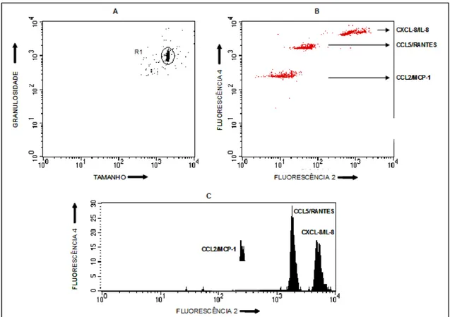
Os níveis plasmáticos de citocinas e quimiocinas foram quantificados utilizando-se o sistema Cytometric Bead Array (CBA) (Becton Dickinson-BD) (Figura 3), que emprega uma mistura de esferas de poliestireno, de intensidades de fluorescência discretas e distintas, recobertas com anticorpos específicos para as citocinas e quimiocinas humanas que são detectadas no canal de fluorescência 3 (FL-3). Essa metodologia permite a avaliação simultânea de várias citocinas e quimiocinas no mesmo ensaio, empregando pequenos volumes de amostras séricas.
Figura 3: Representação esquemática da técnica de CBA.
Nota: Adaptado do bulário do Cytometric Bead Array Flex Set System BD- Biosciences, 2004-2005. Com micro-esferas marcadas, foram adicionados aos anticorpos anti-citocinas/quimiocinas, amostras de soro. Em seguida, a amostra foi marcada por imunofluorescência (ficoeritrina) e analisada no citômetro de fluxo. Os resultados foram obtidos por software específico para CBA (FCAP Array TM Software, BD, Pharmingen, EUA). Os anticorpos utilizados foram anti: IL-1, IL-2, IL-6 IL-10, IL-12, IL- 17A, TNF, IFN- gamma, CXCL8, CXCL-9, CXCL
Neste estudo, a metodologia de CBA foi adaptada dos protocolos originais propostos por Chen et al. (1999), como descrito a seguir:
Alíquotas de 25mL de plasma diluído 1:5 com diluente G (reagente do kit CBA), alíquotas de 25mL dos padrões de citocinas/ quimiocinas, submetidos à diluição seriada com diluente G (“Top Standard” – 2500 pg/mL, 1:2 – 1250 pg/mL, 1:4 – 625 pg/mL, 1:8 – 312.5 pg/mL, 1:16 – 156 pg/mL, 1:32 – 80 pg/mL, 1:64 – 40 pg/mL, 1:128 – 20 pg/mL e 1:256 – 10 pg/mL) e 25mL de diluente G apenas (Controle Negativo), foram transferidas para tubos de poliestireno de 5mL (Falcon – BD, E.U.A). Posteriormente, a cada tubo foram adicionados 15mL da mistura de esferas de captura, conjugadas com anticorpos monoclonais selecionados para a avaliação (anti- 1, 2, 6 IL-10, IL-12, IL- 17a, TNF, IFN- gamma, CXCL8, CXCL-9, CXCL IL-10, CCL 2, CCL5), com subseqüente incubação por 90 minutos, à temperatura ambiente, ao abrigo da luz. Após a incubação, as esferas de captura foram lavadas com 500mL da solução F (“Wash buffer”, reagente do kit CBA), centrifugadas a 600g, por 10 minutos a 18oC e o sobrenadante foi cuidadosamente aspirado e
descartado. As esferas foram então re-incubadas na presença de 20mL do reagente B, que corresponde a um coquetel de anticorpos monoclonais anti-citocinas/quimiocinas humanas, conjugados com o fluorocromo PE (FL-2) por 90 minutos, temperatura ambiente, ao abrigo da luz. Após incubação, as esferas de captura foram novamente lavadas com 500mL da solução F, centrifugadas a 600g, por 10 minutos a 18oC e o sobrenadante foi
3.2.4 – Aquisição e análise dos dados de citocinas e quimiocinas plasmáticas por citometria de fluxo
de citocinas/quimiocinas em concentrações conhecidas (10 pg/mL – 2500 pg/mL) e empregada para determinar as concentrações de cada citocina /quimiocina no plasma teste. Um modelo de ajustamento através da curva do 5º parâmetro logístico, que permite o ajuste da melhor curva não linear para dados detectáveis, foi utilizado. Dessa forma, foi possível extrapolar valores de intensidades de fluorescência de amostras que não caíam dentro dos limites da curva padrão.
Figura 4: Exemplo de perfil dos níveis plasmáticos de quimiocinas através de microsesferas de captura de mulheres com câncer de ovário por citometria de fluxo. (A) Determinação da região da população de microesferas através de gráficos de distribuição puntual de tamanho (FSC) versus granulosidade (SSC). (B) Deslocamento das microesferas, ao longo do eixo X (FL-2) em gráficos de distribuição puntual FL-2 versus FL-4, proporcional à concentração de cada quimiocina presente nos plasmas avaliados. (C) Histograma das microesferas ao longo do eixo X (FL2) em gráficos de distribuição puntual Fluorescência 2 versus Fluorescência 3, proporcional à concentração de cada quimiocina presente nos plasmas avaliados.
3.2.5. Obtenção do plasma livre de plaquetas
Amostras de sangue total coletadas em tubos contendo o anticoagulante citrato de sódio serão submetidas à centrifugação (600 x g) por 15 minutos à TA para obtenção do plasma pobre em plaquetas (PPP). O PPP será novamente centrifugado por 3 min (13.000 x g) a 4oC para obtenção do plasma livre de plaquetas (PLP) que será armazenado a -70oC até o momento do uso.
3.2.6. Obtenção das micropartículas (MPs)
A quantificação das MPs no plasma será realizada pela citometria de fluxo adaptada dos protocolos descritos anteriormente30.
Cem microlitros do PLP serão diluídos em solução de PBS contendo citrato e heparina (1 µg/mL) (diluição de 1:3) e novamente centrifugados por 90 minutos a 14.000 x g a 15°C. O sedimento rico em MPs será ressuspenso no tampão de ligação à anexina 1X (BD Biosciences, Calipornia, US). As MPs serão quantificadas por citometria de fluxo através da calibração com “microbeads” fluorescentes (Spherotech Inc.Libertyville, Illinois, US) de tamanho definido (0,7 a 0,9 µm). Dez microlitros das “beads” serão adicionados à 100 µL de PBS 1X estéril.
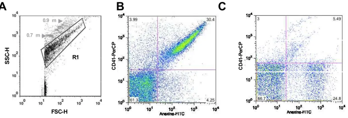
3.2.7. Identificação de micropartículas (MPs) pela Citometria de fluxo Análise fenotípica das MPs por citometria de fluxo
de 100 µL do plasma contendo as MPs com os anticorpos específicos para: neutrófilos (CD66/PE), linfócitos T (CD3/PE), plaquetas (CD41a/PerCP), leucócitos (CD45/APC), monócitos (CD14/PerCP), eritrócitos (CD235a/PECy5) e células endoteliais (CD51/61/PE). Após a adição dos anticorpos, as amostras serão incubadas por 30 min. protegidas da luz. As MPs serão ressuspensas em 100 µL do tampão de ligação de Anexina V (BD Pharmingen) e, finalmente, 5 µL de Anexina V/FITC serão adicionados, já que a Anexina V reconhece resíduos de fosfatidilserina que geralmente estão presentes na superfície das MPs.
As amostras serão levadas ao citômetro de fluxo LSR-Fortessa (Becton Dickson - BD, E.U.A.), onde cerca de 100.000 eventos serão obtidos em cada amostra, sendo pelo menos 2.000 eventos dentro da região específica para MPs. As análises serão feitas utilizando o software FlowJo (Tree Star), onde serão construídos dots plots de todos os marcadores utilizados versus
Anexina/FITC, permitindo assim a identificação e quantificação de cada população de MPs específica.
Figura 5: Representação esquemática da seleção de micropartículas em plasma livre de plaquetas. Foram utilizadas microesferas fluorescentes de tamanho definido (0,7 - 0,9 µm) para delimitar a região (R1) correspondente às MPs em gráficos de tamanho (FSC) versus granulosidade (SSC) (Figura 5A). Além disso, foi utilizada Anexina V conjugada com fluoresceína – FITC versus o marcador de origem das micropartículas. Os resultados foram expressos em frequência percentual do quadrante duplo-positivo referente a presença de micropartículas Anexina V+ e o marcador de população celular+. No exemplo, temos dois exemplos de resultados da combinação Anexina V-FITC x CD41-PerCP (marcador de plaquetas) (Figuras 5B e 5C, respectivamente).
Plaqueta Endotélio Leucócito
15 14 Monócito ⁺ s ⁺
A
SS C-H FSC-H R10.7m m 0.9mm
4. Referências:
1. Estimated Caner Incidence, Mortality and Prevalence Worldwild in 2012.
International Agency for Research on Cancer.
http://globocan.iarc.fr/Pages/fact_sheets_population.aspx (acessado em
20/01/2014).
2. Estimativa de Câncer no Brasil 2014. Instituto Nacional do Câncer.
http://www.inca.gov.br/estimativa (acessado em 20/01/2014).
3. Anthoulakis C, Nikoloudis N. Pelvic MRI as the "gold standard" in the
subsequent evaluation of ultrasound-indeterminate adnexal lesions: A
systematic review. Gynecol Oncol. 2013(13):1271-7.
4. Ueland FR, et al. Preoperative differentiation of malignant from benign
ovarian tumors: the efficacy of morphplogy indexing and Doppler flow
sonography. Gynecol Oncol 2003; 91: 46-50.
5. Jian-Lun Xu, et al. Longitudinal evaluation of CA-125 velocity and prediction
of ovarian cancer. Gynecologic Oncology .2012; 125: 70–74.
6. Cancer Facts and Figures, 2013. American Cancer Society, Surveillance
Research. 2013; p:1-64.
7. Kurman, R J; Shih. The Origin and Pathogenesis of Epithelial Ovarian
8. A. P. Heintz, F. Odicino, P. Maisonneuve et al., ―Carcinoma of the ovary.
FIGO 26th annual report on the results of treatment in gynecological cancer,‖
International Journal of Gynaecology and Obstetrics. 2006; 95:161–192.
9. Benedet, J.L., et al. FIGO staging classifications and clinical practice
guidelines in the management of gynecologic cancers. FIGO Committee on
Gynecologic Oncology. Int J. Gynaecol Obstet. 2000. 70 (2): 209-62.
10. Chang SJ et al. Survival impact of complete cytoreduction to no gross
residual disease for advanced-stage ovarian cancer: A meta-analysis.
Gynecologic Oncology. 2013; 130: 493–498.
11. Trimbus JP, et al. Impact of Adjuvant Chemotherapy and Surgical Staging in
Early-Stage Ovarian Carcinoma: European Organisation for Research and
Treatment of Cancer–Adjuvant ChemoTherapy in Ovarian Neoplasm Trial. J
Natl Cancer Inst 2003;95: 113–25.
12. Harmandayan, G.Z. et al. Ovarian cancer patient surveillance after
curative-intent initial treatment. Gynecologic Oncology .2011; 120: 205–208.
13. Visser, K.E., A. Eichten, and L.M.Coussens, Paradoxical roles of the
immune system during cancer development. NatRev Cancer, 2006. 6(1):
p.24-37.
14. Chen, Yu-Li, et al. Interferon-gamma in ascites could be a predictive
biomarker of outcome in ovarian carcinoma. Gynecologic Oncology .2013; 131:
15. Wajant H. The role of TNF in cancer. Results Probl Cell Differ. 2009;
49:1-15.
16. Szlosarek PW, Grimshaw MJ, Kulbe H, Wilson JL, Wilbanks GD, et al.
Expression and regulation of tumor necrosis factor alpha in normal and
malignant ovarian epithelium. Mol Cancer Ther. 2006; 5: 382–390.
17. Aggarwal BB .Signalling pathways of the TNF superfamily: a double- edged
sword. Nat Rev Immunol. 2003; 3: 745–756.
18. Pinciroli et al. An IL6-correlated signature in serous epithelial ovarian cancer
associates with growth factor response. BMC Genomics 2013, 14:508.
19. Son DS, Kabir SM, Dong YL, Lee E, Adunyah SE. Inhibitory effect of tumor
suppressor p53 on proinflammatory chemokine expression in ovarian cancer
cells by reducing proteasomal degradation of IkB. PLoS One. 2012. 7:e51116.
20. E Chargaff, R. West. The biological significance of the thromboplastic
protein of blood. Journal of Biological Chemistry. 1946; 166: 187-196.
21. Weng, X., et al. Membrane microparticles and diseases. European Review
for Medical and Pharmacological Sciences. 2013; 17: 2420-2427.
22. Valenti, R. Tumor-released microvesicles as vehicles of
immunosuppression. Cancer research. 2007; 67: 2912-2915.
23. Hussein, M.N., et al. Inhibition of microparticle release triggers endothelial cell
24. Angelucci, A. et al. Vesicle associated urokinase plasminogen activator
promotes invasion in prostate cancer cell lines. Clinical and Experimental
Metastasis. 200; 18: 163-170.
25. Al-Nedawi, K. et al. Intercellular transfer of the oncogenic receptor EGFRvIII
by microvesicles derived from tumours cells. Nature Cell Biology. 2008; 10:
619-624.
26. Graves,E.L. Pro invasive properties in ovarian cancer ascites-derived
membrane vesicles. Cancer Research. 2004; 64: 7045-7049.
27. Shedden, K., et al. Expulsion of small molecules in vesicles shed by cancer
cells: association with gene expression and chemosensitivity profiles. Cancer
research. 2003; 63: 4331-4337.
28. Al-Nedawi, K. et al. Endothelial expression of autocrine VEGF upon the
uptake of tumor-derived microvesicles containing oncogenic EGFR.
Proceedings of the National Academy of Sciences of the United States of
America. 2009; 106: 3794-3799.
29. Wang TY, et al. Histologic and immunophenotypic classification of cervical
carcinomas by expression of the p53 homologue p63: a study of 250 cases.
Hum Pathol 2001; 32 (5):479-486.
30. Campos et al. Augmented plasma microparticles during acute Plasmodium
ARTICLE I
Frequency of circulating microparticles in patients with
ovarian cancer
Article Type: Original article
Keywords: ovarian cancer , inflammatory response, microparticles .
1. Introduction
Ovarian cancer comprises a heterogeneous group of neoplasms and is the most lethal gynecologic malignancy in the developed countries. Early detection of disease remains problematic as there is no good screening test and there is a lack of a clearly defined precursor lesion. Thereby, the majority of women are initially diagnosed with tumor metastases beyond the ovary, which results in diminished chances of long-term survival1,2. Surgical cytoreduction and adjuvant chemotherapy are the hallmarks of management for advanced ovarian cancer3.
Currently, the molecular events leading to the development of epithelial ovarian cancer are still unknown. Because of the great heterogeneity in molecular and biological status, ovarian cancer is actually many different diseases, which has different clinical outcomes, and may requires different treatments. Thus, it was proposed a new classification, categorizing ovarian cancer into type I and II tumors. Type I tumors are suggested to behave in an indolent manner, with a stable genome, although somatic mutations are frequently detected in a number of genes4. Type II tumors are proposed to be more aggressive and genetically highly instable; most of them have TP53 mutations, and almost half of the cases have mutation, hypermethylation, or dysfunction of BRCA1/25.These aggressive tumors account for 75% of all ovarian cancer, and are responsible for 90% of deaths from the disease6. Many researches have been conducted to identify a specific biomarker to the ovarian cancer. Therefore, it has been hypothesized that microparticles (MPs) play a fundamental role in the ovarian tumor microenvironment. They are phospholipid vesicles (< 1 μm) released from cells in response to a variety of stimuli, and they have been studied and gained attention due to the large amounts of them releasing from tumor cells. MPs are able to transport cell-specific surface antigens of their cell of origin, such antigens being used to identify the various subtypes of MPs7. It is postulated that MPs releasing from cells may alter the biological activity of the receiver cell, either by transferring receptors that can induce cell signalling, or by transferring second messengers. This binding of MP surface antigens to their specific receptor on cells allows intercellular signaling between distant cells, as well as other processes such as modulation of the immune response8. The purpose of
2. Materials and methods
2.1. Patients and specimens
Patients who were undergoing evaluation for suspected primary ovarian cancer from June 2010 to October 2013 were recruited in a Brazilian clinical center. All patients underwent debulking surgery and received standard platinum-based chemotherapy when indicated. The control group selected
presented no underlying malignancy or infection requiring benign gynecological surgery. Their medical records were obtained until October 2013. The Institutional Review Board approved the study protocol, and all patients provided informed signed consent. Blood sampling was carried intra-operatively, before the anesthetic induction. Flow cytometry was the approach to analyze MPs, cytokines/chemokines, as the method to assess the number and the phenotype of MPs and cytokines/chemokines9. Histological grading and
disease staging were based on the International Federation of Gynecology and Obstetrics (FIGO) classification10,11. In this study, FIGO stage I/II and FIGO stage III/IV ovarian cancers were considered early and advanced disease, respectively.
2.2. Purification of MPs from plasma
MPs were prepared as described elsewhere12,13,14. Briefly, Citrated blood (0.5 mL) was centrifuged at 1,500 × g for 15 min, and plasma was then cooled to -20°C before storage at -80°C. Samples were further centrifuged at 13,000 x g for 3 min to obtain platelet-free plasma. The latter was diluted 1:3 in citrated PBS containing heparin and centrifuged at 14,000 x g for 90 min at 15°C. The resultant MP pellet was then resuspended in 1× annexin V binding buffer (BD Biosciences, California, US).
2.3. Flow cytometry assays
2.3.1. Detection of plasmatic MPs
Unless otherwise stated, all reagentes and mAbs used in the Flow cytometry experiments were provided from BD Biosciences. MPs isolated from plasma were gated (R1) based on their forward (FSC) and side (SSC) scatter distribution in density plots as compared to the distribution of synthetic 0.7 – 0.9 μm SPHERO™ Amino Fluorescent Particles (Spherotech Inc. Libertyville, Illinois, US). Taking into account the presence of phosphatidylserine (PS) residues in MPs surface, events present in R1 were accessed for their positive staining for annexin V (BD Bioscience) – a classical marker for microparticles – using PE-conjugated monoclonal antibodies (mAbs). Mouse IgG PE conjugated isotype control mAbs were used to properly place gates. Annexin V+ events
FITC-conjugated mAbs against the cell markers CD66 (neutrophils), CD41a (platelets), CD51 (endothelial cells), CD235a (erythrocytes), CD45 (leukocytes), CD3 (lymphocytes), and CD14 (monocytes) or the correspondent mouse IgG FITC-conjugated isotype control mABs. The samples were analyzed in a Flow Cytometry FACSCalibur (Becton-Dickinson, California, US). Over 100,000 events were acquired on each sample, to reach at least 2,000 events within the MPs gate.
2.4.Statistical Analyses
Statistical analyses were performed using SPSS software version 13.0 (SPSS Inc., Chicago, IL). Shapiro–Wilk tests were used to verify if the variables were normally distributed. Non-parametric data were compared using the Kruskal–Wallis test followed by the Dunns post-hoc test. Comparison between 2 groups was done using the Mann–Whitney U test with Bonferroni's correction (non-normal data) or t-test (normal data). Normal data are presented as mean and standard deviation, while non-normal variables are presented as median and interquartile range (25th–75th percentiles). Correlations were analyzed using the Pearson or Spearman 2-sided test, if the data were parametric or non-parametri, respectively, and Pearson χ2 test was performed to frequency
3. Results
The phenotype of plasma circulating MPs was investigated with specific mAbs for cell markers (i.e. CD41, CD235, CD45, CD3, CD51, CD14 and CD66) that
were used to discriminate the cellular sources of annexin V+ MPs. The
percentages of annexin V+ MPs stained for each cell marker was compared
between samples from OC patients and control group. Other clinic-pathological data were also analyzed.
The frequency of leukocyte (p < 0.005) derived-microparticles (LMPs) was
A
B
Figure 1: A. Percentage of circulating microparticles (MPs) in patients with
ovarian cancer (OC) and control group according to their specific cellular origin. B. Percentage of circulating microparticles (MPs) in patients with type I and II tumors according to their specific cellular origin.
A
B
C
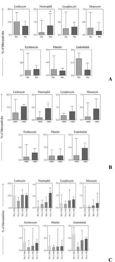
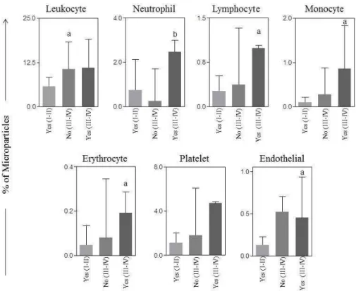
Similarly, when we analyzed the association between MPs with other clinical data, such as stage and cytoreductive operation, we observed that LMPs were higher in patients with advanced stage OC as well as with residual disease > 1cm after surgery when compared with those in early stage. Accordingly, the levels of NMPs were statistically increased in women with stage III/IV who had optimal operation as compared with those presenting the same stage not well debulked. The frequency of lymphocyte, monocyte, erythrocyte and endothelial derived-microparticles were significantly increased in plasma samples from patients with advanced OC and residual disease after surgery lower than 1cm as compared with those with early stage disease and optimal surgical treatment (Figure 3).
Figure 3. Comparison of circulating microparticles (MPs) from patients with
4. Discussion
Although the incidence of OC is low, it is the most lethal gynecologic malignancy and typically has a poor prognosis. Studies have focused on the microparticles released from tumor and from inflammatory cells to identify a biomarker for OC. MPs may represent a potential biomarker because they can be easily isolated from blood, and they have particular features that may correlate with stage and clinical data.
The present study investigated serum levels of neutrophils, platelets, endothelial, erythrocytes, leukocytes, lymphocytes and monocytes derived-microparticles in women with OC and without malignancy. The frequency of LMPs was increased in women with OC as well as was even higher in type II tumors when compared with those without malignancy.
It has been known that tumor microenvironment consists of a variable
combination of tumor cells, stromal fibroblasts, endothelial cells and infiltrating
leukocytes, such as neutrophils, macrophages, lymphocytes and other
inflammatory cells, and they play a fundamental role in tumor modulation and
progression15,16.
Other important consideration is the association between MPs and
communication between cancer cells and various stromal cells infiltrating the tumour interstitium. The actual molecular composition of MPs varies depending on the mechanism of their formation as well as the type and functional state of their cellular origin. MPs are important carriers of membrane components or bioactive molecules, and they can mediate exchange of signaling proteins and genetic material, which altogether may support tumor growth and progression.
A large quantity of MPs has been identified in fluids of patients with
malignant pathologies. Thereby, these study findings corroborate with the
relationship between the immune system and OC inflammation, especially in
tumors with higher aggressiveness, as ovarian type II tumors. These results
enhance the dualistic model that categorizes OC in two types. They suggest a
difference in susceptibility to carcinogenesis in both tumors. On the other hand,
we might not find any explanation for the presence of the decreased levels of
endothelial derived microparticles in the OC group, unless the low number of
patients.
We observed an increased expression of NMPs in patients with higher
levels of CA125. This is an interesting result, and could be correlated with the
ability of the tumor to expresses biomarkers. Moreover, NMPs were statistically
higher in women who had optimally citoreductive surgery, especially those with
participate in the control of immune responses. These NMPs have the particularity to be involved very early in inflammation, which might be crucial for determining later aspects of the cascade responsible for acquired immunity, and may responsible for controlling the immune response as well18,19. The present
study showed that elevated frequency of NMPs could be considered as a prognostic factor for optimal surgical treatment. Nevertheless, additional studies are necessary to support this hypothesis.
Probably, cancer MPs contribute to hypercoagulability, as various types of cancer cells are known to secrete MPs carrying tissue factor VII20. As
mentioned earlier, MPs can interact with the extracellular matrix by depositing paracrine information or facilitating matrix degradation, thereby creating paths of least resistance. Bothey conditions can induce the stromal microenvironment to promote angiogenesis, evasion of the immune response, tumor invasion, and, potentially, metastasis21. Therefore, it is expected to find out a substantial
number of MPs in advanced disease. This study demonstrated that circulating leukocyte, lymphocyte, monocyte, erythrocyte and endothelial derived MPs were higher in patients in advanced stage disease with optimal cytoreductive operation compared to those treated in early ovarian cancer.
A potential limitation of the statistics analysis performed here was that multiple comparison were realized, aiming to find possible associations between MPs and clinical/biological parameters; however, it should be take account that these significant findings were hypothesis-generating, and, consequently, its represent a realistic interpretation of the facts. Moreover, the limited number of patients in the study is explained by the low prevalence of the disease. The median follow-up could also be higher. Additional approaches should also be explored to guarantee the ability to isolate and quantify ovarian cancer MPs from blood and other biological fluids.
This paper describes the initial attempts to characterize MP phenotype
features in ovarian cancer. It was shown here that plasma levels of LMPs were
Conflict of interest statement
None of the authors has any conflicts of interest
Acknowledgments
5. Referências:
1. American Cancer Society. Cancer Facts and Figures 2013. Atlanta: American
Cancer Society; 2013.
2. Chan A, Gilks B, Kwon J, Tinker AV. New insights into the pathogenesis of
ovarian carcinoma. Time to rethink ovarian cancer screening. Obstet
Gynecol. 2012; 120: p.935-40.
3. Chang S J et al. Survival impact of complete cytoreduction to no gross
residual disease for advanced-stage ovarian cancer: A meta-analysis.
Gynecologic Oncology. 2013; 130: p.493–498.
4. Kurman RJ, Shih Ie M. Molecular pathogenesis and extraovarian origin of
epithelial ovarian cancer—shifting the paradigm. Hum Pathol. 2011;
42:918–31.
5. Spellman PT, et al. Integrated genomic analyses of ovarian carcinoma. Nature 2011; 474:609–15.
6. Kurman RJ, Shih IeM. The Origin and Pathogenesis of Epithelial Ovarian Cancer – Proposed Inifying Theory. Am J Surg Pathol. 2010;34(3):433-43.
8.Leroyer AS, Anfosso F, Lacroix R, Sabatier F, Simoncini S, Njock SM, et al.
Endothelial-derived microparticles: biological conveyors at the crossroad of inflammation, thrombosis and angiogenesis. Thromb Haemost 2010; 104(3): 456-463.
9. van der Heyde HC, Gramaglia I, Combes V, George TC, Grau GE. Flow cytometric analysis of microparticles. Methods Mol Biol. 2011; 699:337-54.
10. A. P. Heintz, F. Odicino, P. Maisonneuve et al., ―Carcinoma of the ovary. FIGO 26th annual report on the results of treatment in gynecological cancer,‖ International Journal of Gynaecology and Obstetrics. 2006; 95:161–192.
11. Benedet, J.L., et al. FIGO staging classifications and clinical practice guidelines in the management of gynecologic cancers. FIGO Committee on Gynecologic Oncology. Int J. Gynaecol Obstet 2000. 70 (2): 209-62.
12. Combes V, Taylor T, Juhan-Vague I, Mège J, Mwenechanya J, Tembo M, Grau G, Molyneux M. Circulating endothelial microparticles in malawian children with severe falciparum malaria complicated with coma. JAMA 2004, 291:2542-2544.
14. Campos et al. Augmented plasma microparticles during acute Plasmodium vivax infection. Malaria Journal 2010, 9:327
15. Visser, K.E., A. Eichten, and L.M.Coussens, Paradoxical roles of the immune system during cancer development. Nat Rev Cancer, 2006. 6(1):
24-37.
16. Chen, Yu-Li, et al. Interferon-gamma in ascites could be a predictive biomarker of outcome in ovarian carcinoma. Gynecologic Oncology. 2013;
131:63–68.
17. Weng, X., et al. Membrane microparticles and diseases. European Review for Medical and Pharmacological Sciences. 2013; 17(18): 2420-2427.
18. Eken C; Gasser O; et al. Polymorphonuclear Neutrophil-Derived Ectosomes Interfere with the Maturation of Monocyte-Derived Dendritic Cells . J Immunol. 2008; 180:817-824.
19. Falanga A, Tartari CJ, Marchetti M. Microparticles in tumor progression.
Thromb Res. 2012;129(1):132-136.
20. Yokota N; Koizume S; Self-production of tissue factor-coagulation factor VII complex by ovarian cancer cells. British Journal of Cancer. 2009; 101, 2023 – 2029.
ARTICLE II
Circulating cytokine, chemokine and microparticles signatures in women
with ovarian cancer
Article Type: Original article
Keywords: Ovarian cancer , inflammatory response, cytokines, chemokines, microparticles .
1. Introduction
Ovarian cancer (OC) is still the most frequent cause of death by gynecological malignancy for women. It has the highest fatality-to-case ratio for
all gynecologic malignancies because more than two-thirds of patients have advanced disease at diagnosis. The lethal nature of this pathology is predominantly attributed to its ability to spread extensively with minimal clinical symptoms. In the United States, it is the tenth most common malignancy in women, and accounts for the fifth highest number of deaths, an estimated 14.030 deaths in 20131. OC accounts for about 2.1% of all cancers among
women at Brazil, and around 5,680 new cases are stimated in 20142.. Despite
To date, all attempted OC screening strategies have failed, probably because this disease represents different kinds of cancers4. A new classification
for OC in two different types of cancer has been introduced: Type I tumors involve low grade and indolent tumors. It accounts for many early stage cancers and are characterized by specific mutations including KRAS, BRAF, ERB2, CTNNB1, PTEN, PIK3K, ARID1A and PPP2R1A. Type I tumors rarely harbor TP53 mutations and are stable genetically 5. Type II ovarian tumors are considered the most frequently diagnosed, aggressive, genetically instable, and often disseminated kind of disease. Type II tumors have a very high frequency of TP53 mutations 6.
Molecular biology of oncogenesis in ovarian cancer consists of multiple complex pathways. Previous research has focused on identifying prognostic markers for OC but few definitive results have been robustly validated 7.
Accordingly, malignant cells and stromal cells communicate and exchange information by direct cell-to-cell contacts as well as release of signalling molecules, such as soluble factors10,11. Thereby, there is an
association between MPs, cytokine and chemokine activity during inflammatory responses. Cytokines are small soluble polypeptides released from cells to regulate the activities of other cells via interactions with specific cytokine receptors expressed on their surface12. In OC, there is an increased expression of cytokines/chemokines and/or receptors13. They are able to enhance key
aspects associated with tumor progression, such as proliferation, migration and survival, including chemo-resistance14. This can be due to direct effects on the cancer cells themselves, or via indirect effects on components of the immune system.
The purpose of the study was to evaluate the levels of circulating soluble biomarkers-microparticles, cytokines and chemokines- to characterize the
2. Patients, Materials and methods
2.1. Patients and specimens
Patients who were undergoing evaluation for suspected primary ovarian cancer from June 2010 to October 2013 were recruited in a Brazilian clinical center. All patients underwent debulking surgery and received standard platinum-based chemotherapy when indicated. The control group was selected
as with no underlying malignancy or infection requiring benign gynecological surgery. Their medical records were obtained until October 2013. The Institutional Review Board approved the study protocol, and all of the patients provided informed signed consent. Blood sampling was carried out intra-operatively, before the anesthetic induction. Flow cytometry was the approach to analyze MPs, cytokines/chemokines 15. Histological grading and disease staging were based on the International Federation of Gynecology and Obstetrics (FIGO) classification16,17. In this study, FIGO stage I/II and FIGO
stage III/IV ovarian cancers were considered early and advanced disease, respectively.
2.2. Purification of MPs from plasma
MPs were prepared as described elsewhere 18,19,20. Briefly, citrated blood (0.5 mL) was centrifuged at 1,500 × g for 15 min, and plasma was then cooled to -20°C before storage at -80°C. Samples were further centrifuged at 13,000 x g for 3 min to obtain platelet-free plasma. The latter was diluted 1:3 in citrated PBS containing heparin, and centrifuged at 14,000 x g for 90 min at 15°C. The resultant MP pellet was then resuspended in 1× annexin V binding buffer (BD Biosciences, California, US).
2.3. Flow cytometric assays
2.3.1. Detection of plasma MPs
Unless otherwise stated, all reagents and mAbs used in the flow cytometric experiments were from BD Biosciences. MPs isolated from plasma were gated (R1) based on their forward (FSC) and side (SSC) scatter distribution in density plots as compared to the distribution of synthetic 0.7 – 0.9 μm SPHERO™ Amino Fluorescent Particles (Spherotech Inc. Libertyville, Illinois, US). Taking into account the presence of phosphatidylserine (PS) residues in MPs surface, events present in R1 were accessed for their positive staining for annexin V (BD Bioscience) – a classical marker for microparticles – using PE-conjugated monoclonal antibodies (mAbs). Mouse IgG PE conjugated isotype control mAbs were used to properly define the gates. Annexin V+ events
FITC-conjugated mAbs against the cell markers CD66 (neutrophils), CD41a (platelets), CD51 (endothelial cells), CD235a (erythrocytes), CD45 (leukocytes), CD3 (lymphocytes), and CD14 (monocytes) or the correspondent mouse IgG FITC-conjugated isotype control mABs. The samples were analyzed in a Flow Cytometry FACSCalibur (Becton-Dickinson, California, US). Over 100,000 events were acquired on each sample, to reach at least 2,000 events within the MPs gate.
2.3.2. Detection of plasmatic cytokine/chemokine levels by cytometric bead array immunoassay (CBA)
The secreted cytokine/chemokine analysis by flow cytometry is a methodology in which the simultaneous measurement of multiple biomarkers in a single sample is performed21,22,23. For plasma biomarkers determination,
2.4. Statistical Analysis
Statistical analyses were performed using SPSS software version 13.0 (SPSS Inc., Chicago, IL). Shapiro–Wilk tests were used to verify if the variables were normally distributed. Non-parametric data were compared using the Kruskal–Wallis test followed by the Dunns post-hoc test. Comparison between 2 groups was done using the Mann–Whitney U test with Bonferroni's correction (non-parametric data) or t-test (parametric data). Parametric data are presented as mean and standard deviation, while non-parametric variables are presented as median and interquartile range (25th–75th percentiles). Correlations were analyzed using the Pearson or Spearman 2-sided test, and Pearson χ2 test to frequency differences. Differences were considered significant when P<0.05.
2.4.1 Network Analyses
Cytokine/chemokine/MPs networks were assembled to assess the
association between the cytokine,chemokine and MPs each clinical group.
Spearman’s correlation test was performed to assess the association between
biomarker levels (pg/mL), and frequency of MPs (%). The positive and negative
correlations were significant when the p<0.05. The correlation index (r) were
used to categorize the correlation strength as negative (r<0), moderate
(0.36>r<0.67) and strong (r>0.68). The Graphpad Prism 5.00 software (San
Diego, USA) was used for correlation analyses, and the Cytoscape (version 2.8)
3. Results
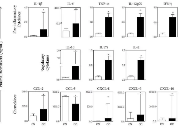
Figure 1. Levels of pro-inflammatory (IL-1β, IL-6, TNF-α, IL-12p70, IFN-γ), regulatory cytokines (IL-2, IL-10, IL-17a), and chemokines (CCL-2, CCL5, CXCL8, CXCL9 E CXCL10) in ovarian cancer (OC) and control (CN) groups. The results are presented in a column chart format and are expressed as the median and interquartile range at pg/mL. Statistical differences between OC and CN were considered significant when p < 0.05.
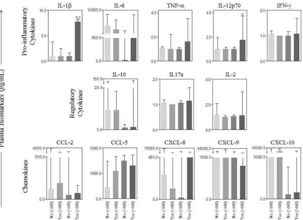
There were no differences in the plasma levels of cytocines and chemokines between type I and type II tumors (Figure 2A). However, when we analyzed the association between these biomarkers with the type of tumor and optimal cytoreductive surgery, we observed that the percentage of pro-inflammatory cytokines IL-1-β and IL-12p70 were statistically higher in patients
A
Figure 3: Levels of serum biomarkers versus optimal cytoreductive surgery and CA125 levels. . The results are presented in a column chart format and are expressed as the median and interquartile range at pg/mL. Statistical differences were considered significant when p < 0.05.
Stage disease were also analysed and we noted the association
Figure 4: Levels of serum biomarkers versus optimal cytoreductive surgery and stage. The results are presented in a column chart format and are expressed as the median and interquartile range at pg/mL. Statistical differences were considered significant when p < 0.05.
In our recent paper, the phenotype of plasma circulating MPs was investigated, and specific mAbs for cell markers i.e. CD41, CD235, CD45, CD3, CD51, CD14 and CD66 were used to discriminate the cellular sources of annexin V+ MPs. The percentages of annexin V+ MPs stained for each cell
We observed that the frequency of leukocyte (LMPs) (p < 0.005) derived-microparticles were significantly increased in plasma samples from OC patients as compared with CN, and in patients with type II tumors as compared with type I tumors. Other association was the higher levels of neutrophil derived MPs (NMPs) in patients presenting optimal surgery and increased levels of CA125
as well (>600 U/mL) (See other findings in the first paper).
To evaluate the supposed relationships among cytokines, chemokines and MPs in OC and CN group, using all data obtained in this study, we built
biological networks, where the nodes represent the cytokines, chemokines and MPs evaluated, and the edges represent positive or negative correlations. We observed that there was a balance among cytokines, chemokines and MPs in the CN group. There were two networks to OC, the first one was composed of twelve cytokines/chemokines, and the other was composed of seven MPs. We found that the first network to OC was characterized by strong and moderate correlations between inflammatory/regulatory cytokines and chemokines, as we found mainly strong correlations in the other network among MPs. Noteworthy,
Thereby, we decided to create a cellular interaction network according to type I and II tumors. We found many moderate and strong correlations among cytokines, chemokines and MPs, and just one negative interconnection in the networks. Interestingly, we also observed a greater number of correlations in type II tumors as compared to type I. This result may reflect the differences between carcinogenesis in both tumors (Figure 6).