Int. J. Electrochem. Sci., 5 (2010) 1399 - 1410
International Journal of
ELECTROCHEMICAL
SCIENCE
www.electrochemsci.org
Approach on the Electrochemical Reactivity of Poly-L-Glutamic
Acid Against Doxorubicin and Its Application in the
Development of a Voltammetric Sensor
Daniela Pereira dos Santos1, Márcio Fernando Bergamini2, Maria Valnice Boldrin Zanoni1,*
1
Institute of Chemistry - University of São Paulo State, UNESP, C.P. 355, 14801-970, Araraquara, SP, Brazil
2
Departamento de Química, Setor de Ciências Exatas, Centro Politécnico, UFPR, 81531-990, Curitiba, PR, Brazil
*
E-mail: [email protected]
Received: 24 June 2010 / Accepted: 19 August 2010 / Published: 1 October 2010
The poly-L-glutamic acid (PGA), was used a biodegradable polymers as conjugated to doxorubicin (DXR) and also in the modification glassy carbon electrode. The interaction operate via terminal amino groups of the drug and the side chain carboxyl groups of branched PGA film and it is the base to develop a simple voltammetric sensor for the determination of doxorubicin in human urine. The preconcentration of DXR on the modified electrode occurs under open circuit and studied by the methods of square wave voltammetry to monitor the drug. Standard curves were linear over the concentration range of 2.20 – 44.5 µmol L-1 giving a detection limit of 0.45 µmol L-1 show that the proposed sensor is suitable for doxorubicin determination in real samples.
Keywords: Doxorubicin, poly-L-glutamic acid, adsorption, semiquinone free radical, urine.
1. INTRODUCTION
Doxorubicin (DXR), Fig. 1, is an antibiotic of the anthracyclines family, widely used in the cancer treatment. But the clinical usefulness of DXR is limited by its dose-related cardiotoxicity. Therefore, much effort has been devoted to define new strategies and biodistribution profile with the aim to reduce their toxicity [1-2].
mechanism of cytotoxicity of DXR [3-5], it is accepted that free radical formation plays an important role in its antitumor effect [6].
Figure 1. Chemical structure of Doxorubicin.
From biological point of view, the doxorubicin has the capacity of generate free radical species from one-electron reduction of ring C, which leads to the formation of a semiquinone free radical following a complex mechanism. This radical is relatively stable under anoxic conditions. On the other hand, the formation of an oxidized semiquinone in ring B is also possible under specific condition, as complexation reaction with iron, for instance. These sequences of reactions are known as “redox -cycling” of doxorubicin, which are potentially important to the knowledge its cytotoxicity [6]. It is known that DXR can promote oxidative damage to DNA in cancerous cells [7-10].
Several methods have been reported for the analysis of DXR based on spectrophotometric and chromatographic techniques [11-14]. Electrochemical studies have been evaluated for both the understanding of DXR action mechanism in vivo and determination in several matrices [15-22].
Doxorubicin is a complex molecule, where both quinone and hydroquinone groups are electroactives and can be used to identify electrochemical reduction and/or oxidation of the drug. The redox process presents two sets of reduction waves, corresponding to the reduction of the quinone group and the carbonyl side chain, respectively. The method is the base for DXR determination via electrochemical reduction [17-19].
The oxidation of DXR involves the oxidation of the hydroquinone group of the dihydroxy-antraquinone moiety after two electrons/two protons transference, which is usually complicated by adsorption process, as shown in the mechanism proposed in the literature (Scheme 1) [20-22].
An important contribution describes the use of electrochemistry detection for in situ oxidative damage to DNA [15, 20]. The proposed mechanism involves reduction and oxidation of DXR in situ when intercalated in double helix DNA by the generation of the mutagenic 8-oxoguanine.
has been investigated to develop new systems of drug released. Within this context, the use of poly-L- glutamic acid (PGA) as model of polyaminoacids could be an ideal system, due its biological and chemical properties [23-24]. This polyaminoacid presents repetitive unities of a specific functional group in a controllable chain, which promotes multiple functions and the availability to be excreted in urine. In addition, the versatile application of the PGA polyaminoacid to coat different materials leads to the possibility to construct different electrochemical sensor previously applied for hydroquinone and catechol [25], hydrazine [26], ascorbic acid [27], uric acid [28], caffeic acid [29], rutin [30] and amoxicillin [31] determination. Thus, the use of poly-L-glutamic acid modified glassy carbon electrode could contribute to try immobilize DXR on the electrode surface, being the promising candidate as carrier-mediated doxorubicin controlled delivery and also to develop a new electroanalytical method for its determination in a first moment.
In the present study we investigated the behavior of the doxorubicin at poly-L-glutamic acid chemically modified electrode, and the possible formation of the semiquinone free radical, which could contribute to decreased its cytotoxicity. It is also reported the optimized conditions to develop a voltammetric sensor for DXR determination in human urine without any pre treatment.
2. EXPERIMENTAL PART
2.1. Apparatus and reagents
Voltammetric measurements were performed with an AUTOLAB PGSTAT 30 potentiostat connected to a microcomputer controlled by software GPES 4.9 for data acquisition. A three electrode system (EG&G PARC) consisting of an SCE as reference electrode, a platinum wire as auxiliary electrode and a glassy carbon electrode as working electrode were used. The glassy carbon electrode (GCE) (3 mm diameter) was polished with alumina (0.3 mm, BUEHLER), washed, and dried at room temperature before use. The UV-vis absorbance analysis were measuring using a Hewlett Packard 8453 spectrophotometer operated at 200–800 nm in a quartz cell. The pH measurements were performed using a Micronal pH meter B222 model with a Micronal combined pH reference electrode. All experiments were done at room temperature.
Suprapur grade reagents supplied by Merck and purified water from a Millipore Milli-Q system (conductivity 0.1 S cm-1) were used in the preparation of all solutions. Phosphate buffer 0.1 mol L-1 were used as supporting electrolyte and prepared by mixing appropriate amounts of sodium phosphate dibasic and sodium phosphate monobasic. Solutions of doxorubicin hydrochloride were prepared using pure analytical grade. Poly-L-glutamic acid (MM = 50,000, Sigma-Aldrich) solution 1% (w/v) in water was utilized in all experiments.
2.2. Preparation of modified glassy carbon electrode
bifunctional nature resulting in its cross-linking capability [32]. Its bifunctional nature facilitates the formation of Schiff base [33], involving the direct reaction between aldehyde groups from glutaraldehyde with the free amino group present in the poly-L-glutamic acid. For this, an aliquot of 4
L of the GLU (0.05% v/v) solution was placed on the polished GCE surface, followed by addition of 12 L of the PGA (1% w/v) solution. The in situ prepared PGA:GLU modified GCE was then set aside and allowed to dry in a sterile atmosphere at room temperature. The voltammetric experiments with the PGA film modified GCE was obtained after submitted it to a conditioning procedure in phosphate buffer 0.1 mol L-1 pH 7.0, where successive voltammetric scan were recorded between -0.8 to +2.0 V (15 cycles, at 100 mV s-1). Then the electrodes were washed and transferred into new solutions containing supporting electrolyte and DXR when necessary.
2.3. Analysis in the commercial sample
The human urine samples were spiked with 1.0 x 10⁻³ mol L-1 of DXR treated in 0.2 mL of methanol for subsequent removal of the proteins. These samples were agitated and placed in a micro centrifuge during 3 min at 6.000 rpm. The supernatant was removed and transferred to a solution of 20 mL of phosphate buffer pH 7.0. All the measurements were carried out using the same PGA/GLU film modified electrode.
3. RESULTS AND DISCUSSION
3.1. Interaction of doxorubicin with poly-L-glutamic acid films on glassy carbon electrode
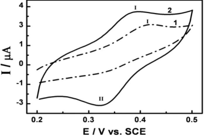
Figure 2 exhibits the electrochemical oxidation of 1.0 x 10⁻⁴ mol L-1 DXR in phosphate buffer pH 7.0 at a bare (curve 1) and PGA:GLU film modified glassy carbon electrode (curve 2), prepared as described in the experimental section.
On bare electrode the DXR is voltammetrically oxidized on the glassy carbon electrode in a process of only one step at around +0.41 V (peak I), attributed to the oxidation of hydroquinone group, in agreement with Scheme 1 described in the literature [20].
Scheme 1. Mechanism for the oxidation of doxorubicin.
A small voltammetric signal for the reverse scan, where the anode-to-cathode peak height ratio (Ipa/Ipc) is around 2 and Epa - Epc= 80 mV, clearly indicating that the process is not completely reversible in this experimental condition. This behavior also agrees with literature [19-22], where it is proposed that the DXR is oxidized by two electrons and two protons transfer involving a complex reaction mechanism dependent of pH.
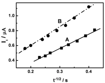
In order to further confirm that the number of electrons transferred is close to 2.0, chronoamperometric experiments were carried out for 1.0 x 10⁻⁴ mol L-1 DXR oxidation in phosphate buffer pH 7.0 and also similar curve was performed for potassium ferricyanide as a control experiment using the same experimental condition. The results very symmetrical chronoamperograms and the plots of net electrolysis current vs. t-1/2 (Fig. 3) show two straight lines, as expected by means of Cottrell equation (equation 1) [34]:
(1)
[35]. Thus, taking the ferrocyanide ion as a standard compound, the number of electrons transferred in DXR oxidation is close to 2
.
Figure 3. Plots of It vs. t-1/2 from chronoamperograms obtained for oxidation of 1.0 x 10⁻⁴ mol L-1 of the ferrocyanide (A) and doxorubicin (B) solutions in phosphate buffer pH 7.0 at a bare glassy carbon electrode.
The effect of glutaraldehyde in the composition of the resulting film of PGA:GLU could interfere in the efficiency of the modified electrode to pre- accumulate the drug. Crosslinking of the PGA with GLU involves a reaction of the amino groups that are on the PGA film along with the aldehyde group of the glutaraldehyde, which mixed together form a Schiff Base [33], and the process increases the adherence of PGA onto the electrode surface. To this end, coating the carbon disk with 16 L samples of a mixture of PGA:GLU giving the following ratios: 97.5%:2.5% and 87.5%:12.5% of PGA:GLU. The reduction peak currents obtained with these two electrodes indicate that the 87.5%:12.5% PGA:GLU coating gives acceptable anion exchange properties whilst giving the required improvement of adhesion to the glassy carbon electrode surface[27, 31].
The modified glassy carbon electrode coated with mixture of poly-L-glutamic acid/glutaraldehyde (PGA:GLU) at a proportion of 87.5%:12.5% was immersed in a solution of 1.0 x 10⁻⁴ mol L-1 DXR in phosphate buffer at pH 7.0 and the obtained cyclic voltammogram is shown in curve 2 of Fig.2. The cyclic voltammogram present redox peak at + 0.38 V (I) and + 0.32 V (II) with higher intensities than those obtained for the bare electrode (curve 1 of Fig.2). In addition, both peaks exhibited the corresponding anodic peak in the reverse scan, which intensity was not verified on bare electrode. This behavior indicates that the product of the reoxidation step is stabilized by the PGA film and presents a defined anodic peak with values of Ipa/Ipc around of 1. Both peaks are separated by Ep = Epa - Epc = 60 mV, indicating transference of one electron.
the peak current values during successive 20 scans. Both cathodic and anodic peak currents increase with successive scans clearly indicating that the DXR is gradually incorporated into the PGA film on the glassy carbon electrode surface along with its reductive product. Taking into consideration that at pH 7.0 the sugar residue of DXR presents positive charged (pKa = 8.2) and the carboxylic group of PGA loaded negatively (pKa = 4.2), the strong affinity that PGA coating exhibited to the DXR seems to be due to electrostatic interaction or even interaction with amine groups available in the DXR and carboxylic group in the film.
In order to investigate the adherence of DXR onto the electrode-surface film coating, cyclic voltammograms were carried out comparing the cathodic response of the glassy carbon electrode modified by PGA:GLU loaded with DXR. This accumulation was obtained under electrochemical cycling (20 cycles) after immersion in 1.0 x 10⁻⁴ mol L-1 of DXR in phosphate buffer at pH 7.0. Then the loaded electrode was transferred to a new supporting-electrolyte solution. For both electrodes the first voltammogram recorded was similar to that obtained in the original solution. On carrying out successive potential cycling applied to the electrode, the peak current is kept almost constant up to 10 cycles, showing a reduction of 2% in the peak intensity. This indicates that the redox process does not form any adsorbed compound that could promote poisoning of the film.
The influence of time in the incorporation of DXR on the electrode response by open circuit was also investigated controlling the time of immersion of the modified electrode in 1.0 x 10⁻⁴ mol L-1 DXR pH 7.0 phosphate buffer solutions from time of 0 to 360s under stirring. The electrode was transferred into a supporting electrolyte phosphate buffer pH 7.0 solution after wash with water. The effect of accumulation time on peak current is increases linearly with the permanence time of the electrode in the solution, reaching a maximum value after 3 min. These results indicate that the DXR is incorporated in the film up to a maximum time where saturation of the active sites is observed. These results show that the DXR adsorption on the modified surface PGA:GLU is more efficient using accumulation “in situ” method, and controlled accumulation time of 3 min. should be recommended.
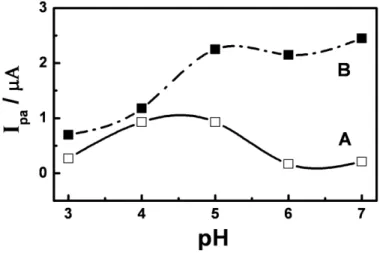
The influence of the pH on the DXR accumulation process onto the PGA:GLU films was examined over a range of pH values from 2 to 12. Once the PGA:GLU-coated electrode was loaded in 1.0 x 10⁻⁴ mol L-1 DXR at different pH values, it was transferred to a new solution of supporting electrolyte. The analyte was pre-concentrated on the film at all pH values investigated during 3 min. These procedures were also conducted using bare electrodes. At both electrodes there is no peak for DXR oxidation at pH 3.0 and pH 7.0, due instability of DXR as previously described in literature [36]. The cyclic voltammograms presents a reversible pair of peaks in all interval of pH 3.0 to 7.0, with ipa/ipc values close to 1 and values of Epa - Epc close to 59 mV [34]. This behavior is not observed at the bare electrode, were the voltammograms exhibit characteristics of a reversible process for DXR only at values of pH 4.0 – 5.0.
As shown Fig. 4, it can also be observed that the values of the anodic peak potentials shifts to less positive values with the increase of the pH for both modified and bare electrode. A linear relationship is observed for Epa vs. pH following the equations: (GCE): Epa (mV) = 887 – 67.0 pH; R = 0.993 (n = 5) and (PGA:GLU): Epa (mV) = 957 – 83.0 pH; R = 0.997 (n = 5).
voltammetric response exhibits a single, reversible, two-electron wave [17, 20]; H2Q Q + 2e- + 2H+ on bare electrode. But, the mechanism seems similar on GCE/PGA:GLU modified electrode, although the transferred number of electrons is close to 1 and also the number of protons involved in the chemical reaction is close to 1 for modified electrode, as obtained by equation Ep/pH = 59 mH+/ne-, defined in literature [34].
Figure 4. Effect of the pH variation on (A) bare and (B) PGA:GLU film modified glassy carbon electrode to oxidation of 1.0 x 10⁻⁴ mol L-1 DXR in phosphate buffer pH 7.0 onto anodic peak potential. Scan rate = 50 mV s-1.
Figure 5. Effect of pH on anodic peak currents (Ipa) for oxidation of 1.0 x 10⁻⁴ mol L-1 DXR in phosphate buffer at a bare (A) and (B) glassy carbon electrode modified by films of PGA:GLU. Scan rate = 50 mV s-1.
for NH3+ group of daunosamine moiety. Taking into consideration that carboxylic group of PGA film is negatively charged (pKa = 4.2) in the pH region above pH 4, seems that DXR is pre-accumulating on electrode surface due electrostatics interaction among the amine group of the sugar residue of DXR and the non protonated group of carboxylic function of the polyaminoacid.
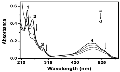
In order to test the interaction between DXR and amine group from poly-L-glutamic acid spectrophotometric curves was recorded for DXR in the absence and presence of poly-L-glutamic acid. The UV-Vis spectra for both experimental conditions are shown in Fig. 6. Typical absorption spectrum of DXR in original form in pH 7.0 exhibits four absorption bands at 233, 253, 293, 488 nm, respectively [37], as shown curve a. In the presence of PGA all the peak are slightly decreased in height and promotes a shift to longer wavelengths near 190 nm. So, it is possible to conclude that the accumulation of DXR on the modified electrode surface with PGA involves the protonated amine group of DXR and non-protonated carboxilate group of the PGA. Therefore, the oxidation of the conjugated occur via stabilization of the semiquinone intermediate on modified electrode, and the number of electrons transferred in the electrodic process is close to 1 and not 2 as diagnosed on bare glassy carbon electrode. After analysis of these parameters, pH 7.0 was chosen as the appropriate pH value for of DXR on modified electrode with PGA films by voltammetric techniques.
Figure 6. Absorption spectra obtained for 1.0 x 10⁻⁴ mol L-1 of doxorubicin (a) in phosphate buffer pH 7.0 in the presence of (b) 0.5 l; (c) 1.5 l and (d) 10 l of poly-L-glutamic acid 1% (w/v).
( = mV s-1) 1/2 (correlation coefficient = 0.993, n = 9). This indicates that the DXR oxidation is controlled by diffusion through the film on electrode surface.
In order to develop a voltammetric sensor able to determine DXR at low concentration in complex matrices it was further optimize the best experimental condition to get best analytical application of the modified electrode.
3.2. Analytical Determination of DXR on GCE/PGA/GLU modified electrode
The analytical properties of the PGA:GLU film modified glass carbon electrode was evaluated for oxidation of 1.0 x 10⁻⁴ mol L-1 DXR in 0.1 mol L-1 phosphate buffer pH 7.0 using square wave voltammetry (SWV). The optimization parameters evaluated by SWV technique were frequency, , (from 8 to 75 Hz), pulse amplitude, E, (from 10 to 100 mV) and scan increment, Es, (from 2 to 10 mV) were carried out. The experimental conditions were adopted, frequency of 20 Hz, 50 mV and scan increment, 2.0 mV, which was used further for analytical purposes.
Figure 7. (A) Square wave voltamograms obtained for DXR in 0.1 mol L-1 phosphate buffer solution pH 7.0 in different concentrations on glassy carbon electrode modified by PGA:GLU films: (a) = 2.23; (b) = 9.16; (c) = 17.9; (d) = 34.0; (e) = 37.5 and (f) = 44.5 (µmol L-1). (B) Analytical curve. = 10 Hz, E = 50 mV and Es = 2.0 mV.
analytical curve. The value of LD was 4.5 x 10⁻⁷ mol L-1, which indicated good sensitivity for the proposed method.
3.3. Analytical Applicability
In order to investigate its analytical application, the proposed SWV procedure based on PGA:GLU film modified electrode was applied for determination of DXR in human urine samples. The DXR quantization was carried out in triplicates using the methodology described in the experimental section submitting the sample to standard addition method of DXR 1.0 x 10⁻³ mol L-1 solution. A mean recovery of 99.2 ± 5.6% was obtained for 3 repetitions and the results were compared with a developed spectrophotometer method (Table 1).
Table 11. Results (3 determinations) obtained for the determination of DXR in human urine by square wave voltammetric procedure in comparison with the spectrophotometer method.
Method DXR add DXR found Recovery*(%)
Proposed
Spectrophotometer
5.00 µmol L-1
20.0 µmol L-1
4.9 µmol L-1
20.1 µmol L-1
99.2 ± 5.6
100.5 ± 3.5
The application of spectrophotometic method requires higher concentration of DXR, but both methodologies were compared and presented good recovery levels, showing to be able for quantification of DXR in urine sample. The good agreement between the two methodologies indicates absent of the matrix effect in the spiked urine sample.
4. CONCLUSIONS
Our findings indicate the potential use of PGA:GLU film, which could be a tool for study mechanism of DXR in cell tumors. Therefore, it could be used as voltammetric sensor based on square wave technique. The preconcentration of DXR on the PGA:GLU film modified glassy carbon electrode could offers a promising analytical way to determine DXR in human urine without any pre treatment step.
ACKNOWLEDGEMENT
References
1. S. Brahim, D. Narinesingh, and A. G-Elie, Biosens.Bioelectron. 17 (2002) 973. 2. Z. Dai, J. Yin, S. Yan, T. Cao, J. Ma, and X. Chen, Polym. Int. 56 (2007) 1122. 3. V.G.S. Box, J. Mol. Graphics Modell. 26 (2007) 14.
4. F. Fukuda, M. Kitada, T. Horie, and S. Awazu, Biochem. Pharmacol. 44 (1992) 755. 5. E. Feinstein, E. Canaani, and L.M. Weiner, Biochemistry 32 (1993)13156.
6. H.G.Keizer, H.M. Pinedo, G.J. Schuurhuis, and H. Joenje, Pharmac. Ther. 47 (1990) 219. 7. S.Y. Zhou, A. Starkov, M.K. Froberg, R.L. Leino, and K.B.Wallace, Cancer Res. 61 (2001) 771. 8. G. Minotti, G. Cairo, and E. Monti, FASEB J. 13 (1999) 199.
9. E.L. Kostoryz, and D.M.Yourtee, Mutat. Res. 490 (2001) 131.
10.K. Kiyomiya, S. Matsuo, and M. Kuruebe, Cancer Chemother. Pharmacol. 47 (2001) 51. 11.U.R. Tjaden, and E.A. Bruijn, J. Chromatogr. Biomed. Appl. 531 (1990) 235.
12.C.S.P. Sastry, and J.S.V.M. Lingeswara Rao, Talanta 43 (1996) 1827.
13.M. Nakao, C. Yamazaki, H. Tominaga, T. Yamaguchi, and Y. Fujita, Anal. Sci. 22 (2006) 313. 14.G. Zagotto, B. Gatto, S. Moro, C. Sissi, and M. Palumbo, J. Chromatogr. B 764 (2001) 161. 15.A.M.O. Brett, M. Vivan, I.R. Fernandes, and J.A.P. Piedade, Talanta 56 (2002) 959.
16.H.M. Zhang, and N.Q. Li, J. Pharm. Biomed. Anal. 22 (2000) 67.
17.J. Vacek, L. Havran and M. Fojta, Collect. Czech. Chem. Commun.74 (2009) 1727. 18.J. Fei, X. Wen, Y. Zhang, L. Yi, X. Chen and H. Cao, Microchim Acta 164 (2009) 85. 19.S. Komorsky-lovrić, and M. Lovrić, Collect. Czech. Chem. Commun. 72 (2007) 1398. 20.A.M.O. Brett, J.A.P. Piedade, and A.M. Chiorcea, J. Electroanal. Chem. 538 (2002) 267. 21.R.P. Baldwin, D. Packett, and T.M. Woodcock, Anal. Chem. 53 (1981) 540.
22.J. Vacek, L. Havran, M. Fojta, Electroanalysis 2009, 21, 2139 – 2144. 23.A. Richard, and A. Margaritis, Crit. Rev. Biotechnol. 21 (2001) 219.
24.O. Gilbert, U. Chandra, B.E.K. Swamy, M.P. Char, C. Nagaraj, and B.S. Sherigara, Int. J. Electrochem. Sci. 3 (2008) 1186.
25.L. Wang, P. Huang, J. Bai, H. Wang, L Zhang and Y Zhao, Int. J. Electrochem. Sci. 2 (2007) 123. 26.M.F. Brugnera, D.P. Santos, and M.V.B. Zanoni, Eclet. Quím. 31 (2006) 63.
27.D.P. Santos, M.V.B. Zanoni, M.F. Bergamini, A-M Chiorcea-Paquim, V.C. Diculescu, and A.M.O. Brett, Electrochim. Acta 53 (2008) 3991.
28.L. Zhang, and X. Lin, Analyst 126 (2001) 367.
29.D.P. Santos, M.F. Bergamini, A.G. Fogg, and M.V.B. Zanoni, Microchim. Acta 151 (2005) 127. 30.D.P. Santos, M.F. Bergamini, V.A.F.F.M. Santos, M. Furlan, and M.V.B. Zanoni, Anal. Lett. 40
(2007) 3430.
31.D.P. Santos, M.F. Bergamini, and M.V.B. Zanoni, Sens. Actuators B 133 (2008) 398. 32.D.R. Walt, and V.I. Agayn, Trends Anal. Chem. 13 (1994) 425.
33.F.C. Pereira, E.P. Bergamo, N.R. Stradiotto, M.V.B. Zanoni, and A.G. Fogg, Electroanalysis 16 (2004) 1439.
34.A.J. Bard and L.R. Faulkner, Electrochemical methods: fundamentals and applications, New York: John Wiley & Sons (1980).
35.A.S. Baransky, W. R. Fawcett, and C. M. Gilbert, Anal. Chem. 57 (1985) 166.
36.L. Keusters, L.M.L. Stolk, R. Umans, and P. van Asten, Pharm. World Sci. 8 (1986) 194. 37.S. Çakír, E. Biçer, E. Coskun, and O. Çakír, Bioelectrochemistry 60 (2003) 11.