R E S E A R C H
Open Access
Positron emission tomography in the detection of
occult primary head and neck carcinoma: a
retrospective study
Gabriel Pereira
1*, Joaquim Castro Silva
2and Eurico Monteiro
2Abstract
Background:The management of cervical lymph node metastases from an unknown primary tumor remains a controversial subject. Recently, Positron Emission Tomography (PET) has proved useful in the detection of these tumors, even after an unsuccessful conventional diagnostic workup. This study was performed to assess the role of PET in the detection of occult primary head and neck carcinomas.
Methods:A retrospective analysis of a four year period at a tertiary referral oncology hospital was conducted. Results:Of the 49 patients with cervical metastases of carcinoma from an unknown primary, PET detected a primary in 9 patients and gave 5 false positive and 4 false negative results. Detection rate, sensitivity, specificity and accuracy were of 18.4%, 69.2%, 86.1% and 81.6%, respectively. PET was also of substantial benefit in detecting distant metastatic disease and, thus, altered therapeutic strategies in a significant amount of patients.
Conclusions:Therefore, PET is a valuable tool in the management of patients with occult primary head and neck carcinoma, not only because it provides additional information as to the location of primary tumors, but also due to the fact that it can detect unexpected distant metastases.
Keywords:Positron emission tomography, Unknown primary tumor, Head and neck carcinoma, Fluorodeoxyglucose, Metastases
Background
Positron emission tomography (PET) is a functional image modality that characterizes the different tissues of the body according to perfusion and metabolic activity. 18 F-fluoro-2-deoxy-D-glucose (FDG), a radioactively la-beled glucose analogue, is utilized due to its capacity to emit positrons that can be accurately localized by PET imaging. As tumor cells have an increased uptake of glu-cose, FDG accumulates within these cells, producing a “hot spot”on the PET image that can, therefore, be dis-tinguished from surrounding normal tissue [1,2].
The utility of PET imaging has been demonstrated in the diagnosis and initial staging of head and neck tumors as well as in the evaluation of persistent or recurrent dis-ease following radiotherapy [3,4]. Others have shown the benefit of PET in the detection of unknown primary
head and neck cancer or synchronous primary tumors [5,6]. One advantage of PET over other imaging modal-ities, such as computed tomography (CT) or magnetic resonance imaging (MRI), is that, since PET imaging visualizes metabolic processes in vivo, relatively small tumors can be detected before structural changes have taken place, as long as they are metabolically active [7]. In fact, previously unapparent tumors, as small as 3 mm, have been detected by PET imaging [8]. PET can also dif-ferentiate normal from metastatic lymph nodes, sinus malignancy from secretions and tumor from fibrosis [7]. Furthermore, it is a non invasive technique that supplies full body information with only one session [9].
However, there are physiological areas of increased up-take in a normal PET scan which are prone to misinter-pretation and can lead to false positive results. In the head and neck, these sites include the thyroid and saliv-ary glands, muscles, Waldeyer’s ring and the brain [7]. False positive results can also be caused by inflammation * Correspondence:[email protected]
1Department of Otorhinolaryngology, Braga Hospital, Sete Fontes, 4710-243,
Braga, Portugal
Full list of author information is available at the end of the article
[7]. Other disadvantages include the limited spatial reso-lution of PET scan which produces an anatomically in-accurate image. Significant improvement has been made at this level with PET/CT fusion technology, where PET imaging is supplemented by an overlay of a CT scan image, with improved sensibility and specificity [2,10].
Metastatic carcinoma in cervical lymph nodes of un-known primary origin is rare and accounts for only 3 to 5% of all head and neck tumors [2]. The most frequent histological finding is squamous cell carcinoma. Certain theories propose that there may not actually be a pri-mary tumor in the aerodigestive tract and rather that the carcinoma has developed within a branchial cleft cyst or may have suffered spontaneous regression [11]. Although intriguing, little or no evidence exists to support these theories and it is more likely that there is, in fact, a sub-clinical primary tumor that cannot be detected by con-temporary methods [2].
Traditional diagnostic evaluation for an unknown primary tumor consists of a thorough clinical examination including fiberoptic endoscopy of all the mucosa of the superior aero-digestive tract, CT and/or MRI followed by panendoscopy with directed biopsies and tonsillectomy [12]. Contrast-enhanced CT scans should cover the area from the skull base to the level of the thoracic inlet and either chest radi-ography or thoracic CT should be performed [2]. More recently, attention has been focused on PET using FDG in the diagnostic workup of these patients, although contro-versy still exists as to the real benefit [1,5,8,9,11,12]. This study was performed to clarify the potential role of PET in the detection of occult primary head and neck carcinoma.
Methods
A retrospective study of all the patients diagnosed with an occult primary carcinoma of the head and neck re-gion was conducted at the Francisco Gentil Portuguese Institute of Oncology of Porto (IPOPFG), a tertiary refer-ral oncology hospital, within a 4 year period (between 2006 and 2009). All patients had histopathological proof of carcinoma of the cervical lymph nodes and had also undergone a comprehensive head and neck physical examination including fiberoptic endoscopy. Only patients, that had previously undergone CT evaluation of the head and neck and either chest radiography or thor-acic CT to rule out a primary tumor, were eligible for this study. After the initial diagnostic evaluation had been negative for primary tumor, these patients had then undergone total-body PET imaging using FDG at the IPOPFG. Patients without confirmation of PET-positive findings (through tissue biopsies) were not included.
Results
A total of 49 consecutive cases of occult primary head and neck carcinoma at the IPOPFG, between 2006 and
2009, met the above inclusion criteria. The medical records of these 49 patients were reviewed (Table 1). Forty-four (89.8%) were men and 5 (10.2%) were women. Overall, the mean age of the study group was 57.3 years, with a range between 36 and 81 years. The results of the neck staging included N1 in 8 patients, N2a in 10 patients, N2b in 10 patients, N2c in 1 patient and N3 in 20 patients. The histological diagnosis was of squamous cell carcinoma in 30 patients, poorly differentiated car-cinoma in 12 patients, undifferentiated carcar-cinoma in 3 patients and adenocarcinoma in 4 patients. With respect to topographical distribution, the upper and middle cer-vical lymph nodes were most frequently involved. Neck level II was involved in 75.5% of patients, whilst levels III and V were involved in 42.9% and 30.6% of cases, re-spectively. Levels IV and I were seldom found to harbor metastatic lymph nodes (16.3% and 10.2%, respectively) and no patient presented with involvement of level VI.
The PET scan was positive for cervical lymph node metastases in all 49 patients. In 14 patients, a possible primary tumor site was indicated by PET. Of these 14 patients, 9 were confirmed histopathologically through tissue biopsies as being squamous cell carcinomas. In the other 5 cases, directed biopsies were negative for tumor (false positive PET findings). Of the 9 primary tumors detected by PET, 4 were situated in the oropharynx (2 base of tongue, 1 palatine tonsil and 1 other oropharyn-geal site), 1 in the hypopharynx, 1 in the sinonasal re-gion, 1 in the parotid gland, 1 in the lung and 1 in the esophagus. Our overall detection rate was 18.4%. Of the 5 false positive results, 2 were located in the nasophar-ynx, 1 in the palatine tonsil, 1 in the hypopharynx and 1 in the supraglottis (Figure 1). Our overall false positive rate was 35.7%. On the other hand, PET detected pos-sible distant metastases that had not been previously documented in 18 patients, which corresponds to a total of 36.7%. These sites included bone metastases in 10 patients, extracervical lymph nodes in 9, hepatic metas-tasis in 5 and pulmonary metastases in 4. Interestingly, 3 out of the 4 patients with adenocarcinoma had infraclavi-cular disease and patients with only lower neck involve-ment (areas IV or V) were also associated with a higher percentage of disease below the clavicles (47.4%).
In addition, 4 patients had false-negative PET findings with positive tissue biopsies. The mean follow-up time period surpassed between PET and primary tumor diag-nosis was 10 months (with a range between 3 and 17 months). These were found to be squamous cell car-cinomas of the palatine tonsil (2 cases) and piriform sinus (2 cases).
Table 1 Patient demographics.
Patient no.
Sex Age (yrs)
Tumor stage
Diagnosis (tumor type)
Localization of cervical lymph node metastases
PET result Result of directed biopsy
Did PET result alter treatment strategy?
1 M 55 N3 PDC II, III negative n/a No
2 M 57 N1 SCC III negative n/a No
3 M 65 N1 SCC II negative n/a No
4 M 65 N2a SCC II negative n/a No
5 F 72 N2a UC II negative n/a No
6 M 72 N3 SCC V pulmonary metastases n/a Yes
7 M 50 N1 SCC II hypopharynx SCC Yes
8 M 55 N3 SCC II, III, V negative n/a No
9 M 40 N1 SCC II oropharynx; bone and
hepatic metastases
SCC Yes
10 M 69 N2b UC II,III bone and hepatic
metastases
n/a Yes
11 M 44 N3 SCC II, III negative n/a No
12 M 48 N2b SCC II negative n/a No
13 M 70 N2a SCC II, III negative n/a No
14 F 57 N1 PDC I base of tongue SCC Yes
15 M 48 N2a PDC II nasopharynx; bone and
extracervical lymph node metastases
negative Yes
16 M 48 N3 Adenocarcinoma II, III negative n/a No
17 M 54 N3 SCC II, III negative n/a No
18 M 47 N2c SCC III, IV hypopharynx negative No
19 M 57 N2b PDC II, III, V hepatic metastases n/a Yes
20 M 68 N2a SCC III supraglottis negative No
21 M 36 N3 Adenocarcinoma II, III, IV, V extracervical lymph node
metastases
n/a Yes
22 M 68 N3 SCC II, V negative n/a No
23 M 44 N3 SCC II, III, IV, V palatine tonsil negative No
24 M 45 N2a PDC II extracervical lymph node
metastases
n/a Yes
25 F 56 N2b Adenocarcinoma V bone and pulmonary
metastases
n/a Yes
26 M 53 N3 PDC II, III bone metastases n/a Yes
27 M 68 N3 SCC II, III lung (primary) SCC Yes
28 M 47 N2a SCC II negative n/a No
29 M 46 N3 SCC II, V negative n/a No
30 M 75 N2b PDC I, II, V parotid gland SCC Yes
31 M 55 N3 SCC II, III negative n/a No
32 M 56 N2b PDC II negative n/a No
33 M 61 N2a SCC IV negative n/a No
34 F 51 N3 SCC II palatine tonsil SCC Yes
35 M 48 N1 PDC I bone metastases n/a Yes
36 M 61 N3 SCC II, III negative n/a No
37 M 58 N2b SCC II, III nasopharynx negative No
38 F 81 N1 SCC IV negative n/a No
39 M 78 N1 SCC V sinonasal primary and
extracervical lymph node metastases
the detection of previously unknown metastases or both. Total PET results determined a sensitivity of 69.2%, a speci-ficity of 86.1% and an accuracy of 81.6%.
Discussion
Despite advanced radiological imaging methods, be-tween 3 and 5% of all head and neck tumors will be diagnosed as being of unknown primary origin [2]. The vast majority are squamous cell carcinomas. Some authors have demonstrated that most of these tumors, when identified, are located in the palatine tonsil or the base of tongue area [13] and most ad-vocate systematic bilateral tonsillectomy [14-16]. However, the real benefit of new imaging modalities and the validity of management strategies remain contreversial [2,11].
In order to reduce the heterogeneity of our study, we selected only those patients that had proven carcinoma
in a cervical lymph node, excluding patients, for ex-ample, with melanomas and tumors of hematopoietic origin. With the intention of evaluating the additional benefit provided by PET over conventional radiologic imaging and workup, all patients included in this study had previously undergone complete clinical and endo-scopic office examination, head and neck CT and, at least, chest radiography.
After conventional workup, our primary tumor detec-tion rate with PET was found to be 18.4%. This corre-sponds to 9 patients with a histopathologically confirmed positive PET result out of a total 49 patients with un-known primary head and neck carcinoma. Detection rates vary in the literature from 5 to 73%, including a mean detection rate of 24.5% suggested by a large review [1]. Besides the ability to detect occult primary tumors, PET can also serve as a screening tool for distant syn-chronous primaries or metastatic disease [12]. Our PET
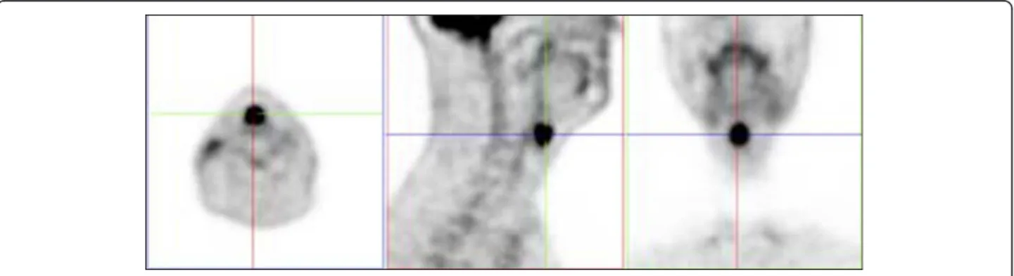
Figure 1Case example of a false positive FDG-PET finding. Axial, sagittal and coronal views of the PET scan are displayed and demonstrate an uptake in the supraglottic region. The area was free of tumor when examined through careful panendoscopy and multiple deep directed biopsies were negative for neoplasia.
Table 1 Patient demographics.(Continued)
40 M 69 N2a PDC II hepatic metastases n/a Yes
41 M 60 N3 SCC II, III, IV bone and extracervical lymph
node metastases
n/a No
42 M 52 N3 PDC II, V pulmonary and hepatic
metastases
n/a Yes
43 M 73 N3 SCC II, V negative n/a No
44 M 56 N3 PDC II, III bone and extracervical
lymph node metastases
n/a No
45 M 69 N3 SCC IV, V esophageal primary, bone and
extracervical lymph node metastases
SCC Yes
46 M 45 N2a Adenocarcinoma V extracervical lymph node
metastases
n/a Yes
47 M 60 N2b SCC I, II pulmonary, bone and
extracervical lymph node metastases
n/a No
48 M 54 N2b SCC I, II base of tongue SCC Yes
49 M 44 N2b UC II, III, IV, V negative n/a No
Suspicious neck mass
Full head and neck examination
Fiberoptic endoscopy
Primary found Primary not found
Work-up & treat accordingly FNA or open biopsy
Carcinoma Melanoma, hematopoietic or thyroid tumors
Work-up & treat accordingly Head and neck CT
Chest X-ray or CT
Primary found Primary not found
Work-up & treat accordingly PET-CT scan
Possible primary Primary not found
Endoscopic examination under general anesthesia
with directed biopsies
Endoscopic examination under general anesthesia
with multiple biopsies
Primary found Primary not found
Treat accordingly Treatment protocols for occult primary tumors
results detected a primary squamous cell carcinoma of the lung and another in the esophagus. Although these could possibly be labeled as synchronous primaries with the true head and neck carcinoma remaining unknown, they were undetected by other imaging techniques and treatment strategies were significantly altered in these patients. Furthermore, possible distant metastases were identified in 18 patients (36.7%). This result is quite higher than that found in the literature [1,17], although this is probably due to the large number of advanced tumor stage (N3) cases in our series. The importance of whole body PET in the detection of distant disease should be stressed, especially when the lower neck is involved. In our study almost half (47.4%) of these patients had unexpected infraclavicular primaries or me-tastases and three of four patients with adenocarcinoma (75%) had pathology below the clavicles.
PET sensitivity in the current review (69.2%) was slightly lower than in most studies [1]. The 4 false negative results that were found emphasize the fact that a negative PET scan does not necessarily rule out the presence of a primary tumor and discard the need for further investigation [12]. Possible causes for such a low sensitivity in our review may be a low tumor uptake of FDG due to tumor differentiation or small size or a high background signal of Waldeyer’s ring [1]. This could, perhaps, justify the failure to detect the two cases of palatine tonsil carcinoma. However, there is always the possibility that there is no primary tumor to begin with, which would significantly reduce the number of true posi-tives [9].
One major weakness of PET in the detection of occult primary tumors is the high false positive rate and low specificity [1]. Due to the high percentage of false posi-tives, some authors have found that there is a lack of benefit in using PET, but suggest that meticulous biopsy sampling and new tracers may ameliorate this aspect [11]. We obtained, in our series, a false positive rate of 35.7% and a specificity of 86.1%. This is consistent with or even somewhat better than previous reviews [1]. Nevertheless, the percentage of false positives is still fairly high. Proposed reasons include high physiologic uptake by the tonsils and muscles of mastication, inflam-mation and benign tumors [11,17]. Overall, PET helped identify 9 primary tumors which had previously gone un-detected. It guided the surgeon to a potential primary tumor site for deep tissue biopsies. 31 patients were con-sidered as having true negative PET scans, as no primary tumor was detected during a mean follow-up period of 22.3 months. This gives a total accuracy of 81.6%. A pro-posed management flow chart is shown in figure 2.
Among other factors that must be considered when opting for PET imaging in the management of patients with unknown primary head and neck car-cinoma are economic issues and availability. Most
reports indicate that perhaps PET scanning is not cost effective [2]. Nonetheless, costs are comparable to whole body MRI [1]. On the other hand, no more than a few PET scans exist nationwide in Portugal, limiting availability to major referral centers.
Conclusions
PET imaging is thus a valuable tool in the detection of occult primary head and neck carcinomas. Not only does it provide additional information as to the location of primary tumors, but it can also help detect unexpected distant metastases. As a result, therapeutic strategies and long-term prognosis are influenced by PET in a substan-tial number of these patients.
Competing interests
The authors declare that they have no financial or non-financial competing interests.
Author details
1
Department of Otorhinolaryngology, Braga Hospital, Sete Fontes, 4710-243, Braga, Portugal.2Department of Otorhinolaryngology, Portuguese Institute of
Oncology of Porto, Rua Dr António Bernardino de Almeida, 4200-072, Porto, Portugal.
Authors’contributions
The authors contributed equally to this study. GP drafted the manuscript. All authors critically reviewed, read and approved the final manuscript.
Authors’information
This study was carried out at the Portuguese Institute of Oncology of Porto.
Received: 21 February 2012 Accepted: 18 June 2012 Published: 18 June 2012
References
1. Rusthoven KE, Koshy M, Paulino AC (2004) The role of
fluorodeoxyglucose positron emission tomography in cervical lymph node metastases from an unknown primary tumor. Cancer 101:2641– 2649
2. Mahoney EJ, Spiegel JH (2005) Evaluation and management of malignant cervical lymphadenopathy with an unknown primary tumor. Otolaryngol Clin N Am 38:87–97
3. Di Martino E, Nowak B, Hassan HA, Hausmann R, Adam G, Buell U, Westhofen M (2000) Diagnosis and staging of head and neck cancer: a comparison of modern imaging modalities (positron emission tomography, computed tomography, color-coded duplex sonography) with panendoscopic and histopathologic findings. Arch Otolaryngol Head Neck Surg 126:1457–1461
4. Greven KM, Williams DW 3rd, McGuirt WF Sr, Harkness BA, D’Agostino RB Jr, Keyes JW Jr, Watson NE Jr (2001) Serial positron emission tomography scans following radiation therapy of patients with head and neck cancer. Head Neck 23:942–946
5. Jungehulsing M, Scheidhauer K, Damm H, Pietrzyk U, Eckel H, Schicha H, Stennert E (2000) 2[F]-Fluoro-2-deoxy-D-glucose positron emission tomography is a sensitive tool for the detection of occult primary cancer (carcinoma of unknown primary syndrome) with head and neck lymph node manifestation. Otolaryngol Head Neck Surg 123:294–301 6. Stokkel MP, Moons KG, ten Broek FW, van Rijk PP, Hordijk GJ (1999)
18 F-Fluorodeoxyglucose dual-head positron emission tomography as a procedure for detecting simultaneous primary tumors in cases of head and neck cancer. Cancer 86:2370–2377
7. Rankin SC (2006) PET in face and neck tumours. Cancer Imaging 6:S89–S95 8. Aassar OS, Fischbein NJ, Caputo GR, Kaplan MJ, Price DC, Singer MI, Dillon
9. Kole AC, Nieweg OE, Pruim J, Hoekstra HJ, Koops HS, Roodenbburg JL, Vaalburg W, Vermey A (1998) Detection of unknown occult primary tumors using positron emission tomography. Cancer 82:1160–1166
10. Schoder H, Yeung HW, Gonen M, Kraus D, Larson SM (2004) Head and neck cancer: clinical usefulness and accuracy of PET/CT image fusion. Radiology 231:65–72
11. Greven KM, Keyes JW Jr, Williams DW 3rd, McGuirt WF, Joyce WT 3rd (1999) Occult primary tumors of the head and neck: lack of benefit from positron emission tomography imaging with 2-[F-18]fluoro-2-deoxy-D-glucose. Cancer 86:114–118
12. Miller FR, Hussey D, Beeram M, Eng T, McGuff HS, Otto RA (2005) Positron emission tomography in the management of unknown primary head and neck carcinoma. Arch Otolaryngol Head Neck Surg 131:626–629 13. Mendenhall WM, Mancuso AA, Parsons JT, Stringer SP, Cassisi NJ (1998)
Diagnostic evaluation of squamous cell carcinoma metastatic to cervical lymph nodes from an unknown head and neck primary site. Head Neck 20:739–744
14. Koch WM, Bhatti N, Williams MF, Eisele DW (2001) Oncologic rationale for bilateral tonsillectomy in head and neck squamous cell carcinoma of unknown primary source. Otolaryngol Head Neck Surg 124:331–333 15. McQuone SJ, Eisele DW, Lee DJ, Westra WH, Koch WM (1998) Occult
tonsillar carcinoma in the unknown primary. Laryngoscope 108:1605–1610 16. Chepeha D, Koch W, Pitman K (2003) Management of unknown primary
tumor. Head Neck 6:499–504
17. Fogarty GB, Peters LJ, Stewart J, Scott C, Rischin D, Hicks RJ (2003) The usefulness of fluorine 18-labelled deoxyglucose positron emission tomography in the investigation of patients with cervical lymphadenopathy from an unknown primary tumor. Head Neck 25:138–145
doi:10.1186/1758-3284-4-34
Cite this article as:Pereiraet al.:Positron emission tomography in the detection of occult primary head and neck carcinoma: a retrospective study.Head & Neck Oncology20124:34.
Submit your next manuscript to BioMed Central and take full advantage of:
• Convenient online submission
• Thorough peer review
• No space constraints or color figure charges
• Immediate publication on acceptance
• Inclusion in PubMed, CAS, Scopus and Google Scholar
• Research which is freely available for redistribution