Protein Interaction Analysis in a Model of Steatohepatitis
with Mallory-Denk Bodies
Bernhard Zatloukal1, Iris Kufferath1, Andrea Thueringer1, Ulf Landegren2, Kurt Zatloukal1,
Johannes Haybaeck1*
1Institute of Pathology, Medical University Graz, Graz, Austria,2Department of Immunology, Genetics and Pathology, Uppsala University, Uppsala, Sweden
Abstract
The in situ proximity ligation assay (isPLA) is an increasingly used technology forin situdetection of protein interactions,
post-translational modifications, and spatial relationships of antigens in cells and tissues, in general. In order to test its
performance we comparedisPLA with immunofluorescence microscopy by analyzing protein interactions in cytoplasmic
protein aggregates, so-called Mallory Denk bodies (MDBs). These structures represent protein inclusions in hepatocytes typically found in human steatohepatitis and they can be generated in mice by feeding of 3,5-diethoxy-carbonyl-1,4-dihydrocollidine (DDC). We investigated the colocalization of all three key MDB components, namely keratin 8 (K8), keratin
18 (K18), and p62 (sequestosome 1) byisPLA and immunofluorescence microscopy. Sensitivity and specificity ofisPLA was
assessed by usingKrt82/2andKrt182/2mice as biological controls, along with a series of technical controls.isPLA signal
visualization is a robust technology with excellent sensitivity and specificity. The biological relevance of signals generated critically depends on the performance of antibodies used, which requires careful testing of antibodies like in
immunofluorescence microscopy. There is a clear advantage of isPLA in visualizing protein co-localization, particularly
when antigens are present at markedly different concentrations. Furthermore,isPLA is superior to confocal microscopy with
respect to spatial resolution of colocalizing antigens. Disadvantages compared to immunofluorescence are increased costs and longer duration of the laboratory protocol.
Citation:Zatloukal B, Kufferath I, Thueringer A, Landegren U, Zatloukal K, et al. (2014) Sensitivity and Specificity ofIn situProximity Ligation for Protein Interaction Analysis in a Model of Steatohepatitis with Mallory-Denk Bodies. PLoS ONE 9(5): e96690. doi:10.1371/journal.pone.0096690
Editor:David R. Borchelt, University of Florida, United States of America
ReceivedJanuary 28, 2014;AcceptedApril 10, 2014;PublishedMay 5, 2014
Copyright:ß2014 Zatloukal et al. This is an open-access article distributed under the terms of the Creative Commons Attribution License, which permits unrestricted use, distribution, and reproduction in any medium, provided the original author and source are credited.
Funding:This work was funded by the Austrian Genome Programme GEN-AU (to KZ) and the Christian Doppler Laboratory BRBT. The funders had no role in study design, data collection and analysis, decision to publish, or preparation of the manuscript.
Competing Interests:Co-author Johannes Haybaeck is a PLOS ONE Editorial Board member. This does not alter the author’s adherence to PLOS ONE Editorial policies and criteria.
* E-mail: [email protected]
Introduction
In order to obtain insights in molecular mechanisms of diseases it is increasingly important to investigate not only levels of expression of proteins and their subcellular localization, but also their interactions with other proteins. This is particularly important because proteins may exert different functions depend-ing on their presence in multi-protein complexes and their localization in cellular compartments. Techniques available for investigating the colocalization of proteins within cells and tissues include double-label indirect immune fluorescence (DIIF) micros-copy and Fo¨rster resonance energy transfer (FRET) microsmicros-copy [1]. More recently, a new technique for visualizing proteins and their interactions based on the proximity ligation mechanism, was added to the spectrum of microscopy techniques [2].
The in situ proximity ligation assay (isPLA) is a technology that extends the capabilities of traditional immunoassays to also include protein interactions, post-translational modifications, and spatial relationships of antigens in cells and tissues. Protein targets can be readily detected and localized with single molecule resolution and objectively quantified in unmodified cells and tissues. Utilizing only a few cells, even transient or weak interactions are revealed in situ and sub-populations of cells can be distinguished. Within
hours, results from conventional immunoprecipitation and co-localization techniques can be independently confirmed byisPLA. isPLA can make use of pairs of primary antibodies raised in different species to recognize the target protein or protein complex of interest. Species-specific secondary antibodies, called PLA probes, each with a unique short DNA strand attached to it, bind the primary antibodies. When the PLA probes are in close proximity, the DNA strands can interact through a subsequent addition of two other circle-forming DNA oligonucleotides. After joining of the two added oligonucleotides by enzymatic ligation, the DNA circles are copied via rolling circle amplification using a polymerase. In the amplification reaction, long DNA strands, each containing several hundred complements of the DNA circle, are formed. These products are easily visualized by microscopy as brightly fluorescent spots using fluorescence-labeled complemen-tary oligonucleotide probes (Fig. 1) [2].
isPLA has been used for investigation of a wide variety of disease conditions, for instance for quantification of HER2-protein complexes in breast cancer [3], and for detecting osteopontin-interactions in resectable gastric cancer.
to that of DIIF confocal microscopy. For this purpose, we used experimentally induced multi-protein aggregates in mouse liver as a test system. This model is based on chronic feeding of mice with a diet containing 3,5-diethoxy-carbonyl-1,4-dihydrocollidine (DDC, also known as diethyl 1,4-dihydro-2,4,6-trimethyl-3,5-pyridinedicarboxylate), which leads to the formation of histopath-ological structures called Mallory-Denk bodies (MDBs) in hepa-tocytes and to ballooning of the hepahepa-tocytes, which are associated with a disruption of the keratin intermediate filament cytoskeleton. MDBs are protein aggregates in the cytoplasm of hepatocytes, which occus in alcoholic and non-alcoholic steatohepatitis (ASH, NASH), copper storage diseases, chronic cholestasis, some types of drug induced liver injuries, and in hepatocytic neoplasms [4]. The MDBs are composed of keratin 8 (K8), keratin 18 (K18), and p62 (sequestosome 1) as main constituents. In this study, we have performed a comprehensive analysis on factors impacting on the visualization of K8, K18 and p62 in MDBs byisPLA and confocal microscopy by using this well established experimental animal model. Since for this model mice knocked out for production of K8 and K18 (krt82/2 and krt182/2, respectively) were available
[5,6], we used the knockout mice as biological controls to demonstrate the specificity of isPLA in visualization of protein co-localization.
Materials and Methods
Male Swiss Albino mice (strain Him OF-1; Institute of Laboratory Animal Research, Medical University of Vienna, Himberg, Austria), Krt82/2129ola, and Krt182/2129ola mice
were kept under SPF conditions and fed a standard diet (Ssniff Spezialdia¨ten GmbH, Soest, Germany). For induction of MDBs, mice were fed a diet containing 0.1% 3,5-diethoxycarbonyl-1,4-dihydrocollidine (DDC; Sigma-Aldrich, Vienna, Austria) for 12 weeks [6]. Mice were killed by cervical dislocation and liver tissues were snap frozen in methyl-butane cooled to the temperature of liquid nitrogen. The animal experiments have been approved by the Austrian Federal Ministry of Science and Research, Division of Genetic Engineering and Animal Experiments (Vienna, Austria) (BMBWF-66.010/0060-II/3b/2013).
For DIIF and isPLA, mouse livers tissue sections (2mm) were fixed with acetone for 10 min at220uC. After air drying, slides were incubated with the DUOLINK Blocking Stock reagent (Olink, Uppsala, Sweden) diluted 1:5 in Antibody Diluent for 30 min at 37uC. Slides were incubated with primary ABs for isPLA (Keratin 8 [Ks 8.7 Progen] 1:50 or mouse-anti-Keratin18 [18.4 Progen] 1:50, and guinea pig anti-p62 [p62CT] 1:200 in Antibody Diluent) for 30 min at room temperature in a humidified chamber (both primary antibodies were applied together) and then washed 365 min in TBS-T. Thereafter slides
were incubated with PLA Probes from DUOLINK anti-mouse PLA PLUS and anti-guinea pig PLA MINUS diluted 1:10 in Antibody Diluent for 30 min at 37uC and washed 265 min at 37uC in TBS-T. For hybridization, slides were incubated with DUOLINK hybridization stock diluted 1:5 in AB diluent for 15 min at 37uC and washed 161 min in TBS-T at 37uC. For the
ligation, slides were incubated with DUOLINK ligation reagent (Ligation Stock 1:5 in AB diluent plus ligase [1:40]) for 15 min at 37uC and washed 262 min in TBS-T at 37uC. For signal amplification, DUOLINK Amplification Stock was diluted 1:5 in AB diluent and polymerase (1:80) was added and slides were incubated for 90 min at 37uC followed by 262 min washings in
TBS-T at 37uC. For the detection, DUOLINK Detection Stock (Texas red; 613 nm) was diluted 1:5 in AB diluent and slides were incubated for 60 min at 37uC. For the final washing, four sequential washing steps were performed with 26SSC, 16SSC, 0.26SSC and 0.026SSC for 2 min each at room temperature.
For the additional DIIF staining, rabbit-anti-K8+18 (50K160; for review on MDB-reacting antibodies see [7] or rat-anti-K8 (TROMA1 tissue culture supernatant, neat) were used as primary antibodies. Primary antibodies were incubated for 30 min at room temperature, followed by 365 min washings in PBS and
incubation with secondary antibodies [Alexa 488-conjugated anti-rabbit (Invitrogen), diluted 1:100 or Alexa 488-conjugated anti-rat (Invitrogen), diluted 1:100] for 30 min at room temper-ature. After 365 min washes in PBS slides were rinsed in 100% ethanol, air-dried and mounted in MowiolR (Sigma-Aldrich). Fluorescence signals fromisPLA and DIIF were detected by Zeiss LSM 510 META scanning laser confocal microscope. Excitation wavelength for Alexa 488 was 488 nm (Argon Laser), and for the isPLA reagent 594 nm (HeNe Laser) (analysis software: ZEN 2009) was used. A summary of all reaction steps for the combination ofisPLA and DIIF is shown in Figure 1.
As controls for specificity of isPLA a series of technical and biological negative controls were performed. Technical controls
Figure 1. Schematic drawing of different antibody binding and visualization steps ofisPLA combined with IF.(A) Primary ABs of
the isPLA bind to two different proteins, which are expected to
colocalize. (B) Secondary ABs conjugated with oligonucleotides bind to the primary ABs (PLA Probe PLUS and PLA Probe MINUS). (C) Two more oligonucleotides (blue) hybridize to the two PLA probes. (D) Ligase (yellow) joins the two added oligonucleotides to form a closed circle. (E) Polymerase (yellow) induces a rolling circle amplification (RCA) using the ligated circle as a template. (F) Fluorescence labeled oligonucle-otides (red) hybridize to the RCA product. (G) Primary ABs of the IF
(purple) bind to one of the proteins, which is also targeted byisPLA. (H)
Secondary ABs of the IF bind to the primary ABs of the IF. They are
fluorescence labeled in red while theisPLA signals are green.
were based on the omission of first antibodies (to exclude cross-reactivity of secondary antibodies or non-specific PLA-signal generation). Biological controls involved knock-out mice for two of the antigens present in MDBs. In this case all primary and secondary antibodies were applied, however because of the absence of one of the antigens detected in PLA, no PLA-related signals should be detected. Sensitivity of PLA was assessed by comparison with single and double-label immunofluorescence analyzed by confocal microscopy. A summary of all controls performed is shown in Table 1.
Results
In a first approach, the reactivity of the antibodies was tested. All antibodies (ABs) used in this study were applied for single and double-label indirect immunofluorescence (IF) microscopy in different sequential orders. Additionally, the primary ABs were tested by using different fluorochrome-labeled secondary ABs in various combinations to ensure equivalent reactivity (data not shown). Irrespective of the experimental setup identical results were observed for antibodies raised in different species but directed to the same antigen (i.e., K8 and K18, respectively) (Fig. 2).
This was essential because two different AB pairs, raised in different species (e.g., mouse and rat), were used in the combined isPLA and IF staining. Only if it was ensured that both ABs, namely the keratin AB used in IF and the keratin AB used in isPLA had identical staining properties, could differences between IF and isPLA be evaluated. Otherwise any observed differences between IF and isPLA could depend on different staining properties of the primary ABs.
Figure 2 shows a double–label IF of wild type Swiss Albino mice before and after DDC treatment using the optimal AB combina-tions as identified in the pre-testing phase. Normal mouse livers did not show any protein aggregations positive for p62, but exhibited a filamentous architecture of the keratin intermediate filament cytoskeleton in the hepatocytes. After DDC treatment ballooned hepatocytes were present with a disrupted keratin cytoskeleton and MDBs involving K8, K18 and p62 were observed (Fig. 3). In addition to the classical MDB, which is positive for keratin and p62, small p62 positive but K8/K18 negative structures were present [8]. However, the nature of these structures has not been characterized yet. Furthermore, small keratin positive MDB-like aggregates were seen in which p62 was only weakly present or even undetectable. It was not possible to draw clear conclusions about p62 positivity in these structures, since the observed results were influenced by the threshold and amplification settings for the different fluorescence channels during confocal microscopy (Fig. 3 B).
The same liver used in the experiment shown in Fig. 3, and the same combinations of ABs were applied for isPLA alone, and isPLA in combination with IF (Fig. 4). For theisPLA, two ABs (m-a-K8 and gp-a-p62) were used to detect protein aggregations in which p62 and K8 co-localized. A third AB (rat-a-K8) was applied on the same tissue sample as the primary AB for the IF. Since isPLA only gives a positive signal if both components (keratin, p62) were present, every MDB detected by the isPLA would also be expected to be positive for keratin 8 in the IF stain. The combined isPLA and IF stain confirmed the presence of K8 in everyisPLA positive MDB. Moreover, it was notable thatisPLA detected the smallest MDBs, visible by DIIF. However, some keratin aggregates resembling MDBs were negative for p62 both in isPLA and IF (Fig. 3 and Fig. 4). In summary, the experiments demonstrated that the sensitivity ofisPLA is at least as high as the sensitivity of Table
DIIF. As technical negative controls for theisPLA the first ABs to p62 and keratin, respectively, were omitted. All negative controls constantly showed negative isPLA signals (negative controls not shown).
Reactivity of ABs used was further assessed inKrt8andKrt18 knock-out mice. In both knock-out mice hepatocytes were negative for keratin intermediate filament cytoskeleton because, in the absence of one partner (e.g., K8) the other still expressed partner (e.g., K18 in Krt82/2 mice) is unstable and does not form intermediate filaments (Fig. 5). In the absence of a polymerization partner, K8 or K18 is quickly degraded. The only structures that were stained positive for the expressed keratin partner in mice fed a standard diet were bile ducts since in bile duct epithelia additional keratins are expressed that form intermediate filaments [8,9]. DIIF microscopy of DDC-fedkrt8knock-out mice using ABs to p62 and K8, respectively, demonstrated the absence of MDBs as previously published [8]. krt82/2 mice showed small p62
positive cytoplasmic structures which were negative for keratin. In contrast,krt182/2mice developed MDBs that were composed of
K8 and p62 [6] (Fig. 5). These mice were then tested by applying combinedisPLA and IF for p62 and K8 or K18 (Figs. 6 and 7). Thekrt82/2 mice were negative both in the K18
+p62 and K8+ p62 isPLA, which underlines the absence of keratin in the small p62 positive structures observed in DIIF (Fig. 3). Furthermore immunofluorescence demonstrated keratin positive aggregates with typical MDB structure which were negative in isPLA. This is in line with previous results demonstrating by double-label immunofluorescence microscopy that some MDB-like aggregates were p62-negative [7]. Thus our isPLA results support these previous findings with an independent technology. In thekrt182/2
miceisPLA revealed positive MDBs only in case of using K8 and p62 ABs as primary ABs for theisPLA. In contrast, theisPLA stain with ABs to K18 and p62 was negative. In krt182/2 mice all
components of theisPLA (K18 and p62 ABs, amplification system, detection system) were applied but no reaction products were
observed. This is consistent with the fact that only one of the two antigens was present, namely p62, while K18 was absent, demonstrating that isPLA is highly specific. Furthermore, these data demonstrated that no cross reactivity occurred between the ABs used forisPLA and those used in IF (in the DIIF a polyclonal rabbit-AB to K8+18 was used, whereas for isPLA a mouse monoclonal AB to K18 was used). This underlined the species
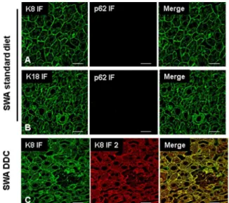
Figure 2. Testing of ABs to keratin and p62 in mouse liver and demonstration of equivalent binding properties of ABs used in IF andisPLA.IF staining of normal mouse liver tissues (Swiss Albino, SWA fed a standard diet) with antibodies to K8 (green), p62 (red) (A) and for K18 (green) and p62 (red) (B). Image (C) shows that the two different ABs directed to K8 (rat-anti-K8 [Troma I] and mouse-anti-K8 [Ks
8.7 Progen]) used forisPLA and IF or DIIF recognize identical structures
in liver from a DDC fed SWA mouse. Scale bars: 20mm.
doi:10.1371/journal.pone.0096690.g002
Figure 3. Testing of ABs to keratin and p62 in mouse liver cells after DDC treatment. Immunofluorescence stain on SWA mouse livers after 12 weeks DDC treatment using ABs to K8 (green), p62 (red) (A), and K18 (green), p62 (red) ABs (B). For image (A), note the presence of small (arrowhead) and large (empty arrow) MDBs. Additionally, small granules positive for p62 only can be observed (arrow). For image (B), note that small MDB-like keratin aggregates can be observed that are
negative for p62 (empty arrow). Scale bars: 20mm.
doi:10.1371/journal.pone.0096690.g003
Figure 4.isPLA for 12 week-DDC treated mice. Images (A, B)
showisPLA signal (red) using ABs to p62 and K8 (A) and p62 and K18
(B). (C) shows a combined visualization of p62 and K8 inisPLA (red) and
K8 in IF (green). (D) shows a combined visualization of p62 and K18 in
isPLA (red) and K18 IF (green). In images (C) and (D) note that even very
smallisPLA positive structures are visible (arrowhead), demonstrating
the high sensitivity ofisPLA. However, there were also larger MDB-like
keratin aggregates that are negative for isPLA stain (empty arrow),
suggesting the absence of p62. Scale bars: 20mm.
specificity of the ABs used in theisPLA system and that there is no cross reactivity betweenisPLA and DIIF.
Discussion
In this study we have performed a comprehensive investigation of clinically relevant morphological alterations by usingisPLA. We have chosen a mouse model for steatohepatitis and MDB formation for testing various methodologies because the main molecular components and interaction partners of MDBs, namely K8, K18, and p62, are well known and characterized, and a broad spectrum of antibodies and models were available. In order to demonstrate the sensitivity and specificity of isPLA we have performed technical as well as biological controls, by including gene knock-out mice for the MDB components K8 and K18.
We appliedisPLA in combination with IF and compared results with DIIF. Signals of isPLA and IF or DIIF were analyzed by confocal microscopy. For these studies it was essential to ensure that ABs used for isPLA and DIIF had equivalent staining properties, and that secondary ABs did not cross-react with other primary ABs. Since performance ofisPLA critically relies on the performance of the ABs used, the scheme of technical controls performed in this study can be recommended for establishing isPLA essentially for any application (see Table 1). Under the prerequisite of proper performance of ABs, results obtained demonstrated thatisPLA is a robust, sensitive and specific tool for
investigating the relationship between proteins and their associa-tion with each other in tissues. In general, findings ofisPLA were similar to those of DIIF. However, isPLA led to much clearer results when one antigen was more abundant than the second one becauseisPLA only yields a signal in case of antigen colocalization (e.g., small MDB-related keratin aggregates in the presence of the keratin intermediate filament cytoskeleton). In contrast, results of DIIF analyzed by confocal microscopy depended on the settings of the different fluorescent channels. Therefore we recommend isPLA as a preferred technique for visualization of protein colocalization in small structures, particularly when larger,
Figure 5. Demonstration of keratin and p62 in DDC treated
krt82/2andkrt182/2mice.IF staining onkrt82/2(A, B) andkrt182/2 (C, D) mouse livers after 12 weeks DDC treatment. Images (A) and (C) show the localization of K8 (green) and p62 (red). Images (B) and (D) show the localization of K18 (red) and p62 (red). A positive
colocalization of p62 and K8 in krt182/2 mice was observed, which
indicates presence of MDBs. In contrastkrt82/2mice did not develop
MDBs after DDC feeding, therefore neither K18 nor p62 accumulated.
ABs to K18 show positivity in bile ducts (BD) inkrt82/2mice (B). The
green reaction in image (D) relates to plasma cells. Scale bars: 20mm.
doi:10.1371/journal.pone.0096690.g005
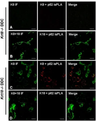
Figure 6.isPLA and IF for 12 week-DDC treated krt82/2 and
krt182/2mice.(A, B)krt82/2mouse liver. (A) CombinedisPLA (K8+
p62, red) and IF (K8, green). (B) CombinedisPLA (K18+p62, red) and IF
(K8+K18, green). (C, D)krt182/2mouse liver. (C) combinedisPLA (K8+
p62, red) and IF (K8, green). (D) CombinedisPLA (K18+p62, red) and IF
(K8+K18, green). Note that krt82/2 do not form MDBs after DDC
feeding and there is noisPLA positive signal. In DDC fedkrt182/2mice
isPLA yielded only positive signals with K8 ABs demonstrating
specificity of the wholeisPLA system. Scale bars: 20mm.
doi:10.1371/journal.pone.0096690.g006
Figure 7. Influence of channel amplification on demonstration of antigen colocalization in isPLA and IF in 12 week-DDC treated krt182/2 mice. (A, B, C) Show different levels of digital amplification of the green channel (IF for K8) and constant amplification
of the red channel showingisPLA for K8 and p62. Arrows indicate a
MDB-like aggregate that was constantly positive for K8 but negative for
p62. Arrowheads indicate MDBs which were positive inisPLA for p62
and K8 but IF showed only presence of K8 (yellow merged signal) in
isPLA positive MDBs after maximal amplification of the green channel.
Scale bars: 20mm.
strongly positively stained structures are present as well (this situation causes problems in setting the appropriate background thresholds in DIIF). For instance, because of these properties of isPLA our analysis provided evidence that p62 was lacking in some MDB-like keratin aggregates. This new observation may shed new light on the role of p62 in MDB pathogenesis.
Furthermore, since isPLA signals depend on the molecularly defined maximal distance between the antigens, isPLA provides better defined resolution for protein colocalization than DIIF isPLA is estimated to permit identification of pairs of epitopes located within less than approximately 40 nm. This is a 5-fold higher resolution compared to DIIF, which allows the detection of distinct signals with a distance of approximately 0.2mm (along the x- and y-axes) due to limitations of standard light microscopy. Since the optical section thickness of confocal microscopy is in the range of 0.5mm (z-axis), isPLA is of particular advantage for demonstrating colocalization of antigens along the z-axis. How-ever, the resolution ofisPLA is lower than that of FRET which detects molecular interactions at a distance of even below 0.5 nm [10].
In recent years other techniques like Array Tomography with High-Resolution Three-Dimensional Immunofluorescence have also been developed. This is a volumetric microscopy method based on physical serial sectioning where ultrathin sections are cut, stained and imaged. The resulting two-dimensional images are then further reconstructed by using computational methods in order to generate three-dimensional volume images, suitable for quantitative analysis [11]. This method is more difficult to realize thanisPLA.
In conclusion, we found that theisPLA provides more accurate results compared to DIIF because of the signal specificity, the
better spatial resolution and the independence of the results and exposure time and digital amplification of microscope channels used (Fig. 7; pros and cons ofisPLA are summarized in Table 2). As the function of proteins critically depends on their interactions with other molecules and the localization of multi component complexes in specific subcellular components, techniques forin situ detection of protein complexes become important. Furthermore, personalized medicine application does not only depend on the expression of the drug target but also on the activation of the target-related pathway, which is characterized by specific interac-tions between cellular components. Therefore application ofisPLA might become increasingly relevant for molecular diagnosis and research requiring a carefully controlled and robust protocol, for which examples and practical guidance is provided in this study.
Acknowledgments
We thank Assoc. Prof. Ola So¨derberg from the Department of Immunology, Genetics and Pathology, Uppsala University, Sweden for
teaching us theisPLA and for his very helpful advice. We are very grateful
to R. Oshima and H. Baribault for providing theKrt82/2mice, and to T.
Magin for providing the Krt182/2mice. This work was funded by the
Austrian Genome Programme GEN-AU (to KZ) and the Christian Doppler Laboratory BRBT (KZ).
Author Contributions
Conceived and designed the experiments: BZ KZ JH. Performed the experiments: BZ IK AT. Analyzed the data: BZ IK AT UL KZ JH. Contributed reagents/materials/analysis tools: KZ JH. Wrote the paper: BZ UL KZ JH.
References
1. Pietraszewska-Bogiel A, Gadella TW (2011) FRET microscopy: from principle to routine technology in cell biology. J Microsc 241: 111–118.
2. Weibrecht I, Leuchowius KJ, Clausson CM, Conze T, Jarvius M, et al. (2010) Proximity ligation assays: a recent addition to the proteomics toolbox. Expert Rev Proteomics 7: 401–409.
3. Aubele M, Spears M, Ludyga N, Braselmann H, Feuchtinger A, et al. (2010) In situ quantification of HER2-protein tyrosine kinase 6 (PTK6) protein-protein complexes in paraffin sections from breast cancer tissues. Br J Cancer 103: 663– 667.
4. Zatloukal K, French SW, Stumptner C, Strnad P, Harada M, et al. (2007) From Mallory to Mallory-Denk bodies: what, how and why? Exp Cell Res 313: 2033– 2049.
5. Zatloukal K, Stumptner C, Lehner M, Denk H, Baribault H, et al. (2000) Cytokeratin 8 protects from hepatotoxicity, and its ratio to cytokeratin 18 determines the ability of hepatocytes to form Mallory bodies. Am J Pathol 156: 1263–1274.
6. Haybaeck J, Stumptner C, Thueringer A, Kolbe T, Magin TM, et al. (2012) Genetic background effects of keratin 8 and 18 in a DDC-induced hepatotoxicity and Mallory-Denk body formation mouse model. Lab Invest 92: 857–867. 7. Stumptner C, Fuchsbichler A, Heid H, Zatloukal K, Denk H (2002) Mallory
body–a disease-associated type of sequestosome. Hepatology 35: 1053–1062. 8. Zatloukal K, Stumptner C, Fuchsbichler A, Heid H, Schnoelzer M, et al. (2002)
p62 is a common component of cytoplasmic inclusions in protein aggregation diseases. Am J Pathol 160: 255–263.
9. Magin TM, Vijayaraj P, Leube RE (2007) Structural and regulatory functions of keratins. Exp Cell Res 313: 2021–2032.
10. Holden SJ, Uphoff S, Hohlbein J, Yadin D, Le Reste L, et al. (2010) Defining the limits of single-molecule FRET resolution in TIRF microscopy. Biophys J 99: 3102–3111.
11. Micheva KD, O’Rourke N, Busse B, Smith SJ (2010) Array tomography: high-resolution three-dimensional immunofluorescence. Cold Spring Harb Protoc 2010: pdb to p89.
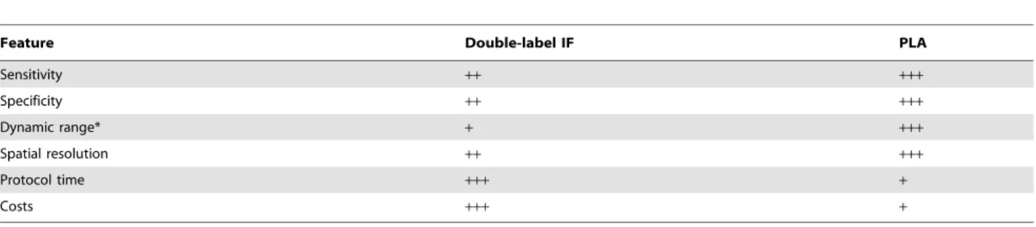
Table 2.Comparison of DIIF microscopy andisPLA for analyzing the colocalization of antigens.
Feature Double-label IF PLA
Sensitivity ++ +++
Specificity ++ +++
Dynamic range* + +++
Spatial resolution ++ +++
Protocol time +++ +
Costs +++ +
Relative advantages are indicated in+to+++.