Submitted25 May 2016 Accepted 23 July 2016 Published24 August 2016
Corresponding author
Hamid Niksirat, niksirat@frov.jcu.cz
Academic editor Jean-Lou Justine
Additional Information and Declarations can be found on page 11
DOI10.7717/peerj.2363
Copyright 2016 Yazicioglu et al.
Distributed under
Creative Commons CC-BY 4.0
OPEN ACCESS
Fine structure of the spermatozoon in
three species of Cambaridae (Arthropoda:
Crustacea: Decapoda)
Cambarus robustus,
Orconectes propinquus
and
Orconectes
rusticus: a comparative biometrical study
Buket Yazicioglu1, Přemek Hamr2, Pavel Kozák1, Antonín Kouba1,* and Hamid Niksirat1,*
1University of South Bohemia in České Budějovice, Faculty of Fisheries and Protection of Waters, South
Bohemian Research Centre of Aquaculture and Biodiversity of Hydrocenoses, Vodňany, Czech Republic
2Upper Canada College, Toronto, ON, Canada
*These authors contributed equally to this work.
ABSTRACT
The ultrastructure of spermatozoa in three species of cambarid crayfish,Cambarus robustus,Orconectes propinquus, andOrconectes rusticus, were studied and compared with eight previously studied species from different crayfish families using morpholog-ical features and biometrmorpholog-ical data. The ultrastructure of spermatozoa show a generally conserved pattern including an acrosome and nucleus in the anterior and posterior parts of the cell, respectively, radial arms that wrap around the nucleus, and the whole cell is enclosed by an extracellular capsule. The most outstanding morphological feature in spermatozoa of three studied cambarid crayfish is the crest-like protrusions in the anterior part of the acrosome that can be used as one of the features for distinguishing the members of this family. Results of biometrical data reveal that acrosome size in the representatives of Parastacidae are the smallest, while representatives of Astacidae show the biggest acrosome. The acrosome size in species belonging to Cambaridae occupy an intermediate position between the two other families of freshwater crayfish. In conclusion, a combination of morphological features and biometrical data of spermatozoa can help distinguishing different species of the freshwater crayfish.
SubjectsBiodiversity, Zoology
Keywords Decapoda, Acrosome, Extracellular capsule, Radial arms, Nucleus, Ultrastructure
INTRODUCTION
Non-motile spermatozoa of decapods are very diverse in their morphology and that makes them suitable cases for phylogenetic studies (Jamieson, 1991;Jamieson & Tudge, 2000;
640 known species (Crandall & Buhay, 2007). In crayfish, aflagellate spermatozoa bear a relatively large acrosome in the anterior part and a nucleus in the posterior containing extensions of microtubular radial arms (Moses, 1961a; Moses, 1961b; Dudenhausen & Talbot, 1982). Spermatozoa of the freshwater crayfish have already been the subject of many ultrastructural studies (Table 1). The different dimensions of the most prominent organelle in crayfish spermatozoa, the acrosome, have been used for taxonomic studies, as well as the presence of some morphological features, especially in the anterior part of the spermatozoon, such as the spike, apical zone and crest can help distinguishing spermatozoa from different species (Yasuzumi & Lee, 1966;Anderson & Ellis, 1967;Niksirat et al., 2013a;
Niksirat et al., 2013b;Kouba, Niksirat & Bláha, 2015).
The objective of the present study is to compare spermatozoal ultrastructure in three cambarid crayfish with other members of crayfish via morphological features and biometrical data.
MATERIALS AND METHODS
Samplings were carried out in Credit River, Norval, Ontario for Cambarus robustus
and Orconectes propinquus, and Cavan Creek, Ontario for Orconectes rusticus during spawning season in May (Licence No. #1082971, MNR, Ontario). Five specimens of each species were anesthetized on ice for at least 10 min, and dissected to obtain the terminal portion of vasa deferentia near to gonopore containing the most developed spermatozoa. Samples for transmission electron microscopy (TEM) were fixed in 2.5% glutaraldehyde in 0.1 M phosphate buffer for 48 h at 4 ◦
C, washed in buffer, and post-fixed in 4% osmium tetroxide for 2 h, washed in buffer, dehydrated through an acetone series (30, 50, 70, 90, 95, and 100% for 15 min each), and embedded in resin (EPON). A series of ultra-thin sections were cut using an UCT ultramicrotome (Leica Microsystems, Wetzlar, Germany), mounted on the copper grids, double-stained with uranyl acetate and lead citrate (Niksirat, Kouba & Kozák, 2015a), and examined with a 1010 transmission electron microscope (JEOL Ltd., Tokyo, Japan) operating at 80 kV. The length (L) and width (W) of the acrosome and the L: W ratio (Jamieson, 1991; Klaus, Schubart & Brandis, 2009; Klaus & Brandis, 2011) were determined in C. robustus(n=322 spermatozoa),
O. propinquus(n=120 spermatozoa),O. rusticus(n=140 spermatozoa) using ImageJ
software (U.S. National Institutes of Health, Bethesda, MD, USA). For further comparison within different crayfish families, extra data were obtained from earlier published research describing spermatozoa ofAstacus astacus(n=128 spermatozoa),A. leptodactylus
(n=98 spermatozoa),Austropotamobius torrentium(n=86 spermatozoa),Pacifastacus
leniusculus(n=54 spermatozoa),Orconectes limosus(n=139 spermatozoa),Procambarus
clarkii (n=115 spermatozoa), Cherax quadricarinatus (n=91 spermatozoa) and C.
destructor (n=111 spermatozoa) (Niksirat et al., 2013a;Niksirat et al., 2013b; Kouba,
Niksirat & Bláha, 2015). The non-parametric Kruskal–Wallis test with subsequent pairwise comparison post-hoc statistical analysis were carried out using R statistical package version 3.2.5. For all statistical tests,p<0.05 was considered significant. Data are expressed as the
Table 1 Published literature about male gamete morphology in the freshwater crayfish species including four species of Astacidae, nine species of Cambaridae, and five species of Parastacidae.
Family Species References
Astacus astacus Pochon-Masson, 1968;López-Camps et al., 1981;Niksirat et al., 2013b;Niksirat & Kouba, 2016
Astacus leptodactylus Eliakova & Goriachkina, 1966;Niksirat et al., 2013b Austropotamobius torrentium Niksirat et al., 2013b
Astacidae
Pacifastacus leniusculus Dudenhausen & Talbot, 1979;Dudenhausen & Talbot, 1982; Du-denhausen & Talbot, 1983;Yazicioglu et al., 2014;Niksirat et al., 2013a
Cambaroides japonicus Kaye et al., 1961;Yasuzumi et al., 1961;Yasuzumi & Lee, 1966
Cambarus sp. Anderson & Ellis, 1967
Cambarus robustus Present study*
Orconectes limosus Niksirat et al., 2013a
Orconectes propinquus Present study*
Orconectes rusticus Berrill & Arsenault, 1982;Snedden, 1990; Present study* Procambarus clarkii Moses, 1961a;Moses, 1961b;Niksirat et al., 2013a;Dong, Hou &
Yang, 2014
Procambarus leonensis Felgenhauer & Abele, 1991 Cambaridae
Procambarus paeninsulanus Hinsch, 1992;Hinsch, 1993a;Hinsch, 1993b
Cherax albidus Beach & Talbot, 1987
Cherax destructor Jerry, 2001;Kouba, Niksirat & Bláha, 2015
Cherax quadricarinatus López-Greco, Vazquez & Rodríguez, 2007,López-Greco and Lo Nos-tro, 2008;Kouba, Niksirat & Bláha, 2015
Cherax cainiia Beach & Talbot, 1987;Jamieson, 1991
Parastacidae
Parastacus defossus Noro, López-Greco & Buckup, 2008
Notes.
aWe assume that the smooth marron, a common and widespread species largely involved in aquaculture formerly calledC. tenuimanuswas examined. SeeAustin & Ryan (2002)
for details.
RESULTS
Morphological features
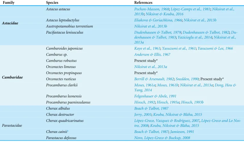
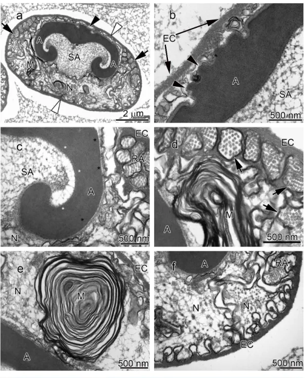
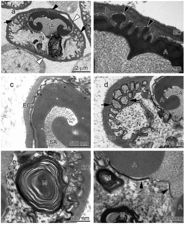
The acrosome complex, as the most prominent organelle, is located in the anterior part of the spermatozoon. It consists of two distinct components including the main body of the acrosome vesicle and the subacrosomal zone (Figs. 1A,2Aand3A). The anterior-most central portion of the acrosome vesicle is folded into a series (usually 2–5) of protrusions resembling a crenulated crest (Figs. 1B,2Band3B). The main body of the acrosome vesicle appears to be divided into two, sometimes indistinct, zones. In the innermost zone some filaments are visible while those filaments are not present in the outer layer (Figs. 1C,2Cand3C). The space posterior to the main body of the acrosome vesicle is filled by a flocculent electron lucent subacrosomal zone. The density of the subacrosomal zone is less in the vicinity of the posterior-most part of the main body of the acrosome vesicle (Figs. 1A,2Aand3A). Radial arms consisting of microtubules wrap around the main body of acrosome vesicle, but remain contained within the extracellular capsule (Figs. 1D,2Dand
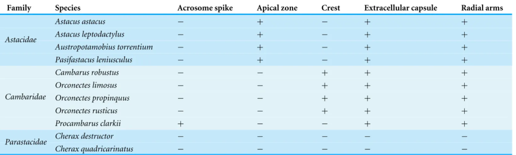
Table 2 Summarizes comparative morphological features among eleven species of the freshwater crayfish.
Family Species Acrosome spike Apical zone Crest Extracellular capsule Radial arms
Astacus astacus − + − + +
Astacus leptodactylus − + − + +
Austropotamobius torrentium − + − + +
Astacidae
Pasifastacus leniusculus − + − + +
Cambarus robustus − − + + +
Orconectes limosus − − + + +
Orconectes propinquus − − + + +
Orconectes rusticus − − + + +
Cambaridae
Procambarus clarkii + − − + +
Cherax destructor − − − − −
Parastacidae
Cherax quadricarinatus − − − − −
spermatozoon containing nuclear materials (Figs. 1F,2D,2F, and3F). The spermatozoon is tightly enclosed by an extracellular capsule (Figs. 1A,2Aand3A).Table 2summarizes comparative morphological features among eleven species of freshwater crayfish, eight from literature and three from this study.
Biometrical data
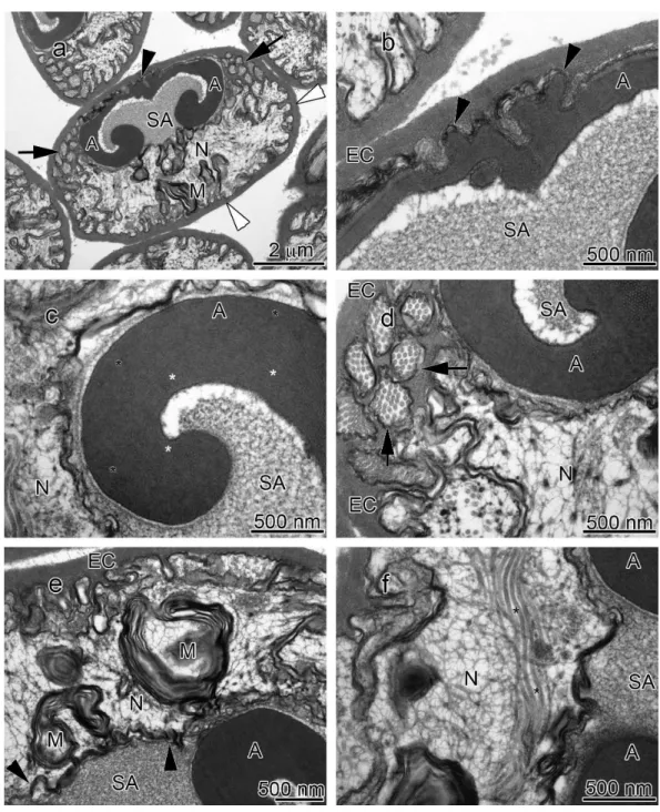
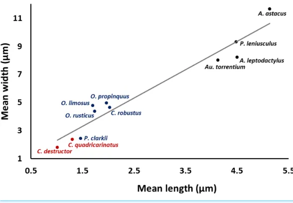
A significant correlation (p<0.0001, r=0.97) was observed between the length and
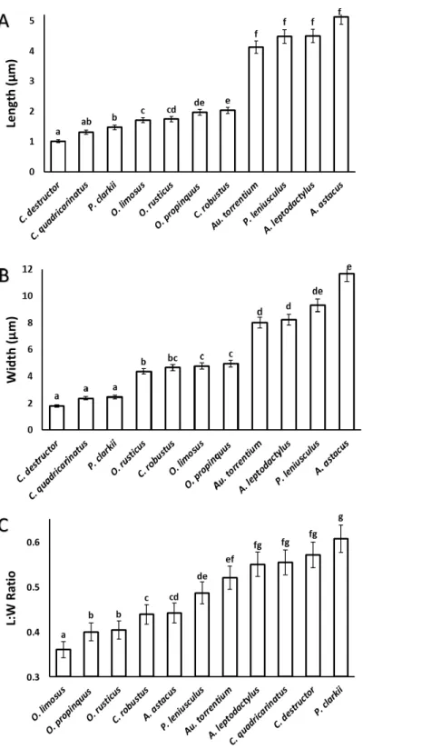
width of the acrosome vesicle in all studied species (Fig. 4). The Kruskal–Wallis test showed significant differences among length, width and L: W of studied groups (p<0.05).
The smallest and largest acrosome length were recorded inC. destructor andA. astacus, respectively. Significant differences were observed in the acrosome length among studied species (p<0.05) except between these species (1)C. destructor andC. quadricarinatus,
(2)C. quadricarinatus andP. clarkii, (3)O. limosusandO. rusticus, (4)O. rusticus, and
O. propinquus, (5)O. propinquusandC. robustus, and (6)Au. torrentium,P. leniusculus,
A. leptodactylusandA. astacus(Fig. 5A).
The smallest and largest acrosome width were recorded inC. destructor andA. astacus, respectively. Significant differences were observed in the acrosome width among studied species (p<0.05) except between these species (1)C. destructor,C. quadricarinatus, and
P. clarkii, (2)O. rusticus, andC. robustus, (3)C. robustus,O. limosus, andO. propinquus, (4)Au. torrentium,A. leptodactylus, andP. leniusculus, (5)P. leniusculusandA. astacus
(Fig. 5B).
The smallest and largest acrosome length to width ratio were recorded inO. limosusand
P. clarkii, respectively. Significant differences (p<0.05) were observed in the acrosome
length to width ratio among studied species (p<0.05) except between these following
groups: (1)O. propinquusandO. rusticus, (2)C. robustusandA. astacus, (3)A. astacusand
P. leniusculus, (4)P. leniusculusandAu. torrentium, (5)Au. torrentium,A. leptodactylus,
C. quadricarinatus, andC. destructor, (6)A. leptodactylus,C. quadricarinatus,C. destructor
Figure 4 Correlation between length and width of the spermatozoon acrosome in eleven species of freshwater crayfish.(p<0.0001,r=0.97).
DISCUSSION
Morphological features
The results of the present study show that the general morphology of spermatozoa in studied members of Cambaridae is similar to other crayfish including a relatively large acrosome, nucleus and radial arms that are enclosed by an extracellular capsule (Jamieson & Tudge, 2000;Vogt, 2002). Radial arms are usually the extensions of the nucleus that wrap around the acrosome vesicle. These arms are present in Astacidae and Cambaridae, but not in studiedCherax species (Beach & Talbot, 1987;Kouba, Niksirat & Bláha, 2015). The radial arms in decapod spermatozoa may be composed of microtubules, nuclear material, or both (Tudge, 2009). Molecular studies identified tubulin proteins, as major units of microtubules in the proteomic profile of the crayfish male gamete that confirms the microtubular nature of radial arms (Niksirat et al., 2014a;Niksirat et al., 2015b) and as seen in the TEM images in this study. Although, microtubular radial arms undergo protein tyrosine phosphorylation during spermatophore post-mating storage on the body surface of female crayfish (Niksirat et al., in press), the exact role(s) of radial arms in fertilization is yet to be determined. The extracellular capsule seems to be an envelope for tight compaction of long organelles such as radial arms. This hypothesis is further supported by the absence of a capsule in the studiedCheraxspermatozoa, where radial arms are not present (Beach & Talbot, 1987;Vogt, 2002;Kouba, Niksirat & Bláha, 2015).
lose their internal matrix and are transformed into membranous lamellae during the early spermatid stage of the crayfishCambarussp. which is still able to provide energy to the cell (André, 1962;Anderson & Ellis, 1967). A positive staining of Janus green B, an indicator of active mitochondria, in the same area of the crayfish spermatozoon has been reported (André, 1962). Several proteins related to metabolism and energy production were identified in the protein profile of the crayfish male gamete that may confirm the presence of an energy supply center in the sperm cell (Niksirat et al., 2014a;Niksirat et al., 2015b).
A set of electron lucent pores, a unique morphological feature, have only been reported at the margins of the main body of the acrosome inP.leniusculus(Niksirat et al., 2013b). The anterior-most margin of the main body of the acrosome vesicle in different crayfish species showed a diversity in shape that can be used as an important morphological feature for distinguishing different species of freshwater crayfish (Niksirat et al., 2013a;
Niksirat et al., 2013b;Kouba, Niksirat & Bláha, 2015; present study). For example, in Cambaridae, a horn-like spike was observed in the anterior part of the fully developed spermatozoon ofCambaroides japonicus(Yasuzumi & Lee, 1966). A similar spike-shaped structure has been reported in spermatozoa of Cambarussp. (Anderson & Ellis, 1967) andProcambarus leonensis (Felgenhauer & Abele, 1991). While several other studies on spermatozoal ultrastructure and spermatogenesis inProcambarus(Moses, 1961a;Moses, 1961b;Hinsch, 1992;Hinsch, 1993a;Hinsch, 1993b) did not report an acrosomal spike, development of a spike in the anterior part of the acrosome vesicle has been observed inProcambarus clarkiiwhen the spermatozoa are inside the vas deferens (Niksirat et al., 2013a). An apical zone, an area filled with bundles of curled filaments has been reported inA. astacus,A. leptodactylus,A. torrentium,P. leniusculus(Pochon-Masson, 1968;López-Camps et al., 1981;Niksirat et al., 2013a;Niksirat et al., 2013b). Those filaments and some material originating from the acrosome are released outside the spermatozoon and form a new formation called filament-droplet structure that could facilitate egg-spermatozoon binding during fertilization in crayfish (Niksirat, Kouba & Kozák, 2014b). In the present study, crest-like protrusions observed in the anterior part of the acrosome vesicle of spermatozoa can be used as one of the morphological features for distinguishing cambarids from other species of freshwater crayfish.
Biometrical data
The size of the acrosome vesicle in the representatives of Parastacidae (Cherax) are the smallest within studied crayfish species. The representatives of Astacidae including
Astacus,Pacifastacus, andAustropotamobiusshowed the largest acrosome vesicles. The acrosome size in species belonging toOrconectesandProcambarusas representatives of Cambaridae occupy an intermediate position among the above mentioned families of freshwater crayfish.
In conclusion, despite conserved general pattern of the crayfish spermatozoon, combination of morphological features such as apical zone, crest and spike in the anterior part of the acrosome, and biometrical data of the acrosome dimensions can provide a tool to distinguish different species of freshwater crayfish families.
ADDITIONAL INFORMATION AND DECLARATIONS
Funding
This project was supported by the Ministry of Education, Youth, and Sports of the Czech Republic CENAKVA (No. CZ.1.05/2.1.00/01.0024), CENAKVA II (No. LO1205 under the NPU I program) and the Grant Agency of the University of South Bohemia (017/2016/Z). The funders had no role in study design, data collection and analysis, decision to publish, or preparation of the manuscript.
Grant Disclosures
The following grant information was disclosed by the authors:
Ministry of Education, Youth, and Sports of the Czech Republic CENAKVA: CZ.1.05/2.1.00/01.0024.
CENAKVA II: LO1205 (NPU I).
Grant Agency of the University of South Bohemia: 017/2016/Z.
Competing Interests
The authors declare there are no competing interests.
Author Contributions
• Buket Yazicioglu performed the experiments, analyzed the data, wrote the paper,
prepared figures and/or tables, reviewed drafts of the paper.
• Přemek Hamr wrote the paper, reviewed drafts of the paper.
• Pavel Kozák conceived and designed the experiments, reviewed drafts of the paper. • Antonín Kouba performed the experiments, reviewed drafts of the paper.
• Hamid Niksirat conceived and designed the experiments, performed the experiments,
analyzed the data, wrote the paper, prepared figures and/or tables, reviewed drafts of the paper.
Field Study Permissions
The following information was supplied relating to field study approvals (i.e., approving body and any reference numbers):
Data Availability
The following information was supplied regarding data availability: The raw data has been supplied as aSupplemental Dataset.
Supplemental Information
Supplemental information for this article can be found online athttp://dx.doi.org/10.7717/ peerj.2363#supplemental-information.
REFERENCES
Anderson WA, Ellis RA. 1967.Cytodifferentiation of the crayfish spermatozoon: acrosome formation, transformation of mitochondria and development of mi-crotubules.Zeitschrift für Zellforschung und mikroskopische Anatomie77:80–94
DOI 10.1007/bf00336700.
André J. 1962.Contribution à la connaissance du chondriome.Journal of Ultrastructure Research6:1–185DOI 10.1016/S0889-1605(62)80002-0.
Austin CM, Ryan SG. 2002.Allozyme evidence for a new species of freshwater crayfish of the genusCheraxErichson (Decapoda: Parastacidae) from the south-west of Western Australia.Invertebrate Systematics16:357–367DOI 10.1071/IT01010.
Beach D, Talbot P. 1987.Ultrastructural comparison of sperm from the crayfishes
Cherax tenuimanusandCherax albidus.Journal of Crustacean Biology 7:205–218
DOI 10.2307/1548602.
Berrill M, Arsenault M. 1982.Spring breeding of a northern temperate crayfish, Or-conectes rusticus.Canadian Journal of Zoology60:2641–2645DOI 10.1139/z82-339.
Braga A, Nakayama CL, Poersch L, Wasielesky W. 2013.Unistellate spermatozoa of decapods: comparative evaluation and evolution of the morphology.Zoomorphology 132:261–284DOI 10.1007/s00435-013-0187-2.
Crandall KA, Buhay JE. 2007.Global diversity of crayfish (Astacidae, Cambari-dae, and Parastacidae—Decapoda) in freshwater.Hydrobiologia595:295–301
DOI 10.1007/s10750-007-9120-3.
Dong W-L, Hou C-C, Yang W-X. 2014.Mitochondrial prohibitin and its ubiquitination during crayfishProcambarus clarkiispermiogenesis.Cell and Tissue Research
359:679–692DOI 10.1007/s00441-014-2044-0.
Dudenhausen E, Talbot P. 1979.Spermiogenesis in the crayfish,Pacifastacus leniusculus
[Abstract].Journal of Cell Biology83:225a.
Dudenhausen EE, Talbot P. 1982.An ultrastructural analysis of mature sperm from the crayfishPacifastacus leniusculus, Dana.International Journal of Invertebrate Reproduction and Development 5:149–159DOI 10.1080/01651269.1982.10553464.
Dudenhausen EE, Talbot P. 1983.An ultrastructural comparison of soft and hardened spermatophores from the crayfishPacifastacus leniusculusDana.Canadian Journal of Zoology61:182–194DOI 10.1139/z83-023.
Felgenhauer B, Abele L. 1991.Morphological diversity of decapod spermatozoa Crustacean sexual biology. New York: Columbia University Press, 322–341.
Hinsch GW. 1992.Junctional complexes between the sertoli cells in the testis of the crayfish,Procambarus paeninsulanus.Tissue and Cell 24:379–385
DOI 10.1016/0040-8166(92)90054-B.
Hinsch GW. 1993a.Ultrastructure of spermatogonia, spermatocytes, and sertoli cells in the testis of the crayfish,Procambarus paeninsulanus.Tissue and Cell25:737–742
DOI 10.1016/0040-8166(93)90054-O.
Hinsch GW. 1993b.The role of sertoli cells in spermatid maturation in the testis of the crayfish,Procambarus paeninsulanus.Tissue and Cell 25:743–749
DOI 10.1016/0040-8166(93)90055-P.
Jamieson BGM. 1991.Ultrastructure and phylogeny of crustacean spermatozoa.Memoirs of the Queensland Museum31:109–142DOI 10.1016/S0044-8486(01)00511-7.
Jamieson BGM, Tudge CC. 2000. Crustacea–Decapoda reproductive biology of inverte-brates. In: Jamieson BGM, ed.Progress in male gamete ultrastructure and phylogeny, vol. 9, 1–95. Part C.
Jerry DR. 2001.Electrical stimulation of spermatophore extrusion in the freshwater yabby (Cherax destructor).Aquaculture200:317–322
DOI 10.1016/S0044-8486(01)00511-7.
Kaye GI, Pappas GD, Yasuzumi G, Yamamoto H. 1961.The distribution and form of the endoplasmic reticulum during spermatogenesis in the crayfish,Cambaroides japonicus.Zeitschrift für Zellforschung und mikroskopische Anatomie53:159–171
DOI 10.1007/bf00339439.
Klaus S, Brandis D. 2011.Evolution of sperm morphology in potamid freshwater crabs (Crustacea: Brachyura: Potamoidea).Zoological Journal of the Linnean Society 161:53–63DOI 10.1111/j.1096-3642.2009.00625.x.
Klaus S, Schubart CD, Brandis D. 2009.Ultrastructure of spermatozoa and
spermatophores of old world freshwater crabs (Brachyura: Potamoidea: Gecarcin-ucidae, Potamidae, and Potamonautidae).Journal of Morphology 270:175–193
DOI 10.1002/jmor.10678.
Kouba A, Niksirat H, Bláha M. 2015.Comparative ultrastructure of spermatozoa of the redclawCherax quadricarinatusand the yabbyCherax destructor(Decapoda, Parastacidae).Micron69:56–61DOI 10.1016/j.micron.2014.11.002.
López-Camps J, Bargalló R, Bozzo MG, Durfort M, Fontarnau R. 1981.The sper-matogenesis of crustaceans. VII. Review of spermatozoon of the crayfishAstacus astacus(Malacostraca, Decapoda, Macrura, Reptantia).Gamete Research4:65–82
DOI 10.1002/mrd.1120040110.
López Greco LS, Lo Nostro FL. 2008.Structural changes in the spermatophore of the freshwater ‘red claw’ crayfishCherax quadricarinatus(Von Martens, 1898) (Decapoda, Parastacidae).Acta Zoologica89:149–155
DOI 10.1111/j.1463-6395.2007.00303.x.
quadricarinatus(Von Martens, 1898) (Decapoda, Parastacidae).Acta Zoologica 88:223–229DOI 10.1111/j.1463-6395.2007.00269.x.
Moses MJ. 1961a.Spermiogenesis in the crayfish (Procambarus clarkii) I. Structural characterization of the mature sperm.Journal of Biophysical and Biochemical Cytology 9:222–228.
Moses MJ. 1961b.Spermiogenesis in the crayfish (Procambarus clarkii). II. Description of stages.Journal of Biophysical and Biochemical Cytology10:301–333.
Niksirat H, Andersson L, James P, Kouba A, Kozák P. 2014a.Proteomic profiling of the signal crayfishPacifastacus leniusculusegg and spermatophore.Animal Reproduction Science149:335–344DOI 10.1016/j.anireprosci.2014.07.024.
Niksirat H, James P, Andersson L, Kouba A, Kozák P. 2015b.Label-free protein quan-tification in freshly ejaculated versus post-mating spermatophores of the noble cray-fishAstacus astacus.Journal of Proteomics123:70–77DOI 10.1016/j.jprot.2015.04.004.
Niksirat H, Kouba A. 2016.Subcellular localization of calcium deposits in the noble crayfishAstacus astacusspermatophore: implications for post-mating spermatophore hardening and spermatozoon maturation.Journal of Morphology 277:445–452
DOI 10.1002/jmor.20509.
Niksirat H, Kouba A, Kozák P. 2014b.Post-mating morphological changes in the spermatozoon and spermatophore wall of the crayfishAstacus leptodactylus: insight into a non-motile spermatozoon.Animal Reproduction Science149:325–334
DOI 10.1016/j.anireprosci.2014.07.017.
Niksirat H, Kouba A, Kozák P. 2015a.Ultrastructure of egg activation and cortical reaction in the noble crayfishAstacus astacus.Micron68:115–121
DOI 10.1016/j.micron.2014.09.010.
Niksirat H, Kouba A, Pšenička M, Kuklina I, Kozák P. 2013a.Ultrastructure of sperma-tozoa from three genera of crayfishOrconectes,ProcambarusandAstacus(Decapoda: Astacoidea): new findings and comparisons.Zoologischer Anzeiger252:226–233
DOI 10.1016/j.jcz.2012.06.002.
Niksirat H, Kouba A, Rodina M, Kozák P. 2013b.Comparative ultrastructure of the spermatozoa of three crayfish species:Austropotamobius torrentium,Pacifastacus leniusculus, andAstacus astacus(Decapoda: Astacidae).Journal of Morphology 274:750–758DOI 10.1002/jmor.20132.
Niksirat H, Vancová M, Andersson L, James P, Kouba A, Kozák P. 2016.Protein modification in the post-mating spermatophore of the signal crayfishPacifastacus leniusculus: insight into the tyrosine phosphorylation in a non-motile spermatozoon.
Animal Reproduction ScienceIn PressDOI 10.1016/j.anireprosci.2016.07.009.
Noro C, López-Greco LS, Buckup L. 2008.Gonad morphology and type of sexuality inParastacus defossusFaxon 1898, a burrowing, intersexed crayfish from southern Brazil (Decapoda: Parastacidae).Acta Zoologica89:59–67
DOI 10.1111/j.1463-6395.2007.00294.x.
Macroures.Discussion et conclusions’ Annales des Sciences Naturelles Zoologie Paris 12e serie 10:367–454.
Snedden WA. 1990.Determinants of male mating success in the temperate crayfish
Orconectes Rusticus: Chela size and sperm competition.Behaviour115:100–113
DOI 10.1163/156853990X00301.
Tudge C. 2009. Spermatozoal morphology and its bearing on decapod phylogeny. In: Martin JW, Crandall KA, Felder DL, eds.Decapod crustacean phylogenetics. CRC Press, 101–119.
Tudge CC, Scheltinga DM, Jamieson BG. 2001.Spermatozoal morphology in the ‘‘symmetrical’’ hermit crab, Pylocheles (Bathycheles) sp.(Crustacea, Decapoda, Anomura, Paguroidea, Pylochelidae).Zoosystema23:117–130.
Vogt V. 2002. Functional anatomy. In: Holdich DM, ed.Biology of freshwater crayfish. Oxford: Blackwell Science, 53–151.
Yasuzumi G, Kaye GI, Pappas GD, Yamamoto H, Tsubo I. 1961.Nuclear and cytoplasmic differentiation in developing sperm of the crayfish,Cambaroides japonicus.Zeitschrift für Zellforschung und mikroskopische Anatomie53:141–158
DOI 10.1007/bf00339438.
Yasuzumi G, Lee KJ. 1966.Spermatogenesis in animals as revealed by electron mi-croscopy.Zeitschrift für Zellforschung und mikroskopische Anatomie73:384–404
DOI 10.1007/bf00329018.
Yazicioglu B, Linhartova Z, Niksirat H, Kozak P. 2014.First report of intersex in the signal crayfishPacifastacus leniusculus(Dana, 1852).Crustaceana87:1559–1566