ISSN 1517-7076 http://www.materia.coppe.ufrj.br/sarra/artigos/artigo10916 Revista Matéria, v. 12, n. 4, pp. 574 – 582, 2007
Autor Responsável: Nelson Heriberto Almeida Camargo Data de envio: 18/05/07 Data de aceite: 29/06/07
Synthesis and characterization of nanostructured ceramics powders for
biomedical applications
CAMARGO, Nelson Heriberto de Almeida; BELLINI, O. J.; GEMELLI, Enori; TOMIYAMA, M. Santa Catarina State University – UDESC, Center of Technological Science – CCT
Department of Mechanical Engineering – DEM Campus Universitário - Bairro Bom Retiro, P.O. Box 631 e-mail: [email protected], [email protected],
[email protected], [email protected]
RESUMO
Os materiais nanoestruturados são promissores e vêm tomando espaço em diferentes áreas de aplicação termomecânica, aeroespacial, nuclear e biomédica. Na área biomédica, as biocerâmicas na composição (Ca/P), os fosfatos de cálcio têm se destacado, em razão das características mineralógicas destes biomateriais serem semelhantes a da apatita estrutura óssea do esqueleto humano e por apresentarem boa biocompatíbilidade. A síntese de pós cerâmicos nanoestruturados de fosfato de cálcio e materiais nanocompósitos são promissores em aplicações cirúrgicas médico-odontológicas na fixação de próteses, enchimento ósseo, em revestimentos, na estabilização de implantes e como elemento matricial na reconstituição da estrutura óssea. Este trabalho teve como objetivo a otimização do método síntese de pós biocerâmicos nanoestruturados em fosfato de cálcio e nanocompósito fosfato de cálcio/SiO2n, nas composições percentuais de sílica nanométrica (SiO2n) de 5%, 10% e 15% em volume. Foram sintetizadas quatro composições, a matriz fosfato de cálcio e nanocompósitos fosfato de cálcio/SiO2n. Os resultados apresentados estão relacionados ao método de síntese, da caracterização mineralógica, morfológica e do comportamento térmico para as diferentes composições de pós nanoestruturados sintetizados.
Palavras chaves: Síntese, fosfato de cálcio, nanoestrutura, nanocompósitos, biocerâmicas.
ABSTRACT
Nanostructured materials have been largely studied in the last few years because they have a great potential to applications in different fields like physics, chemistry, biology, mechanic and medicine. Synthesis and characterization of nanostructured materials is a subject of great interest involving science, market, politicians, government and society. The nanostructured materials are in demand in biomedical area, mainly the bioceramics composed of calcium phosphates (Ca/P), which have an excellent biocompatibility and mineralogical characteristics similar to those of bones. The aim of this work was to optimize the method of powder synthesis of nanostructured calcium phosphate and of nanocomposites composed of calcium phosphate//SiO2n, containing 5, 10 and 15% (in volume) of nanometric silica (SiO2n). The results are expressed according to the method of synthesis, mineralogical and morphological characterization, and thermal behavior for the different compositions of the nanostructured powder synthesized.
Keywords: Synthesis, calcium phosphate, nanostructured, nanocomposites, bioceramics.
1 INTRODUCTION
calcium phosphate nanostructured bioceramic powders and calcium phosphate/SiO2n nanocomposites containing 5%, 10% and 15% in volume of nanometric silica (SiO2n). Therefore, it was synthesized four compositions, one of calcium phosphate as a base material and three of nanocomposites (calcium phosphate/SiO2n).
2 MATERIALS AND METHODS
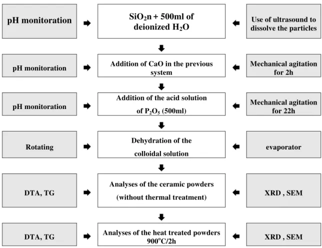
It was used the method based on dissolution-precipitation involving a solid-liquid reaction of phosphoric acid dissolving CaO to form calcium phosphate powders and nanocomposites. The raw materials used were calcium oxide powder (CaO) obtained from calcination of CaCO3 at 900oC for 2 hours. The CaCO3 powder (code number 24996) with 98% purity was provided by LABSYNTH LTDA laboratory, and the phosphor oxide (P2O5) was provided by RIEDEL-DE HAËN - Germany laboratory (99.5% purity, code number 1807). During the synthesis process, the molar factor used between calcium and phosphate (Ca/P) was 1.67 for the four compositions: calcium phosphate and calcium phosphate nanocomposites containing 5, 10 and 15% of nanometric silica (SiO2n). Figure 1 shows the method of synthesis and characterization for the four ceramic powders.
During the synthesis process, the pH was monitored at approximately each 15 min. The obtained materials from the synthesis were dried in a rotating evaporator and then investigated by scanning electron microscopy, X-ray diffraction, differential thermal analysis and thermogravimetry in order to observe the morphology, to identify the mineralogy composition and to study their thermal behaviour, respectively. The characterization study was performed on the nanostructured powders after drying in a rotating evaporator and after being treated at 900oC/2h.
pH monitoration
SiO
2n
+ 500ml of
deionized H
2O
Use of ultrasound to dissolve the particles
pH monitoration Addition of CaO in the previous system
Mechanical agitation for 2h
pH monitoration
Addition of the acid solution
of P2O5 (500ml)
Mechanical agitation for 22h
Rotating Dehydration of the
colloidal solution evaporator
DTA, TG
Analyses of the ceramic powders
(without thermal treatment) XRD , SEM
DTA, TG Analyses of the heat treated powders
900oC/2h XRD , SEM
Figure 1: Method of synthesis and characterization of the four ceramic powders.
3 RESULTS AND DISCUSSIONS
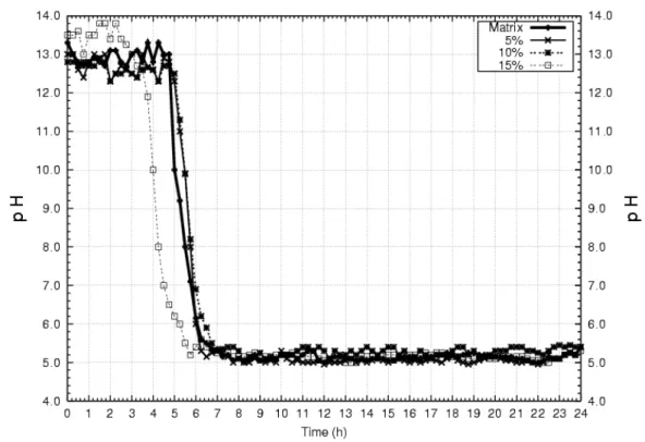
The measures of pH for the four compositions have shown that it increases with the incorporation of calcium oxide powder in the aqueous solution. On the other hand, the addition of acid solution decreases the pH value. Figure 2 illustrates the pH curves as a function of time for the different compositions, which shows that pH decreases after 4 hours of mechanical agitation due to the addition of the acid solution into the colloidal solution.
Figure 2: Behaviour of pH for the different compositions.
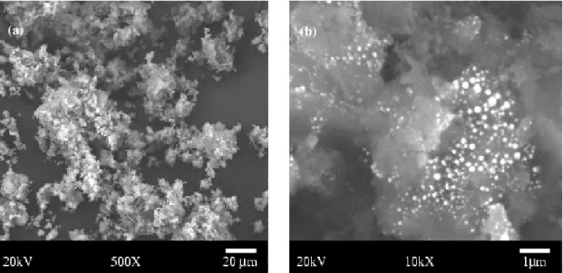
Morphological characterization, investigated by scanning electron microscopy (SEM), was performed on the calcium phosphate powder, on the calcium phosphate/SiO2n nanocomposite obtained after drying in a rotating evaporator and after calcination at 900oC for 2 hours. After drying the powders were ground and sieved through a 200 ABNT sieve. Figures 3a and 3b show that their morphology is constituted by clusters of small particles; figure 3b indicates the morphology of the nanocomposite powder with 15% (in volume) of SiO2n. In this figure it is possible to see the nanoparticles of SiO2n dispersed into the bioceramic powder.
The results obtained with the calcium phosphate, after treatment at 900oC for 2 hours, indicate the presence of clusters of small equiaxial particles with a size below 150 nm, as we can see in figure 4a.
(a) (b)
Figure 3: (a) Morphology of the calcium phosphate powder after drying in a rotating evaporator; (b) Morphology of the nanocomposite powder calcium phosphate/15% SiO2n
after drying in a rotating evaporator.
For the nanocomposite powders it was also observed that the morphology is composed of clusters of very small equiaxial particles, probably due to the presence of a second phase (SiO2n) well dispersed in the calcium phosphate powder (figure 4b). The presence of nanoparticles of SiO2n in the material, in inter-intragranular sites, inhibits the process of superficial diffusion associated to the nucleation and growth of the calcium phosphate’s grains, controlling the formation of β-calcium phosphate phase, and influencing the process of densification of the nanomaterial. Another observation was the presence of silica in the compositions, which can be explained by the difference in the thermal expansion coefficient of calcium phosphate matrix, which has a higher value (α = 14,85.10-6 ºC-1) compared with that of silica (α = 0,5. 10-6C -1
). This may lead to a modification in the superficial diffusion kinetic of the species toward the nucleolus of calcium phosphate, and probably inhibited the formation of crystobalite, as it can be observed in the X-ray spectra (figure 6) by the absence of peaks of crystobalite.
(b) (a)
Figure 4: (a). Morphology of the calcium phosphate powders after calcination at 900oC/2hours; (b) Morphology of the calcium phosphate/10%SiO2n nanocomposite powder after calcination.
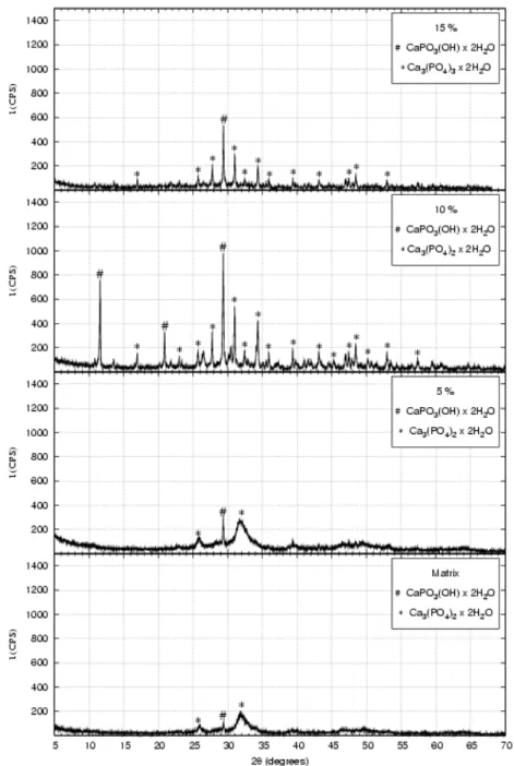
Figure 5: X-ray diffraction patterns of the powders after drying in a rotating evaporator.
The results from X-ray diffraction (XRD), performed on the powders of calcium phosphate and nanocomposites, after drying in the rotating evaporator (figure 5), have shown the presence of hydrated calcium phosphate (Brushite) with the composition CaPO3(OH).2H2O, and hydrated tri-calcium phosphate with the composition Ca3(PO4)2.2H2O for all the samples, indicating better crystallisation of brushite phase and of calcium phosphate for the nanocomposite powders. The second phase, formed of nanometric particles and uniformly dispersed in the material in inter-intragranular region, is more likely the responsible for improving the crystallisation.
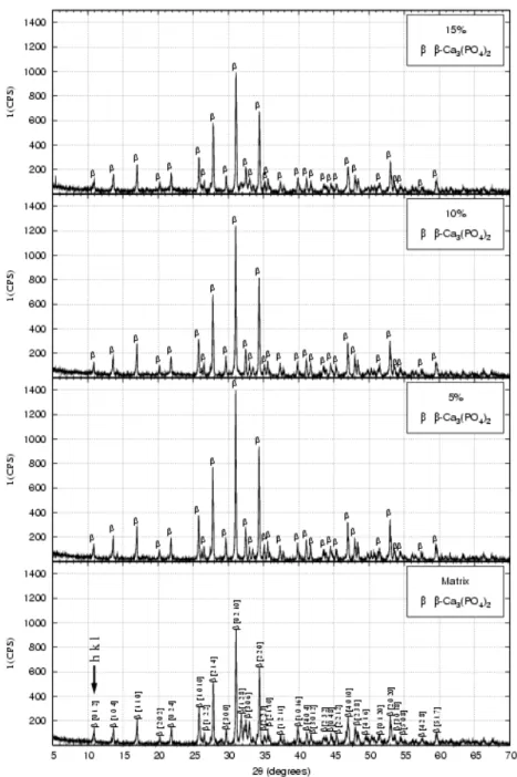
For the powders heat treated at 900oC/2h it was observed the crystallised structure of β − tri-calcium phosphate, for all the different compositions, as illustrated in figure 6.
Figure 6: XRD patterns of the powders after calcination at 900oC/2h.
It was observed that the major peak (2θ = 31.1o), represented by the main plain of diffraction [0 2 1 0], indicates the presence of β−tri-calcium phosphate.
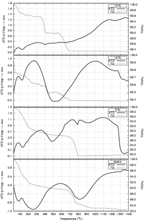
Evidences from thermogravimetry (TG) showed a steady decrease in mass loss between 25 and 800oC for all the nano-powders after drying in the rotating evaporator (figure 7). The investigations from differential thermal analyses (DTA) for the calcium phosphate indicated two peaks; one at approximately 130oC due to water evaporation without modification of the structure of the nanostructured powder, and the second at approximately 580oC, indicating water loss (dehydrogenation) from the bioceramic powder with modification of the structure of calcium phosphate and brushite [9-10].
Figure 7: Thermogravimetry and differential thermal analyses of the nano-powders after drying in the rotating evaporator.
Also, another peak was observed at approximately 1350oC showing the allotropic transformation from β to α tri-calcium phosphate. For the nanocomposites with 5%, 10% and 15% (in volume) of SiO2n, DTA curves displayed a peak at approximately 95oC indicating water evaporation without any modification in the structure of the nano-powder, and a second peak at approximately 350ºC, for the material with 5% of SiO2n, indicating the dehydrogenation with modification of the structure of the nanocomposite powder. For the materials containing 10% and 15% of SiO2n this peak appears at approximately 250oC. The presence of nanometric silica in the inter-intragranular region in the calcium phosphate might provide the water loss (dehydrogenation) due to the high surface area of the nanometric silica (≈80m2
/g), improving slightly the kinetic of superficial diffusion of the species toward the bulk of the calcium phosphate. Between 1000ºC and 1150 ºC there is another peak indicating the formation of β− tri-calcium phosphate phase (figure 7)
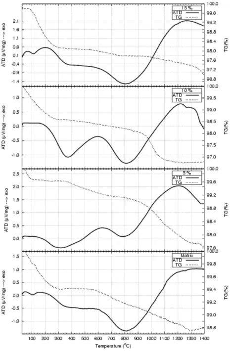
Figure 8: DTA and TG curves for the nano-powders obtained after calcination at 900oC/2h.
Evidences from DTA and TG performed on the nanostructured powders after calcination at 900oC/2h showed that the first peak at approximately 130oC is due to the water evaporation (figure 8). The second peak, at approximately 600ºC is due to the dehydrogenation and to the allotropic transformation from
α to β phase. The third peak, at approximately 1200ºC is due to the allotropic transformation of calcium phosphate from β to α.
4 CONCLUSION
The method of dissolution-precipitation with solid-liquid reaction led to the production of nanostructured powders of calcium phosphate and nanocomposites with particles size bellow 100 nm. The measure of pH showed that it takes 4 hours, after addition of acid solution, to reach a steady value of approximately 5.4.
582 The use of ultrasound to disperse the nanometric silica (SiO2n) into the calcium phosphate base material demonstrated to be efficient, leading to a homogeneous distribution of these nanoparticles, in the microstructure of the ceramic.
Investigations by SEM on the nanostructured powders, obtained after calcination, showed that the morphology is composed of equiaxial particles, with a size bellow 200nm, disposed in clusters.
XRD allowed us to identify the hydrated calcium phosphate – brushite (CaPO3(OH) 2H2O), and the hydrated tri-calcium phosphate - Ca3(PO4)2.2H2O (figure 5), on the powders after drying in the rotating evaporator. On the powders treated at 900ºC/2h it was identified β-calcium phosphate and hydroxyapatite phases.
DTA analysis showed that the dehydrogenation occurs at 600ºC and 300ºC for the calcium phosphate and for the nanocomposites, respectively, after drying in rotating evaporator (figure 7). After heat-treated, DTA curves exhibited a peak at approximately 600ºC for the nanocomposite powders, indicating the dehydrogenation and the allotropic transformation of α to β. For all the nanostructured powders the allotropic transformation of the calcium phosphate from α to β occurs at approximately 1200ºC.
5 BIBLIOGRAPHY
[1] HENCH, Larry L., “Medical and scientific products”, In: Ceramics and Glasses, SCHNEIDER, S.J., United States: ASM International, 1991.
[2] HENCH, L.L., “Bioceramics”, Journal of American Ceramic Society, v. 81, n. 7, pp. 1705-1728, 1998. [3] HENCH, L.L., “Bioceramics: From concept to clinic”, Journal of American Ceramic Society, v. 74, n. 7,
pp. 1487-1510, 1991
[4] RIEGER, W., “Medical applications of ceramics”, In: High-Tech Ceramics, KOSTORZ, G., Zurique: Academic Press, 1988.
[5] MAMANI GILAPA, Leonidas Cayo, Elaboração e caracterização de um material biocerâmico àmatriz de fosfato de cálcio produzido a partir de um pó cerâmico reciclado. Dissertação (Mestrado em Ciência e Engenharia de Materiais) – PGCEM/UDESC, 1995.
[6] CAMARGO, N.H.A., SOARES, C., GEMELLI, E., “Síntese e caracterização de biocimentos nanoestruturados para aplicações cirúrgicas otopédicas-odontológicas”, In: 50º Congresso Brasileiro de Cerâmica, Blumenau – SC, pp. 1-14, maio de 2006.
[7] CAMARGO, N.H.A., KARVAT, F., GEMELLI, E., “Elaboração e caracterização de uma cerâmica de fosfato de cálcio e compósitos, fosfato de cálcio/Al2O3-a para aplicação como implante e restituição óssea”, In: 50º Congresso Brasileiro de Cerâmica, Blumenau – SC, pp. 1-12, maio de 2006.
[8] PRASHANT, N. KUMTA, CHARLES SFEIR, DONG-HYUN LEE, DANA OLTON, DAIWON CHOI., “Nanostructured calcium phosphates for biomedical applications: novel synthesis and characterization”, Acta Biomaterialia, v.1, pp. 65-83, 2005.
[9] SOARES, C., Síntese e Caracterização de Biocimentos Nanoestruturados para Aplicações Biomédicas, Dissertação de mestrado - UDESC/Joinville, pp. 91, 2006.