BrazJOtorhinolaryngol.2014;80(5):451---452
Brazilian
Journal
of
OTORHINOLARYNGOLOGY
www.bjorl.org
CASE
REPORT
Right
ectopic
sphenoid
sinus
pituitary
adenoma
夽
Adenoma
hipofisário
ectópico
de
seio
esfenoidal
direito
Lara
Bonani
de
Almeida
Brito,
Paulo
Tinoco,
Túlio
Tinoco,
Flavia
Rodrigues
Ferreira
∗,
Vânia
Lúcia
Carrara
HospitalSãoJosédoAvaí,Itaperuna,RJ,Brazil
Received24November2012;accepted21April2013 Availableonline3July2014
Introduction
Adenomas are the most common pituitary tumors, corre-spondingfrom10%to20%ofallbraintumors.1Occasionally, these tumors can extend out of the pituitary fossa, and, onrareroccasions,theycanalsobefoundinectopicsites, havinguncertainoriginanddiverselocation.2
Ectopic pituitary adenomas are clinically detected because of their local mass effect and/or hormone hypersecretion.2Paranasalsinuscomputedtomographyand magnetic resonance imaging are used to study these conditions preoperatively, with diagnosis confirmed by histopathologyandimmunohistochemistry.3
Treatmentincludessurgicalresection,whichmayormay notbeassociatedwithradiationtherapy,andtheprognosis isgood.3
Clinical
case
E.S.,an82-year-oldfemalehadahistoryofheadacheand nasalcongestionforoneyear.Shehadpreviouslyundergone treatmentforrhinosinusitiswithnoclinicalimprovement.
夽 Pleasecitethisarticleas:BritoLB,TinocoP,TinocoT,Ferreira FR,CarraraVL. Rightectopicsphenoidsinus pituitary adenoma. BrazJOtorhinolaryngol.2014;80:451---2.
∗Correspondingauthor.
E-mail:[email protected](F.R.Ferreira).
Uponpresentation,anasalmassfillingtheright nostril andtheright aspectof thecavumwasdetectedon endo-scopicexamination.
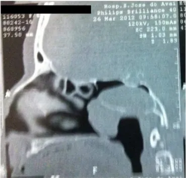
Computedtomographyoftheparanasalsinusesrevealed atumoroccupyingtheright nostrilandtherightaspectof thecavum(Fig.1).
Endonasal endoscopic surgery was conducted, and the entire tumor mass was resected from the right sphenoid sinus.Thespecimenwassentforhistopathological examina-tion,withinconclusiveresults.Immunohistochemicalstudy confirmed that it was an ectopic pituitary adenoma. On follow-up,thepatienthasnotedimprovementofher symp-toms.
Discussion
Anectopicpituitaryadenomaisdefinedasapituitarygland tumorlocated outofthe sellaturcicaand havingno con-nection with the intrasellar gland.4 They were described by Erdhelm in 1909, and may be found in the sphenoid sinus region, clivus, parapharyngeal space, nasal cavity andnasopharynx,hypothalamus,thirdventricle,andinthe suprasellarlocations.3,4
They are considered rare neoplasms originating from embryonic remnants of Rathke’s pouch. Since they were first described, approximately 50 cases of ectopic pitu-itaryadenomahavebeenreported,62%ofwhichoccurred in women at a mean age of 50 years, most commonly locatedinthesphenoidsinus(40%)andinsuprasellarsites (33%).4
http://dx.doi.org/10.1016/j.bjorl.2014.05.022
452 BritoLBetal.
Figure 1 Paranasal sinus tomography showing a sphenoid sinustumorextendingintothecavum.
According to medical literature, about one-third are endocrine-inactivetumorsdiagnosedasanexamination find-ingorfromtheirlocaleffect.Theremainingtwo-thirdshave hormonalactivityandusuallysecreteACTH,elicitinga Cush-ing’sdiseasepicture,althoughtheycanalsobeassociated withacromegalyandhighbloodprolactin.5
Diagnosisshouldbemadethroughhistory,physical exam-ination,paranasalsinuscomputedtomographyandmagnetic resonanceimagingshowingasoft-tissuedensitymassviewed in a paranasal cavity, as in the clinical case described, withnosellarabnormalities.5 Incasesofsuspectedtumor endocrineactivity,thepatientshouldhaveadditionaltests, suchassalivarycortisolestimation, ACTH,andCRHwhen Cushing’s syndrome is suspected; serum random GH and IGF-1testswhenacromegaly issuspected;when consider-inghyperprolactinemia,serumprolactinandTSHshouldbe performed.3,5
Differential diagnosis includes chordomas, nasopharyn-geal carcinoma,or a tumorderived froma minorsalivary gland.However,aclivuslesionshouldbedistinguishedfrom a meningioma, an epidermoid cyst, fibrousdysplasia, and pituitarytumors.6
Management includes surgical resection via either transsphenoidalortranssphenoethmoidalapproachtoreach thesphenoidsinusinadditiontotransfacialandtransnasal transmaxillaryapproachestotheclivus.5,6Malignant trans-formationisunusual;insuchcases,postoperativeradiation therapymaybeaddedifresectionisincomplete.6
Final
comments
Theectopicpituitaryadenomaisofgreatimportance,asit isararecondition.Oncediagnosisismade,complete clini-calimprovementcanbeachievedbysurgicalmanagement, whetherornotthisisassociatedwithradiationtherapy.
Conflicts
of
interest
Theauthorsdeclarenoconflictsofinterest.
References
1.Luk IS, Chan JK, Chow SM,Leung S. Pituitary adenoma pre-sentingas sinonasal tumor:pitfalls in diagnosis. Hum Pathol. 1996;27:605---9.
2.SchteingartDE,ChandlerWF,LloydRV,Ibarra-PerezG.Cushing’s syndromecausedbyanectopicpituitaryadenoma.Neurosurgery. 1987;21:223---7.
3.VanDerMeyA,VanKriekenJ,DulkenH,VanSetersA,Vielvoye J,HulshofJ.Largepituitaryadenomaswithextensionintothe nasopharlnx,reportofthreecaseswithareviewoftheliterature. AnnOtolRhinolLaryngol.1989;98:618---62.
4.LewisH,ToddH,MaieH,VíctorT.Suprasellaradrenocorticotropic hormone-seccretingectopicpituitaryadenoma:casereportand literaturereview.Neurosurgery.2002;50:618---25.
5.CollieRB,CollieMJ.Extracranialthyroid-stimulating hormone-secreting ectopic pituitary adenoma of the nasopharynx. OtolaryngolHeadNeckSurg.2005;133:453---4.