CONSTITUIÇÃO DO JÚRI
Doutora Maria Manuela Rodeia Grave Espada Niza Doutor António Freitas Duarte
Doutora Maria Teresa da Costa Mendes Vitor Villa de Brito
UNIVERSIDADE DE LISBOA
Faculdade de Medicina Veterinária
CANINE PRIMARY HYPERPARATHYROIDISM: CLINICAL APPROACH TO HYPERCALCAEMIA
CATARINA AFONSO GOMES
ORIENTADOR David John Walker, BVetMed(Hons)
DipACVIM DipECVIM-CA MRCVS
CO-ORIENTADOR Doutora Maria Teresa da Costa
Mendes Vitor Villa de Brito
2016
LISBOA
CONSTITUIÇÃO DO JÚRI
Doutora Maria Manuela Rodeia Grave Espada Niza Doutor António Freitas Duarte
Doutora Maria Teresa da Costa Mendes Vitor Villa de Brito
UNIVERSIDADE DE LISBOA
Faculdade de Medicina Veterinária
CANINE PRIMARY HYPERPARATHYROIDISM: CLINICAL APPROACH TO HYPERCALCAEMIA
CATARINA AFONSO GOMES
DISSERTAÇÃO DE MESTRADO INTEGRADO EM MEDICINA VETERINÁRIA
ORIENTADOR David John Walker, BVetMed(Hons)
DipACVIM DipECVIM-CA MRCVS
CO-ORIENTADOR Doutora Maria Teresa da Costa
Mendes Vitor Villa de Brito
2016
LISBOA
Acknowledgement
Ao David, por me ter dado uma oportunidade única e por apesar de constantemente ocupado, estar sempre disponível quando precisava e também pela constante preocupação. À professora Teresa, por toda a ajuda que me deu, pelas ideias e pelos esclarecimentos que ajudaram a construir esta tese pouco a pouco.
A todo o pessoal da Anderson Moores, tanto clínicos como enfermeiros e assistentes que me receberam e ajudaram durante os 6 meses de estágio. Um obrigado especial aos médicos Lucy, Fábio, Mónica, Louise, Maria e Florence pelo trabalho todo e tudo o que aprendi, aos imagiologistas Carolina, Roberto, Petra e Tommaso por tudo o que me ensinaram, ao Ricardo sempre disponível para ajudar e ao Paul, que sempre me explicou tudo sem sequer pedir. Um obrigado também às enfermeiras de medicina, em especial à incansável Julia.
A toda a turma C, em especial à Bia, Neves, Domingues, Di, Sónia, Marlene, Ana Sá, Su, André e Roque, porque vocês para mim foram a faculdade do primeiro ao último dia e sem vocês nada teria sido o mesmo.
Ao grupo da SMMEL, porque convosco todos os tempos eram sempre bem passados, à Lili e à Clarisse e o seu bolo de iogurte.
Aos amigos de sempre de Sesimbra, em especial ao Marco e Mariana. À Krebs, a madrinha mais aluada e à minha afilhada Adriana.
À VETuna, por todas as experiências e pessoas que não teria conhecido sem ela. Um obrigada especial aos meus afilhados Pastelão, Primadonna, Stripper, Snap e Devota, pelo reconhecimento e por me fazerem sentir que a minha presença foi valorizada. Ao Estrela e Pecho, sempre presentes na defesa dos mezzos e ao Silvi, membro do quarteto fantástico, pelas horas de conversas/monólogos.
À MI, parceira de tudo, pela compreensão nos mil momentos, a grande maioria deles passados a rir.
À Sara, pela constante companhia e por tudo o que passámos.
Ao meu avô Manuel, que sempre acreditou que as suas netas eram as melhores e que eram capazes de fazer tudo o que quisessem.
À Eve, por ser sempre a mais entusiasta e incansável cobaia que podia haver.
Mas acima de tudo, aos meus pais, à Inês e à Raquel, porque mais que qualquer um estiveram lá sempre e para tudo. Obrigada por me terem educado como o fizeram e por me terem dado todas as oportunidades que tive para poder e querer ser sempre melhor. Não poderia desejar mais.
Abstract
Canine primary hyperparathyroidism: clinical approach to hypercalcaemia
Canine primary hyperparathyroidism (PHPTH) is an endocrine disorder, where one or more parathyroid glands autonomously produce and secrete parathyroid hormone (PTH), which results in hypercalcaemia (Feldman, 2010).
Diagnosis of PHPTH is achieved when there are inappropriate PTH concentrations (normal or increased) in the presence of elevated ionised calcium (iCa) concentration with no other identifiable cause (Skelly, 2012). iCa is the only active fraction of calcium and does not always correlate to total calcium, which is why iCa should be used to assess serum calcium status (Schenck & Chew, 2008).
PHPTH is usually diagnosed after detection of hypercalcaemia in a blood analysis performed for unrelated reasons, as clinical signs are often not perceived by the owners (Feldman, Hoar, Pollard, & Nelson, 2005; Feldman, 2015a).
Treatment by parathyroidectomy, percutaneous ultrasound-guided ethanol ablation or percutaneous ultrasound-guided heat ablation is curative and prognosis is excellent for treated dogs, but hypocalcaemia is a frequent postoperative complication (Caplan, 2013; Feldman, 2015a; Flanders, 2003; Nelson, 2009; Rasor, Pollard, & Feldman, 2007; Séguin & Brownlee, 2012).
The retrospective study had the objective of characterising a sample of six dogs diagnosed with PHPTH at Anderson Moores Veterinary Specialists and analyse the procedures and tests conducted in the clinical approach to previously identified hypercalcaemia.
Key words: primary hyperparathyroidism, parathyroid hormone, hypercalcaemia, ionised calcium
Resumo
Hiperparatiroidismo primário canino: abordagem clínica à hipercalcémia
O hiperparatiroidismo primário canino (PHPTH) é uma doença endócrina, na qual uma ou mais glândulas paratiróides produzem e secretam hormona paratiroideia ou paratormona (PTH) autonomamente, o que resulta em hipercalcémia (Feldman, 2010).
O diagnóstico de PHPTH é efectuado quando existem concentrações de PTH inapropriadas (normais ou aumentadas) na presença de concentrações aumentadas de cálcio ionizado (iCa) sem outra causa identificável (Skelly, 2012). O iCa é a única fracção activa do cálcio e nem sempre se correlaciona com o cálcio total, motivo pelo qual o iCa deve ser utilizado para avaliar o cálcio em circulação (Schenck & Chew, 2008)
O PHPTH é normalmente diagnosticado após a detecção de hipercalcémia numa análise sanguínea efectuada por motivos não relacionados, uma vez que os sinais clínicos normalmente não são identificados pelos donos (Feldman, Hoar, Pollard, & Nelson, 2005; Feldman, 2015a)..
O tratamento com paratiroidectomia, ablação percutânea com etanol guiada por ultra-som ou ablação percutânea com calor guiada por ultra-som é curativo e tem um prognóstico excelente para animais tratados, embora a hipocalcémia seja uma complicação pós-cirurgica frequente (Caplan, 2013; Feldman, 2015a; Flanders, 2003; Nelson, 2009; Rasor, Pollard, & Feldman, 2007; Séguin & Brownlee, 2012).
O estudo retrospectivo deste trabalho teve como objectivo a caracterização de uma amostra de seis cães diagnosticados com PHPTH na Anderson Moores Veterinary Specialists e analizar os procedimentos e testes efectuados na abordagem clínica à hipercalcémia previamente identificada.
Palavras-chave: hiperparatiroidismo primário, hormona paratiroideia, paratormona, hipercalcémia, cálcio ionizado
Table of contents
Acknowledgement ... iii
Abstract ... iv
Resumo ... v
Table of contents ... vi
Index of Figures ... viii
Index of tables ... ix Abbreviations ... x Placement ... xi 1. Primary Hyperparathyroidism ... 2 1.1. Parathyroids ... 2 1.1.1. Anatomy ... 2 1.1.2. Histology ... 3 1.1.3. Calcium physiology ... 4 1.2. Hyperparathyroidism ... 12
1.2.1. Renal secondary hyperparathyroidism ... 12
1.2.2. Nutritional secondary hyperparathyroidism ... 14
1.3. Primary Hyperparathyroidism ... 16
1.3.1. Definition and aetiology ... 16
1.3.2. Pathophysiology ... 17
1.3.3. Signalment ... 18
1.3.4. Anamnesis - clinical signs ... 19
1.3.5. Physical examination ... 20 1.3.6. Clinical pathology ... 20 1.3.7. Assays ... 25 1.3.8. Imaging ... 27 1.3.9. Differential diagnosis ... 29 1.3.10. Pretreatment considerations ... 29 1.3.11. Treatment ... 30
1.3.12. Posttreatment management – hypocalcaemia ... 36
1.3.13. Prognosis ... 40
1.3.14. Associated diseases ... 41
2. Clinical approach to hypercalcaemia ... 42
2.1. Hypercalcaemia - clinical signs ... 42
2.2. Differential diagnosis ... 43
2.3. Diagnostic approach ... 47
2.3.1. Signalment ... 47
2.3.2. History and physical examination ... 48
2.3.3. Clinical pathology ... 48
2.3.4. Imaging ... 49
2.3.5. Assays ... 50
2.4. Acute treatment ... 51
2.5. Prognosis ... 55
3. Retrospective study: Clinical approach to hypercalcaemia in 6 dogs with PHPTH . 56 3.1. Objective ... 56
3.2. Material and methods ... 56
3.3. Results ... 57 3.3.1. Signalment ... 57 3.3.2. Previous history ... 57 3.3.3. Clinical signs ... 57 3.3.4. Physical examination ... 57 3.3.5. Clinical pathology ... 57
3.3.6. Imaging ... 60 3.3.7. Treatment ... 60 3.3.8. Posttreatment management ... 61 3.3.9. Histopathology ... 61 3.4. Discussion ... 62 3.5. Conclusion ... 67 References ... 69
Index of Figures
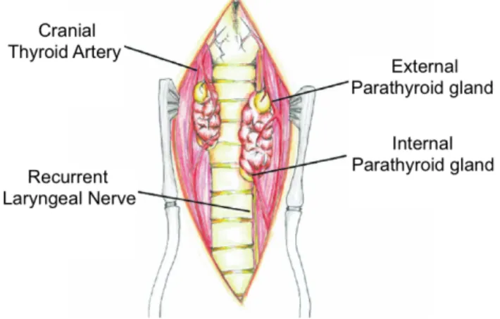
Figure 1 - Internal and external parathyroid glands ... 2
!
Figure 2 - Parathyroid glands and surrounding structures ... 3
!
Figure 3 - Parathyroid chief cells organised in chords around capillaries. Haematoxylin andeosin, 200x ... 3!
Figure 4 - Sites of calcium resorption in the nephron. Passive reabsorption occurs in the
proximal convoluted tubule and PTH regulation occurs in the distal convoluted tubule ... 6!
Figure 5 - Synthesis and secretion of PTH and the regulation sites of PTH biosynthesis by
iCa or calcitriol (1,25 Vit D3) ... 7! Figure 6 - Serum calcium “set point”, the concentration of serum iCa at which PTH
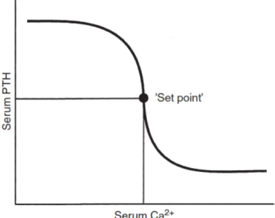
concentration is half maximal ... 9!
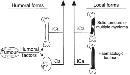
Figure 7 – Humoral and local forms of hypercalcaemia of malignancy caused by increased
stimulation of osteoclastic bone resorption or increased tubular resorption of calcium . 44!
Figure 8 - Humoral factors produced by tumours cause hypercalcaemia of malignancy by
acting as systemic hormones which stimulate osteoclastic bone resorption of increase tubular reabsorption of calcium. ... 45!
Index of tables
Table 1 – Summary of clinical pathology results in dogs with PHPTH. ... 24
!
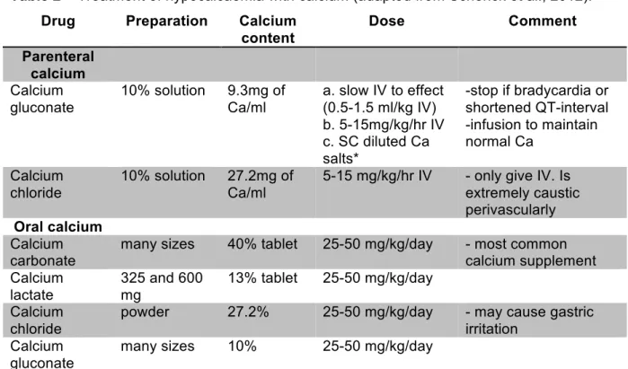
Table 2 – Treatment of hypocalcaemia with calcium ... 40
!
Table 3 – Treatment of hypocalcaemia with Vitamin D ... 40
!
Table 4 – Specific treatment of hypercalcaemia ... 54
!
Table 5 – Successive concentrations of serum tCa measured in the six cases, before treatment. ... 58!
Table 6 – Successive concentrations of serum iCa measured in the six cases, before treatment. ... 58!
Table 7 – Successive concentrations of serum phosphate measured in the six cases, before treatment. ... 58!
Table 8 – Successive PTH serum concentrations measured in the six cases, before treatment. ... 59!
Table 9 – Successive PTHrP serum concentrations measured in five of the cases. ... 59
!
Table 10 – Urea and creatinine serum concentrations in the six cases. ... 59
!
Table 11 – Vitamin D metabolites measured in one case. ... 59
!
Table 12 – Successive USG in five of the cases, before treatment. ... 60
!
Abbreviations
ACTH – adrenocorticotropic hormone ADH – antidiuretic hormone
AKI – acute kidney injury ALP – alkaline phosphatase ALT – alanine aminotransferase ARF – acute renal failure
BARF – biologically appropriate raw food BID – twice daily (bis in die)
BUN – blood urea nitrogen
cAMP - cyclic adenosine monophosphate CaR – calcium-sensing receptor
CBC – complete blood count CKD – chronic kidney disease CO2 – carbon dioxide
CRF – chronic renal failure CT – computed tomography ECF – extracellular fluid ECG – electrocardiogram
EDTA – ethylenediaminetetraacetic acid FGF-23 – fibroblast growth factor-23 FNA – fine needle aspirate
GFR – glomerular filtration rate GH – growth hormone
GPCR – G protein-coupled receptor
HHM – humoral hypercalcaemia of malignancy HPTH – hyperparathyroidism
iCa – ionised calcium
IRIS – International Renal Interest Society IRMA – immunoradiometric assays
IV – intravenous administration MEN – multiple endocrine neoplasia PHPTH – primary hyperparathyroidism PO – oral administration (per os) PTH – parathyroid hormone
RIA – radioimmunoassay
SC – subcutaneous administration SID – once daily (semel in die) tCa – total calcium
TID – thrice daily (ter in die) US – ultrasonography USG – urine specific gravity UTI – urinary tract infection VDR – vitamin D receptor
Placement
The student’s curricular placement was in Anderson Moores Veterinary Specialists in Winchester, United Kingdom for a 6 months externship from October 2nd 2015 to March 18th 2016.
A total of 990 hours were completed during the placement on a schedule basis from Monday to Friday, with 9-hour days from 8.30am to 17.30pm.
The first and last half hour of each day was spent in ward rounds where the medics presented their cases and gave relevant information to both the on call medic and nurses. The student had a rotation between internal medicine consults, imaging, medicine procedures and wards, with one out of four weeks spent in wards. The student had the opportunity to follow the cases from the first consult to the complete investigation by imaging, medical procedures and access to laboratory results.
A total of 204 hours were spent in internal medicine consults, 272 hours in imaging and 170 hours in medicine procedures.
Of the 272 hours completed in imaging included mostly ultrasound exams, ultrasound-guided procedures, radiography but also an adding total of approximately 68 hours in computed tomography (CT) scans and fluoroscopy (swallow studies and cystography). Ultrasound-guided procedures included cystocentesis, fine needle aspirates, tru-cut biopsies (mainly liver, kidney and miscellaneous tumours), abdominal and thoracic fluid sampling, bile sampling and chest drainage.
The medicine procedures included gastrointestinal, respiratory and urinary endoscopy, foreign body endoscopic removal, percutaneous endoscopic gastrostomy, oesophagostomy feeding tubes, cerebrospinal fluid taps, joint taps, bronchoalveolar lavage, bone marrow biopsy and aspiration,
Approximately 34 hours were spent in assisting to miscellaneous activities including: magnetic resonance imaging scans, wound management, chest drains placement, skin biopsies, muscle biopsies, echocardiography, electrocardiography and electromyography.
1. Primary Hyperparathyroidism
1.1. Parathyroids
1.1.1. Anatomy
The parathyroid glands are small ellipsoid purplish endocrine organs, closely related to the thyroid gland, which is located on the external surface of the trachea. These glands generally measure 2-5mm in diameter and 0,5-1mm in width, with no demonstrated correlation to body weight according to a study by Mulligan and Francis in 19511 (Hullinger, 2013; Rosol &
Capen, 1997).
There are usually four structurally independent glands, one embedded (external parathyroid) and one on the surface (internal parathyroid) of each of the thyroid glands (Figure 1). The external parathyroid, also known as parathyroid gland III, is frequently located on the surface of the thyroid gland on its cranial dorsolateral edge. The internal parathyroid, or parathyroid gland IV, is usually found at various depths of thyroid tissue, in the caudal portion of the thyroid. In 1951, Mulligan and Francis also reported common variations in number and location of the parathyroid glands. Marine (1914)2 and Reed et al. (1928)3 reported an
incidence of accessory parathyroid tissue in 3 to 6% of dogs (Hullinger, 2013).
Figure 1 - Internal and external parathyroid glands (modified from Bonczynski, 2007).
The blood supply is related to the supply from the thyroid glands, with the external parathyroid being vascularised by small branches of the cranial thyroid artery and the internal parathyroid by vessels surrounding the thyroid parenchyma. The venous and lymphatic
1 Mulligan, R.M., Francis, K.C. (1951). Weights of thyroid and parathyroid glands of normal male dogs. The Anatomical Record 110:139–143.
2 Marine, D. (1914). Observations on tetany in dogs. Journal of Experimental Medicine 19:89–105.
drainage are also the same as the thyroid glands and it is likely that the innervation of the parathyroids is too (Figure 2) (Hullinger, 2013; Rosol & Capen, 1997).
Figure 2 - Parathyroid glands and surrounding structures (modified from Caplan, 2013).
1.1.2. Histology
The parathyroid tissue is composed by principal or chief cells, small polygonal cells with round nuclei and slightly acidophilic pale staining cytoplasm (Figure 3), that produce and secrete parathyroid hormone (PTH). PTH is stored in granules of irregular shape in the cytoplasm. Oxyphil cells are also present, usually in clusters of cells. These cells are larger than chief cells with very acidophilic cytoplasm, but their function is not fully understood (Mescher, 2013).
Figure 3 - Parathyroid chief cells organised in chords around capillaries. Haematoxylin and
1.1.3. Calcium physiology
1.1.3.1. Function and distribution
Calcium is an inorganic element essential to survival, included in the macrominerals group (Goff, 2015). Calcium has two primary functions: (1) to provide skeletal support and (2) to participate in biochemical intracellular and extracellular functions (Feldman, 2015a; Schenck, Chew, Nagode & Rosol, 2012).
Ionised calcium (iCa or Ca2+) is necessary for several body functions including enzymatic
reactions, membrane transport and stability, blood coagulation, nerve conduction, neuromuscular transmission, muscle contraction, vascular smooth muscle tone, hormone secretion, bone formation and resorption, control of hepatic glycogen metabolism, cell growth and division (Schenck et al., 2012).
The vast majority of calcium (99%) is found in bone, primarily as hydroxyapatite; 0.9% is intracellular; and 0.1% is extracellular. Most of the intracellular calcium is contained in mitochondria, granules of the endoplasmic reticulum or bound to proteins, with only a very low concentration of calcium present in the cytosol (Cunningham & Klein, 2007; Rosol, Chew, Nagode & Capen, 1995).
There is a 10,000-fold concentration gradient of iCa between extracellular fluid and cytosol, which enables iCa to serve as a messenger to activate intracellular processes (Rosol et al., 1995) through a rise and fall during brief cellular responses or during initial phases of sustained responses (Rasmussen, 1989). The iCa role is carried at very low and tightly controlled concentrations, because excess in cytosolic iCa causes toxicity and may lead to cellular death (Rasmussen, Barrett, Smallwood, Bollag & Isales, 1990; Rasmussen, 1989; Schenck et al., 2012).
The calcium in the extracellular fluid (ECF), including intestinal and blood calcium, exists in three fractions: iCa (56%), protein-bound (34%) and complexed (10%). These values refer to healthy dogs (Schenck & Chew, 2012). The variation between ionised and protein-bound calcium is dependent on serum pH, with a rise in acidity causing increased competition between hydrogen ions and iCa for the negative-charged sites in the proteins, resulting in higher concentrations of iCa (Rosol et al., 1995).
The biologically active and actively regulated fraction of calcium is iCa (Feldman, 2015a; Rosol et al., 1995). The protein-bound calcium is mainly bound to albumin, and in a smaller amount to globulins, while complexed calcium is bound to phosphate, bicarbonate, sulfate, citrate and lactate (Goff, 2015; Rosol et al., 1995; Schenck et al., 2012).
1.1.3.2. Calcium homeostasis
Normal homeostasis of serum calcium maintains the concentration within a narrow range, making adjustments within a 5% deviation of the normal values (Cunningham & Klein, 2007). The normal serum concentrations of calcium in dogs are 9.0 - 11.5mg/dL or 2.2 - 3.8mmol/l for total calcium (tCa) and 5.0 - 6.0mg/dl or 1.2 - 1.5mmol/l for iCa (Schenck et al., 2012), though each laboratory has its own reference range.
The calcium serum concentration is mainly regulated by: (1) PTH; (2) calcitriol, the active form of vitamin D; and (3) calcitonin. Under specific conditions other hormones, including adrenal corticosteroids, oestrogens, thyroxine, growth hormone (GH), glucagon and parathyroid hormone-related protein (PTHrP), may also affect the calcium control (Rosol & Capen, 1997).
In fetus the calcium homeostasis is maintained by PTHrP, as low levels of PTH are present (MacIsaac, Caple, et al., 1991; MacIsaac, Heath, et al., 1991).
The calcium regulating hormones act on three target organs: (1) the intestine, the absorption site; (2) the kidneys, the excretion site; and (3) the skeleton, the largest storage site in the body (Favus, Bushinsky & Lemann, 2006; Rosol & Capen, 1996). In normal adult animals the amount of calcium absorbed in the intestine matches the amount excreted by urine and the small losses through sweat and intestinal secretions (Rosol et al., 1995).
Intestinal absorption of calcium can occur in two mechanisms: a passive, nonsaturable, paracellular diffusion or an active, saturable, cell-mediated transport. The passive absorption depends on electrochemical gradients, requiring high concentrations of calcium in the intestinal lumen. This diffusional process depends on the permeability of each intestinal segment, which is highest in the duodenum, jejunum and ileum, intermediate in the colon and lowest in the cecum (Favus et al., 2006). The active transport absorbs calcium down a concentration gradient, from the intestinal lumen to the interstitial fluid. This process is facilitated by transporting proteins including: a calcium channel protein that allows iCa to cross to the cytosol, a vitamin D-dependent calcium-binding protein that transports calcium from the apical surface of cells, which is then pumped by the plasma membrane Ca2+ ATPase pump protein to the extracellular space on the basolateral side of the cell. All three of these proteins are dependent on calcitriol (Goff, 2015).
The amount of calcium absorbed by each mechanism depends on the calcium content of the diet. High calcium intakes result in larger amounts being absorbed passively, which in turn trigger lower concentrations of calcitriol, the hormone responsible for stimulating active absorption. On the other hand, lower calcium dietary content stimulates calcitriol secretion and, consequently, increases active absorption on a longer-term basis. Calcitriol stimulation of the intestinal epithelial vitamin D receptor (VDR) will contribute to the production of proteins that partake in cell-mediated transport (Favus et al., 2006).
The absorption in the intestines depends on: the acidity, the presence of other dietary components or disease in the small intestine, the integrity of the villi and on calcitriol stimulation (Schenck et al., 2012).
The non-protein bound calcium, including ionised and complexed calcium, is the ultrafilterable fraction of calcium, which composes the glomerular filtrate. To maintain calcium homeostasis, the kidney must reabsorb 98% or more of the filtered calcium. The majority of calcium is reabsorbed in the proximal convoluted tubules (70%), mainly by passive mechanisms, and in the thick ascending loop of Henle (20%). Approximately 8% of calcium is actively absorbed in the distal convoluted tubules, the main site for physiologic regulation of its excretion (Figure 4) (Favus et al., 2006; Rosol et al., 1995).
Figure 4 - Sites of calcium resorption in the nephron. Passive reabsorption occurs in the
proximal convoluted tubule and PTH regulation occurs in the distal convoluted tubule (from Rosol et al., 1995).
Legend: PCT - proximal convoluted tubule; DCT - distal convoluted tubule
The bone osteocytes are surrounded by lacunae, which are connected by canaliculi. The fluid in the lacunae and canaliculi is rich in calcium, which can be rapidly transported to the ECF when stimulated by PTH (Goff, 2015).
The calcium in this fluid is present as amorphous crystals or in solution and comprises 0.5% of the bone calcium (Cunningham & Klein, 2007). When larger amounts of calcium are required osteoclasts reabsorb solid bone, dissolving the mineralised matrix in calcium and phosphorus, which can then be mobilised to the ECF (Schenck et al., 2012).
Hypocalcaemia results in the secretion of PTH by the parathyroid glands within seconds. The presence of increased PTH secretion will: increase the renal tubule reabsorption within minutes, decreasing the urinary losses; mobilise bone calcium stores, both osteocytic and osteoclastic-mediated within minutes to hours; indirectly enhance intestinal absorption of calcium by stimulating the synthesis of calcitriol within 24 hours (Favus et al., 2006; Goff, 2015).
Hypercalcaemia, a less frequent occurrence, results in the suppression of PTH secretion and consequently decreases tubular reabsorption and increases renal excretion of calcium. This decline in PTH along with higher iCa concentrations decreases calcitriol synthesis (Schenck et al., 2012). Increased calcitonin secretion by the thyroid, also inhibits renal reabsorption and bone calcium resorption, decreasing the mobilisation of calcium to the ECF (Goff, 2015). Serum phosphate or inorganic phosphorus is also affected by calcium regulating hormones: PTH reduces phosphate renal reabsorption and stimulates phosphate release from the bone; and calcitriol increases phosphate intestinal absorption (Brown & Juppner, 2006; Rosol & Capen, 1996).
1.1.3.3. Parathyroid hormone
PTH is an 84-amino acid straight chain polypeptide produced and secreted by chief cells of the parathyroid glands. The intact PTH (1-84) is the biologically active form of the hormone, which has two functional domains: the N-terminal (PTH 1-34), which has the major biological activity, and the C-terminal (Rosol & Capen, 1996; Rosol et al., 1995).
The ribosomes of the chief cells synthesise the precursor of PTH, preproparathyroid hormone (preproPTH), composed by 115 amino acids (a.a.). PreproPTH penetrates the cisternal space of the rough endoplasmic reticulum where, within a minute, a 25 a.a sequence is cleaved, resulting in proparathyroid hormone (proPTH). Then proPTH, composed by 90 a.a, goes to the Golgi apparatus where enzymes cleavage a 6 a.a. sequence concluding the synthesis of mature PTH (Figure 5) (Rosol & Capen, 1997).
Figure 5 - Synthesis and secretion of PTH and the regulation sites of PTH biosynthesis by
The parathyroid glands’ function is regulated through the stimulation or inhibition of the secretion and synthesis of PTH and parathyroid cellular proliferation. The main regulators are iCa, via extracellular calcium-sensing receptor (CaR), and calcitriol, via the VDR, with iCa being the most important regulator in a minute-to-minute basis (Feldman, 2015a; Schenck et al., 2012). Low iCa concentration stimulates PTH secretion, PTH gene expression and parathyroid cellular proliferation, whilst high iCa concentration inhibits those actions. Calcitriol inhibits PTH gene expression and may reduce the secretion of PTH and parathyroid cellular proliferation. According to Slatopolsky et al., 19961, phosphate also directly stimulates PTH
gene expression and is thought to also indirectly stimulate PTH secretion and parathyroid cellular proliferation (Brown & Juppner, 2006).
The CaR is a G protein-coupled receptor (GPCR) expressed in the parathyroid cells, where it inhibits PTH secretion when calcium binds to it. CaR is also present in the kidneys, where it enhances or reduces the calcium excretion in urine depending on the iCa concentration (Feldman, 2015a).
The different responses to increase PTH secretion depend on how long hypocalcaemia lasts. The initial response is to secrete the PTH contained in the secretory vesicles in the chief cells, which happens over seconds to a few minutes. If the stimulus is maintained for longer periods of time, the following actions take place: (1) the intracellular degradation of PTH is reduced (resulting in more biologically active PTH available for secretion), from minutes to around an hour; (2) there is an increase in the PTH gene expression and consequently an increase in the synthesised PTH, from several hours to days; (3) there is proliferation of parathyroid cells, from days to weeks or longer, eventually leading to enlarged glands (Brown & Juppner, 2006). The amount of PTH available for secretion depends on the synthesised amount and the amount that suffers intracellular degradation (Schenck et al., 2012).
The rate of PTH secretion has a steep inverse sigmoidal relation with iCa, in which small variations in iCa result in marked changes in PTH secretion. This is important to maintain a precise control in the concentrations of iCa (Rosol et al., 1995). According to Brown, 19912, the set point for PTH secretion is the iCa concentration that occurs at serum PTH concentration midway between maximal an minimal values of PTH obtained experimentally (Figure 6). Normal iCa concentrations are slightly higher than the set point (Schenck et al., 2012).
PTH has a short half-life (less than 5 minutes) before it is metabolised peripherically and excreted, mainly by the liver and kidneys. In the liver PTH is metabolised and degraded by Kupffer cells and renal clearance occurs through glomerular filtration (Kronenberg, Bringhurst, Segre & Potts, 2001; Rosol et al., 1995).
1 Slatopolsky, E., Finch, J., Denda, M., Ritter, C., Zhong, M., Dusso, A., Mac-Donald, P., Brown (1996). Phosphorus restriction prevents parathyroid gland growth. High phosphorus directly stimulates PTH secretion in vitro. Journal Clinical Investigation 97: 2534–2540.
Figure 6 - Serum calcium “set point”, the concentration of serum iCa at which PTH
concentration is half maximal (from Rosol, 1995).
The PTH1 receptor or PTH/PTHrP receptor is the main receptor mediating PTH action, binding the N-terminal of PTH and PTHrP equally. It is mostly expressed in renal epithelial cells and osteoblasts, though it is also present in other cells. PTH2 receptor, on the other hand, binds PTH but not PTHrP (Schenck et al., 2012).
PTH is the main hormone controlling iCa on a minute-to-minute basis, with the kidneys and bones as target organs. PTH actions in the kidney are: to increase the reabsorption of calcium, to inhibit phosphate reabsorption and to stimulate the synthesis of calcitriol. In the bones, PTH causes mobilisation of calcium and phosphate from the rapid turning-over pool and after a few hours from an additional slower pool to the blood. Chronically, PTH leads to an increase in the number and on the activity of osteoclast cells and if intermittently administrated enhances the formation of trabecular bone. The overall effect of PTH is to increase iCa concentration, decrease phosphate concentration and increase renal synthesis of calcitriol (Brown & Juppner, 2006).
1.1.3.4. Parathyroid hormone-related protein
PTHrP is a protein composed of 139, 141 or 173 a.a. considered a polyhormone with multiple biologically active regions (Rosol & Capen, 1997; Rosol et al., 1992). The N-terminal region of the three forms of PTHrP (1-34) has significant homology with the N-terminal region of PTH (Broadus & Nissenson, 2006; Philbrick, 2001). This homology explains how PTHrP is able to bind and activate the PTH1 receptors in the kidney and bone with an affinity similar to PTH (Philbrick, 2001; Rosol et al., 1992). PTHrP secreted by tumours can stimulate this receptor and mimic PTH action, causing the paraneoplasic effects on the metabolism of calcium and phosphate that occur in humoral hypercalcaemia of malignancy (HHM) syndrome (Philbrick, 2001; Wysolmerski, 2012).
PTHrP is also produced by several normal tissues including: stratified squamous epithelium, adrenal cortex and medulla, fetal and adult parathyroid glands, adenohypophysis, thyroid, skeletal and smooth muscle, kidney, bone, lactating mammary gland, brain, pancreas, ovary,
testicle, myometrium and placenta. The expression of PTHrP gene in a large number of tissues accounts for the variety of types of neoplasms that can cause HHM (Rosol et al., 1992).
The different actions of PTHrP can be divided in three groups: (1) normal endocrine, (2) normal paracrine and (3) abnormal endocrine in HHM. In most normal tissues PTHrP carries a paracrine action. PTHrP acts and possibly is also metabolised and degraded locally, which explains its low circulating concentrations (Rosol et al., 1992).
Studies in sheep and mice have showed that in fetus PTHrP has an endocrine activity, where the midregion is responsible for the transport of calcium through the placenta, necessary for the fetus to maintain the required calcium concentration and also to keep the fetal-maternal calcium gradient, with the fetus having the highest concentration (Care et al., 1990; Kovacs et al., 1996).
1.1.3.5. Calcitriol
In domestic animals the active metabolites derived from Vitamin D2 and Vitamin D3 have
equal bioactivity, so the generic terms 1,25-dihydroxyvitamin D or 1,25(OH)2D and calcitriol
are used to refer to the metabolites from both Vitamin D2 and Vitamin D3 (Schenck et al.,
2012).
Dogs and cats are dependent on dietary intake of Vitamin D, as they are unable to adequately synthesise Vitamin D in the skin like herbivores and omnivores (How, Hazewinkel & Mol, 1994).
Vitamin D requires metabolic activation to be physiologically functional. The metabolism of vitamin D starts in the liver, where it is hydroxylation into 25-hydroxyvitamin D occurs. This metabolite is then transported to the kidney, where in the renal tubules 1!-hydroxylase produces 1,25-dihydroxyvitamin D (calcitriol) (Bonczynski, 2007; Holick & Garabedian, 2006). The second step in this metabolic activation is closely controlled by ionic and hormonal mechanisms, with serum concentrations of PTH, calcitriol, phosphate and calcium being the main regulators. Renal 1!-hydroxylase is inhibited by hypercalcaemia, hyperphosphataemia, excess of calcitriol and absence of PTH (Bonczynski, 2007; Pike, Meyer & Lee, 2011; Schenck et al., 2012). GH, oestrogen and prolactin are also important in increasing renal synthesis of calcitriol during growth, pregnancy and lactation, respectively (Rosol et al., 1995).
The main function of calcitriol is to maintain calcium and phosphorus homeostasis, in order to sustain adequate levels for bone matrix mineralisation (Pike et al., 2011; Rosol & Capen, 1997). Calcitriol maintains calcium levels predominantly through: (1) stimulation of intestinal calcium absorption, by activating several proteins in the small intestine to facilitate the movement of calcium into the circulation; (2) osteoclastic bone resorption by inducing pre-osteoclasts to become mature pre-osteoclasts, which dissolve bone mineral and matrix releasing
calcium into the extracellular space; (3) inhibition of PTH synthesis; and (4) negative feedback on calcitriol formation in the kidney (Holick & Garabedian, 2006; Rosol et al., 1995). Calcitriol also carries functions not related to calcium homeostasis, including inhibition of cell growth and stimulation of cell differentiation (Rosol & Capen, 1997).
Calcitriol acts on the VDR that is present in larger quantities on its target tissues: bone, kidney, intestine and parathyroid glands; but it is also present in many other tissues (Schenck et al., 2012).
1.1.3.6. Calcitonin
Calcitonin is a 32 amino acid polypeptide synthesised in the thyroid by parafollicular or C cells (Cunningham & Klein, 2007).
This hormone leads to the decrease of serum iCa following rapid rises. Calcitonin is considered an “emergency hormone” to protect against hypercalcaemia, not being one of the primary minute-to-minute regulators of iCa (Rosol & Capen, 1997; Rosol et al., 1995).
The regulation of the secretion by C cells is mediated by CaR, the same receptor as the chief cells in the parathyroid glands. The secretion is continuous in normocalcaemia and markedly increased when iCa rises. Hyperphosphataemia also serves as a stimulant for calcitonin secretion (Rosol & Capen, 1997)
Calcitonin decreases iCa by inhibiting osteoclastic osteolysis and decreasing osteoclasts numbers, inhibiting bone resorption and decreasing calcium movement from the bone to the plasma (Rosol & Capen, 1997). This inhibition, however, is only temporary and chronic hypercalcaemia will result in high concentrations of calcitonin and C cell hyperplasia, having limited biological significance in iCa concentrations (Rosol et al., 1995).
Calcitonin also decreases phosphate reabsorption in renal tubules, stimulates diuresis of sodium, chloride and calcium, and leads to short term increases in bone formation rate (Rosol & Capen, 1997).
1.2. Hyperparathyroidism
Hyperparathyroidism (HPTH) is a condition caused by excessive levels of PTH in the body (Venes, 2013). This condition can be divided in primary and secondary HPTH, with two distinctive forms of secondary hyperparathyroidism: renal secondary hyperparathyroidism and nutritional secondary hyperparathyroidism (Polzin, 2010).
In primary hyperparathyroidism (PHPTH), there are inappropriate high or high-normal levels of PTH despite elevated concentrations of serum calcium, with no other identifiable cause (Skelly, 2012). On the other hand, in secondary hyperparathyroidism, PTH concentrations rise in response to low serum calcium or high serum phosphate levels (Venes, 2013).
1.2.1. Renal secondary hyperparathyroidism
Renal secondary hyperparathyroidism is a common complication of chronic kidney disease (CKD), in which impaired renal function ultimately results in excessive PTH secretion (Polzin, 2010; Rosol & Capen, 1997). The overall frequency of dogs with CKD that have renal HPTH is 76%, with 36% in International Renal Interest Society (IRIS) stage 1, 50% in IRIS stage 2, 96% in IRIS stage 3 and 100% in IRIS stage 4 (Cortadellas, Palacio, Talavera & Bayón, 2010).
The pathophysiology of this disease is complex and multifactorial in its origin (Polzin, 2010). The loss of nephrons due to CKD results in a decrease in the glomerular filtration rate (GFR), which in turn causes phosphate retention (Chew, Dibartola & Schenck, 2011; Cortadellas et al., 2010). This increase in serum phosphate causes a reduction in iCa concentration, through the formation of complexes, and also inhibits renal 1!-hydroxylase activity, which reduces the production of calcitriol (Cortadellas et al., 2010; Polzin, Ross, & Osborne, 2009; Polzin, 2010).
Fibroblast growth factor-23 (FGF-23) is a protein that maintains phosphate homeostasis. When phosphate levels are increased, as it occurs in CKD, FGF-23 causes an increase in its renal excretion and also inhibits renal 1!-hydroxylase, reducing calcitriol production and consequently decreasing the intestinal absorption of phosphate (Brito Galvão, Nagode, Schenck, & Chew, 2013).
The decreased concentrations of calcitriol occur not only because of 1!-hydroxylase inhibition but also, in the later stages of CKD, due to the damage in renal tubular cells, which limits the synthetic capacity of calcitriol (Cortadellas et al., 2010; Polzin, 2010; Rosol & Capen, 1997; Skelly, 2012). The concentrations of iCa are decreased because of the relative calcitriol deficiency, the formation of complexes with phosphate and skeletal resistance to PTH action (Skelly, 2012).
The secondary HPTH is a product of the reduction in calcitriol and iCa and the increase in phosphate, because these changes stimulate the parathyroid glands to increase PTH secretion (Chew et al., 2011; Cortadellas et al., 2010; Grauer, 2009; Polzin et al., 2009; Polzin, 2010; Rosol & Capen, 1997).
Secondary HPTH in the first stages of CKD is often responsible for maintaining normal concentrations of calcitriol (Chew et al., 2011; Cortadellas et al., 2010; Rosol & Capen, 1997), as PTH stimulates renal 1!-hydroxylase and the synthesis of calcitriol (Polzin, 2010). PTH also increases iCa through its direct action in the kidneys and bone and indirectly by increasing intestinal absorption of calcium, which leads to more calcium being mobilised to the blood and also improves the renal excretion of phosphate (Grauer, 2009). Animals with early CKD, usually have normal phosphate and iCa because PTH compensates the hyperphosphataemia and mild hypocalcaemia, though both can be present (Chew et al., 2011; Rosol & Capen, 1997).
Renal secondary hyperparathyroidism is beneficial in short-term, where it maintains phosphate within its normal concentration. In the long-term, however, PTH seizes to be able to prevent hyperphosphataemia and the consequences overweight the benefits (Chew et al., 2011; Polzin et al., 2009).
Decreased concentrations of calcitriol cause skeletal resistance to PTH and elevate the set-point for calcium-induced suppression of PTH secretion, which allows for hyperparathyroidism to persist even with normal or increased iCa concentrations (Polzin et al., 2009; Polzin, 2010). The absence of calcitriol as a parathyroid cell inhibitor, also allows for parathyroid gland hyperplasia to occur (Canalejo et al., 2003; Polzin, 2010; Rosol & Capen, 1997; Skelly, 2012).
Secondary HPTH causes damage to several organs, including: bones, kidneys, brain, heart, smooth muscle, lungs, erythrocytes, lymphocytes, pancreas, adrenal glands and testes (Bro & Olgaard, 1997; Cortadellas et al., 2010; Polzin, 2010). PTH toxicity seems to increase the entry of calcium into cells with PTH1 and/or PTH2 receptors, which can promote cellular death (Polzin, 2010).
PTH stimulates osteoclastic resorption, which chronically results in fibrous osteodystrophy (rubber jaw), and may also cause soft tissue mineralisation (Chew et al., 2011; Grauer, 2009; Polzin et al., 2009). High levels of PTH may cause the progressive loss of renal function by promotion of nephrocalcinosis (Polzin, 2010). PTH is also a uremic toxin that is thought to contribute to nonregenerative anaemia through impairment of erythropoiesis and decrease in the life span of red blood cells (Chew et al., 2011; Grauer, 2009). Other consequences include: carbohydrate intolerance, platelet dysfunction, impaired cardiac and skeletal muscle function, altered B cell proliferation, synaptosome and T cell dysfunction and defective fatty acid metabolism (Polzin, 2010).
The high frequency of dogs with CKD and renal HPTH, along with the facts that PTH is a uremic toxin and both HPTH and hyperphosphataemia correlate with progression of CKD and are associated with a decreased survival time, emphasises the importance of managing both HPTH and hyperphosphataemia (Chew et al., 2011; Cortadellas et al., 2010; Grauer, 2009; Rosol & Capen, 1997; Roudebush, Polzin, Adams, Towell, & Forrester, 2010). The aims of managing ion disturbances and HPTH are: (1) to reduce hyperphosphataemia, (2) to restore normal concentrations of calcitriol and (3) to maintain normal calcium concentrations (Polzin et al., 2009).
The management of renal HPTH comprises phosphate restriction, which includes dietary phosphate restriction and intestinal phosphate binding-agents, and calcitriol administration (Skelly, 2012). These measures are referred as renoprotective therapies, as they slow the progression of CKD, being especially important in IRIS stages 2 and 3 (Brown, 2007).
Phosphate restriction is the most important therapeutic measure in dogs with stable compensated CKD, preventing the progression of the disease by blunting renal HPTH, though PTH may not return to normal (Chew et al., 2011). The aim of phosphate restriction is to maintain a plasma concentration below 4.6mg/dl in IRIS stage 2, below 5.0mg/dl in IRIS stage 3 and below 6.0mg/dl in IRIS stage 4 (IRIS, 2015).
Dietary phosphate restriction can be achieved by feeding clinical renal diet therapy. If phosphate concentrations persist higher than the recommended values for the respective IRIS stage, intestinal phosphate binding-agents such as calcium acetate, calcium carbonate, aluminium hydroxide, aluminium carbonate or lanthanum carbonate can be used (Brown, 2007; Chew et al., 2011; Grauer, 2009; IRIS, 2015).
Calcitriol therapy in patients with controlled phosphate concentrations has been shown to prolong survival in dogs with CKD when administered in stage 3 and stage 4. PTH and iCa must be monitored to prevent iatrogenic complications (IRIS, 2015; Polzin et al., 2009; Roudebush et al., 2010).
1.2.2. Nutritional secondary hyperparathyroidism
Nutritional secondary hyperparathyroidism is a compensatory response to imbalances of calcium and phosphate of dietary origin. This mineral imbalance occurs when dogs are fed a diet with low levels of calcium or vitamin D, or diets with high phosphate content and normal or low levels of calcium (Feldman, 2015a). Frequent sources of mineral imbalances, as the ones referred, are diets composed predominantly of meat (Rosol & Capen, 1997; Skelly, 2012).
The low levels of calcium and high levels of phosphate in the diet cause insufficient absorption of calcium and excessive absorption of phosphate. Phosphate also forms complexes with calcium reducing the amount of this mineral available for intestinal absorption (Rosol & Capen, 1997).
These imbalances result in hypocalcaemia, which stimulates PTH secretion and leads to HPTH and both hypertrophy and hyperplasia of all parathyroid glands (Rosol & Capen, 1997). The rise in PTH secretion causes an increase in iCa concentration, partly due to bone resorption, which returns iCa concentration to normal and lowers serum phosphate (Schenck et al., 2012).
Chronic ingestion of mineral imbalanced diets causes chronic PTH elevations, which leads to the development of metabolic bone disease, that unlike renal HPTH, tends to cause osteopaenia of the long bones and vertebrae (Rosol & Capen, 1997; Schenck et al., 2012). As a result, pathologic fractures can occur and owners commonly observe the development of acute lameness (Feldman, 2015a). After correction of the diet, there is usually resolution of the bone abnormalities but residual deformities may persist after pathologic fractures (Barr, 2006).
According to Kallfelz, 19901, the incidence of nutritional HPTH has decreased substantially
since the feeding of nutritionally complete and balanced diets has increased. On the other hand, biologically appropriate raw food (BARF) and homemade diets are more likely to cause nutritional HPTH (Schenck et al., 2012; Skelly, 2012).
Young dogs are more susceptible but nutritional HPTH can affect dogs of any age that are fed imbalanced diets (Schenck et al., 2012).
1.3. Primary Hyperparathyroidism
1.3.1. Definition and aetiology
As previously referred, PHPTH is defined as inappropriate high or high-normal levels of PTH despite elevated concentrations of serum calcium with no other identifiable cause (Skelly, 2012).
The cause for excessive inappropriate secretion of PTH is the presence of one or more abnormal parathyroid glands functioning autonomously. Usually the cause is a solitary parathyroid adenoma, though it may also be due to adenomas in more than one gland, adenomatous hyperplasia of one or more parathyroid glands or parathyroid carcinoma (Feldman, 2010).
In studies conducted in 130, 110 and 238 dogs (Feldman, 2014; Feldman, Hoar, Pollard & Nelson, 2005; Rasor, Pollard & Feldman, 2007) with PHPTH, approximately 90% had a solitary parathyroid gland enlarged, but in a study by Milovancev (2013) including 62 dogs, only 68% were reported to have solitary gland enlarged and 26% had two enlarged glands. In a large series of dogs with PHPTH caused by a solitary mass, 87% were adenomas, 8% were primary hyperplasia and 5% were carcinomas (Feldman, 2015a). Similarly, Arbaugh et al. (2012) reported 94% of adenomas and 6% of hyperplasia in 17 dogs with a solitary parathyroid gland affected. Wisner et al. (1997), however, reported around 20% of hyperplasia and 20% of adenocarcinomas, values much higher than other studies presented before and after.
Parathyroid carcinomas are more rare, affecting from 5 to 10% of dogs with PHPTH (Berger & Feldman, 1987; Gear et al., 2005).
According to DeVries et al. (1993)1, multiple gland involvement can have any combination of
adenoma, carcinoma or hyperplasia and the histopathology of recurrent PHPTH masses has the same likelihood of being the same as the previous excised or ablated gland as being different (Feldman, 2015a).
Histopathological classification of parathyroid tissue is complicated and inconsistent. Within 17 glands examined by three clinical pathologists there was disagreement on their classification in 35% (Ham et al., 2009). It has been suggested by van Vonderen, Kooistra, Peeters, Rijnberk, & van den Ingh (2003) that adenomatous and hyperplasic tissue have no functional difference and may be a single entity with a continuum of morphologic structures. The histological classification may have no clinical significance other than confirmation of excision of autonomous parathyroid tissue. Its clinical relevance is limited as all tumours act
biologically similar and local invasion and distant metastasis are not a common component (Feldman, 2014, 2015a; Ham et al., 2009).
1.3.2. Pathophysiology
PTH causes an increase in renal synthesis of calcitriol, by stimulation of renal 1!-hydroxylase and a decrease in phosphate concentrations, by inhibition of phosphate renal tubular reabsorption and increased phosphaturia. PTH also increases iCa concentrations, through stimulation of bone resorption, stimulation of renal tubular reabsorption of calcium and indirectly by stimulating calcitriol production, which increases intestinal absorption of calcium (Brown & Juppner, 2006).
In PHPTH there is autonomous secretion of PTH that results in a loss of homeostatic control from its normal suppressors, namely increased iCa, calcitriol and phosphate concentrations. PTH continues to be produced and secreted without responding to the negative feedback by those factors, ultimately leading to hypercalcaemia, hypophosphataemia and hyperphosphaturia (Brown & Juppner, 2006; Feldman, 2010; Klausner, Fernandez, O’Leary, Johnston & Osborne, 1986). The normal mechanism to correct hypercalcaemia is the suppression of PTH secretion, which is not possible in the case of autonomous secretion by the parathyroid glands, so increased PTH secretion and hypercalcaemia persist (Feldman, 2010).
As bone resorption is continuously stimulated, chronically cancellous and cortical bone thinning occur and eventually fibrous osteodystrophy develops (Klausner et al., 1986). Stiff gait and fractures can occur but are uncommon. Only one dog in the 360 from several studies presented with pathologic fractures (Arbaugh, Smeak & Monnet, 2012; Feldman et al., 2005; Gear, Neiger, Skelly & Herrtage, 2005; Ham et al., 2009; Milovancev & Schmiedt, 2013; Pollard, Long, Nelson, Hornof & Feldman, 2001; Sawyer et al., 2011).
According to Arnaud and Strewler (1981) 1, PTH initially reduces the renal excretion of
calcium, but as iCa concentrations increase, renal tubular resorption is surpassed and hypercalciuria develops. As PTH reduces the reabsorption of bicarbonate in the proximal convoluted tubules, this loss may lead to hyperchloraemic metabolic acidosis. The combination of hypercalciuria, increased urine pH and excretion of substances that promote precipitation of calcium (e.g. phosphate and oxalate) facilitates the urolith formation in this disease (Klausner et al., 1986; Skelly, 2012). Additionally, uroliths predispose urinary tract infection (UTI) occurrence (Feldman, 2015a).
The resulting hypercalcaemia is responsible for the development of a reversible form of nephrogenic diabetes insipidus. Calcium acts as an antagonist of antidiuretic hormone (ADH)
1
in renal collecting tubules, disrupting the urine-concentrating mechanism, causing polyuria and consequent compensatory polydipsia (Feldman, 2010; Skelly, 2012).
It is not fully understood if mild PHPTH directly impairs renal function, but in the majority of human patients renal function remains stable (Wysolmerski & Insogna, 2012). In a reduced number of dogs the disease seems to affect renal function, but not all cases with severe kidney disease could be justified by hydronephrosis and function loss caused by uroliths or nephroliths obstruction (Feldman, 2015a).
Hypercalcaemia tends to hyperpolarise membranes, which can cause muscular, neuromuscular and neurological signs (e.g. decreased activity, weakness, shivering, trembling or stiff gait). Increased calcium can also affect gastrointestinal motility by reducing the excitability of smooth muscle, leading to gastrointestinal signs (Feldman, 2015a).
1.3.3. Signalment
PHPTH affects older dogs, with more than 95% being 7 years or older (Feldman, 2010). In a study with 210 dogs the mean age at diagnosis was 11.2 years, with a range from 6 to 17 years, and in a series of 335 dogs it was 10.7 years (Feldman et al., 2005; Feldman, 2014). The mean body weight reported at diagnosis was 22.2kg, ranging from 2.6 to 58.8kg, and 24kg in a study with 210 dogs and in a series of 335 dogs, respectively (Feldman et al., 2005; Feldman, 2014).
There seems to be no apparent gender predisposition for this disease, and Feldman et al. (2005) reported 54% of the dogs were males and 46% were females.
In a study by Feldman et al. (2005) including 210 dogs, 20% were Keeshonds, 14% were mixed breed dogs, 9% were Labrador retrievers, 6% were German Shepherd dogs and 6% were Golden retrievers, with the remaining being from 39 different breeds. It was referred that Keeshonds are not a breed commonly seen in their hospital, which underlines the relevance of PHPTH in this breed. In studies by Gear et al. (2005) and Rasor et al. (2007) which included 110 and 29 dogs with PHPTH, 19% and 14%, respectively were Keeshonds. In a more recent publication, Feldman reported that in a series of 335 dogs, 14% of the dogs were Keeshonds, though a large number of breeds were represented (Feldman, 2014). PHPTH in Keeshonds is heritable and seems to have an autosomal dominant mode of inheritance with a possible age-dependent penetrance (Goldstein et al., 2007).
In a study by Refsal et al. (2001)1 it was found that Keeshonds have an odds ratio of 50.7 for
PHPTH and are the breed most likely to have the disease, with Golden retrievers presenting an odds ratio of 1.6 and Dachshunds with an odds ratio of 2.0 (Goldstein et al., 2007).
There has been one report for neonatal HPTH in German shepherd dogs by Thompson et al. (1984)1, but no other case has been reported since (Skelly, 2012).
1.3.4. Anamnesis - clinical signs
The clinical signs in PHPTH result from the actions of excessive PTH in the body, not the space the tumour itself occupies (Nelson, 2009).
In 20 to 50% of PHPTH cases, the owners do not notice any clinical signs even after being informed of which signs to expect (Feldman, 2015a). In the study by Feldman et al. (2005) conducted in 210 dogs with PHPTH, in 42% of the cases hypercalcaemia was identified in laboratory testing done for reasons unrelated to PHPTH, more commonly for routine screening in geriatric dogs or routine preanaesthetic evaluation for dentistry procedures. When clinical signs are present, they tend to be mild, insidious and nonspecific. Some owners only realise which clinical signs the dog had retrospectively after treatment for PHPTH (Feldman, 2015a). As PHPTH affects older dogs, many owners ascribe clinical signs to the ageing process (Skelly, 2012). As a general rule, dogs with PHPTH present without being ill or not as ill as dogs affected by other diseases that cause hypercalcaemia (Feldman, 2014).
In the study by Feldman et al. (2005), the remaining 58% dogs presented for abnormalities that may be associated with hypercalcaemia or PHPTH. The most observed signs affected 50% of all dogs and were those consistent with urolithiasis or UTI (i.e. stranguria, pollakiuria, and/or haematuria). Other signs reported were: polyuria and polydipsia (48%), weakness (46%), decreased activity (43%), decreased appetite (37%), weight loss or muscle wasting (18%), vomiting (13%) and shivering or trembling (10%) (Feldman et al., 2005). In another study with 110 dogs (Rasor et al., 2007) the following clinical signs were reported: polyuria and polydipsia (43%), weakness (42%), lethargy (38%), decreased appetite (32%), weight loss or muscle wasting (19%), vomiting (10%) and shivering and trembling (6%).
The less frequent signs are constipation, diarrhoea and stiff or painful gait. Central nervous system signs can occur, including mental dullness and in seldom cases obtundation, seizures, collapse or coma (Feldman, 2015a).
In dogs with parathyroid carcinoma the clinical signs and their prevalence is similar to those previously referred. The difference is weakness was the most common sign whereas polyuria and polydipsia are more common in cases of adenoma or hyperplasia (Sawyer et al., 2011). The time between the onset of clinical signs and diagnosis ranged from 0 days (dogs with no clinical signs) to 2.5 years, with a mean of 5 months (Feldman et al., 2005).
1 Thompson, K.G., Jones, L.P., Smylie, W.A. et al., (1984) Primary hyperparathyroidism in German shepherd dogs: a disorder of
1.3.5. Physical examination
Physical examination is generally unremarkable in dogs with PHPTH. In the study conducted in 210 dogs, 71% had no abnormalities detected on their physical exam (Feldman et al., 2005) and in a series of 335 dogs, 76% also had unremarkable physical exams (Feldman, 2014).
In the study by Feldman et al. (2005), the abnormalities detected were muscle wasting, slow to rise or apparent weakness, obesity and thin body condition, each of they occurring in less than 10% of dogs (Feldman et al., 2005).
Bone deformities are extremely rare, but have been reported by Capen and Martin (1983)1
and Gear et al. (2005) (Feldman, 2015a).
Cervical palpation of enlarged parathyroid glands is extremely uncommon in dogs and in total of 320 dogs with PHPTH reported in different studies, only one dog with a parathyroid carcinoma had a palpable mass (Arbaugh et al., 2012; Feldman et al., 2005; Ham et al., 2009; Milovancev & Schmiedt, 2013; Sawyer et al., 2011). The masses are difficult to palpate because of their location dorsolaterally to the trachea, their small diameter of 4 to 8mm and being covered by layers of muscles (Feldman, 2015a).
Though physical examination is generally unremarkable, it is very important that it is done thoroughly in order to rule out other differential diagnosis for hypercalcaemia (e.g. hypercalcaemia of malignancy, hypoadrenocorticism, toxicosis, CKD or acute kidney injury [AKI]) (Feldman, 2010, 2015a).
1.3.6. Clinical pathology
As each laboratory has its own reference range the referred values are those used by the cited authors or the values generally used.
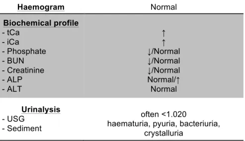
A summary of clinical pathology results in dogs with PHPTH can be found in Table 1.
1.3.6.1. Haemogram
Complete blood count (CBC) in dogs with PHPTH is usually unremarkable, as well as bone marrow aspirates and peripheral blood smears (Feldman, 2010, 2015a). Feldman et al. (2005) reported that there are no consistent abnormalities detected (Feldman et al., 2005).
1.3.6.2. Biochemical profile
1.3.6.2.1. tCa and iCa in PHPTH
Hypercalcaemia (i.e. excessive amount of calcium in the blood), in dogs is usually defined as fasting serum tCa concentrations higher than 12mg/dl with a normal reference range of 9.9 to
11.6mg/dl. Ionised hypercalcaemia is present when iCa concentration is higher than 1.5mmol/l (reference range from 1.12 to 1.41mmol/l) (Feldman, 2014; Schenck et al., 2012; Venes, 2013).
In a study including 210 dogs with PHPTH by Feldman et al. (2005), all had tCa in concentrations above 12mg/dl, as it was a requirement for inclusion in the study. The mean tCa was 14.5mg/dl, ranging from 12.1 to 23.4mg/dl, with 52% of them presenting concentrations between 12 and 14mg/dl, 30% with tCa concentrations between 14 and 16mg/dl, 12% with concentrations between 16 and 18mg/dl and 6% had concentrations above 18mg/dl.
In the same study (Feldman et al., 2005), 91% of the dogs had increased iCa and 9% had iCa within reference range. The authors argue that those 9% were false values, altered by variables such as the collection method or the time between collection and the time when the assay was done, as iCa is more easily altered by those variables than tCa. The mean iCa concentration was 1.71mmol/l, ranging from 1.22 to 2.41mmol/l: 27% had concentrations between 1.42 and 1.65mmol/l, 48% had concentrations between 1.66 and 1.90mmol/l and 16% had concentrations above 1.90mmol/l.
Hypercalcaemia is the hallmark of PHPTH, and in 5 different studies the tCa mean concentrations were between 13.6mg/dl and 14.3mg/dl with a range of 12.1 to 23.4mg/dl (Feldman et al., 2005; Gear et al., 2005; Ham et al., 2009; Milovancev & Schmiedt, 2013; Pollard et al., 2001). The iCa mean concentration in those same studies was between 1.67 and 1.90mmol/l, ranging from 1.43 to 2.55mmol/l except in Feldman et al. (2005) where some dogs had iCa within range, as it has been previously justified (Feldman et al., 2005; Feldman, 2015a; Gear et al., 2005; Ham et al., 2009; Milovancev & Schmiedt, 2013; Pollard et al., 2001).
The magnitude of hypercalcaemia does not help differentiating the cause or the extent of the disease (Schenck & Chew, 2012), though Feldman and Skelly, suggest that calcium progressively increases throughout the course of the disease (Feldman, 2015a; Skelly, 2012).
1.3.6.2.2. Measurement of tCa and iCa
Calcium status is usually assessed using tCa concentration, despite iCa being the only active fraction, because tCa measurement is more readily available. iCa does not correlate directly to tCa and inferring so can lead to errors in the interpretation of the results (Schenck & Chew, 2008). In a study by Schenck and Chew (2005) with 1633 dogs, 27% had diagnosis discordance when tCa was used to predict iCa, and in dogs with chronic renal failure (CRF) the discordance was of 36%. tCa concentration overestimated normocalcaemia and underestimated hypocalcaemia. Adjustment equations were developed in an attempt to improve the correlation between tCa and iCa using total protein and albumin, but these were not verified using iCa. In the same study the diagnosis discordance using adjustment
equations was in 37% for all dogs and of 54% in dogs with CRF. So neither tCa alone nor tCa adjustment equations can accurately predict iCa, and a direct measurement of iCa is required for its accurate assessment (Schenck & Chew, 2005, 2008). tCa can give an indication of calcium status but iCa is a more pertinent and useful (Skelly, 2012).
Serum calcium concentrations can be affected by certain factors. The factors that can falsely increase calcium when assessed in some analysers are: marked lipaemia, haemolysis and contamination by chalkboards in the laboratory and rarely postprandial samples. Usage of glassware and plastic containers washed with detergents for storage of samples can cause false increases or decreases on calcium levels. Haemoconcentration by dehydration rarely causes mild increases in calcium and prolonged storage can cause an artifactual decrease in calcium concentrations (Feldman, 2010, 2015a).
Additionally dogs younger than 3 months have slightly higher concentrations of calcium than dogs over a year old (Schenck & Chew, 2012).
iCa concentration depends of pH, where a more acid pH increases the amount of iCa because the dissociation of calcium bound to proteins is favoured, whilst a more alkaline pH causes decreases in iCa. In samples collected and processed aerobically there is loss of carbon dioxide (CO2), which increases the pH of the sample, favouring protein and calcium
binding and decreasing iCa (Schenck et al., 2012). Anaerobic collection and processing are more precise than those done aerobically, but require more complicated techniques. To surpass this difficulty, mathematical formulae were developed for specific species to correct iCa to a pH of 7.4 in samples collected aerobically, that correlate very well to iCa in anaerobic samples (Schenck et al., 2012; Schenck & Chew, 2008, 2012).
1.3.6.2.3. Phosphorous
Dogs with PHPTH usually present with normophosphataemia or hypophosphataemia, with concentrations below 4mg/dl because of PTH induced urinary loss of phosphorous (Bonczynski, 2007; Feldman, 2015a). If phosphate concentrations are not at the low end of the reference range or below, renal failure could be developing (Skelly, 2012).
Dogs younger than 1 year old have higher normal phosphate concentrations (Feldman, 2015).
In a study with 210 dogs (Feldman et al., 2005), the mean phosphate concentration was 2.8mg/dl ranging from 1.3 to 6.1mg/dl (reference range 3.0 - 6.2mg/dl). In these dogs, 13% had a phosphate concentration below 2.0mg/dl, 52% had a concentration between 2.0 and 2.9mg/dl, 28% had a concentration between 3.0 and 3.9mg/dl, 5% had a concentration between 4.0 and 4.9mg/dl and 2% had a concentration above 4.9mg/dl. The dogs with phosphate concentrations above 4.9mg/dl also had concomitant increased blood urea nitrogen (BUN) and creatinine concentrations. The mean serum phosphate concentration in the dogs from a control group with similar ages was 4.6mg/dl, higher than in dogs with
PHPTH. Feldman (2014) in a series of 335 dogs, reported a mean serum phosphate concentration of 2.7mg/dl.
The mean serum phosphate concentrations in 3 other studies were 2.91mg/dl1, 2.86mg/dl
and 2.4mg/dl (Gear et al., 2005; Milovancev & Schmiedt, 2013; Pollard et al., 2001).
Feldman (2010) recommends evaluation of phosphorous whenever serum calcium concentrations are assessed.
1.3.6.2.4. BUN and creatinine
In the study by Feldman et al. (2005), including 210 dogs with PHPTH, the BUN mean concentration was 16.9mg/dl, ranging from 5 to 92mg/dl (reference range 18-30mg/dl) and the creatinine mean concentration was 0.8mg/dl, ranging from 0.4 to 4.1mg/dl (reference range 0.5-1.5mg/dl). Relatively to BUN concentrations, 3% of dogs had values below 10mg/dl, 60% had values between 10 and 17mg/dl, 23% had values between 18 and 22mg/dl, 10% had values between 23 and 28mg/dl and 3% had values above 30mg/dl. The distributions of creatinine concentrations were: 60% with a concentration or 1.0mg/dl or below, 37% with a concentration between 1.0 and 1.5mg/dl, 2% with a concentration between 1.6 and 2.0mg/dl and 1% with a concentration above 2.1mg/dl. In the 200 dogs from the control group, BUN and creatinine mean concentrations were 24mg/dl and 1.2mg/dl, respectively. These values were considerably higher than those from dogs with PHPTH. In another report on 29 dogs with PHPTH (Gear et al., 2005), 13 dogs had increased BUN and 10 dogs had increased creatinine. However, in these 29 dogs there were more dogs with high renal parameters than in other reports with over 300 dogs with PHPTH where renal failure was rare (Arbaugh et al., 2012; Feldman et al., 2005; Ham et al., 2009; Milovancev & Schmiedt, 2013; Sawyer et al., 2011).
1.3.6.2.5. ALT and ALP
In the study by Feldman et al. (2005), 40% of dogs with PHPTH had an increase in alkaline phosphatase (ALP) levels, with a mean concentration of 241U/l, ranging from 12 to 4431U/l (reference range 5-92U/l). These values are possibly a result from increased osteoblastic activity in bone (Feldman, 2015a). In the study by Gear et al., (2005) 13 out of 27 dogs (48%) were also found to have increased ALP, but two had been treated with glucocorticoids and one was diagnosed with hyperadrenocorticism (HAC).
Alanine aminotransferase (ALT) concentrations are usually normal but can have mild increases (Feldman, 2015a).
1