Article
0103 - 5053 $6.00+0.00
*e-mail: [email protected]
A Comparative Study of Irradiation Systems for Photoinactivation of Microorganisms
Lucas F. de Paula,*,a Renata O. Santos,a Henrique D. Menezes,a Jonas R. de Britto,b
João B. Vieira Jr.,b Paulo P. Gontijo Filhoc and Carlos A. de Oliveiraa
aInstituto de Química, bFaculdade de Engenharia Elétrica and cNúcleo de Imunologia, Microbiologia e
Parasitologia, Universidade Federal de Uberlândia, 38408-100 Uberlândia-MG, Brazil
Estudou-se a construção de sistemas de LED aplicados a processos de fotosensitização. Visando à obtenção de dispositivos mais eicientes avaliou-se a aplicação de LED de alta potência (HPLED), que apresenta maior luxo fotônico, tendendo a excitar uma maior fração de moléculas. Foram descritos detalhes de construção de 3 diferentes sistemas de irradiação completamente distintos (lâmpada halógena iltrada, LED convencional, HPLED), utilizando azul de metileno como fotosensitizador. A comparação entre a eiciência de cada equipamento foi realizada através da cinética de fotooxidação de 1,3-Diphenylisobenzofuran e inativação de Staphylococcus aureus. A emissão de luz de cada equipamento, sobreposta à absorção do fotosensitizador, também se mostrou um importante parâmetro na estimativa da eiciência dos equipamentos. Foi observada uma inibição bacteriana superior a 99% para concentrações de 5 × 10⁻⁶ mol dm-3 de fotosensitizador,
no sistema que emprega HPLED. Os equipamentos baseados em LED, apresentaram resultados satisfatórios além de baixo custo e simples montagem.
The construction of LED systems applied to photosensitization processes was studied. Aiming to obtain more eficient devices it was evaluated the use of high intensity LED (HPLED), that present greater photonic lux, tending to excite a greater fraction of molecules. Construction details for 3 different irradiation systems were described (iltered halogen bulbs, conventional LED, HPLED), using methylene blue as a photosensitizer. A comparison between the eficiency of the equipments was carried out through the kinetics of photooxidation of 1,3-Diphenylisobenzofuran and
Staphylococcus aureus photoinactivation. The overlap between the light emited from each equipment and the photosensitizer absorption, was also an important parameter in estimating the eficiency of the equipment. Bacterial inhibition higher than 99% for concentrations of 5 × 10⁻⁶ mol dm-3
of the photosensitizer was observed, in the system that uses HPLED. The equipments based on LED showed satisfactory results, besides their low cost and simple to assembling.
Keywords: high power LED, Staphylococcus aureus, methylene blue, irradiation systems
Introduction
Photodynamic Inactivation (PDI)1 or Photodynamic
Antimicrobial Chemotherapy (PACT)2 is the name of a
process, derived from Photodynamic Therapy, that causes the death of microorganisms under the action of visible light and a speciic drug, also called photosensitizer.3 The
electronic excitation of the photosensitizer in the presence of oxygen implies in the generation of a large number of reactive oxygen species (ROS), as for example, singlet oxygen,4,5 in a process known as photosensitization.
Since 1976, the oxidative action of singlet oxygen and other ROS on tumor cells has been reported in the
medical and scientiic communities.5,6 Around the world,
several research results have been published on the use of photosensitization to ight diseases caused by fungi, viruses, bacteria and yeasts. 3,5,7-11
Among the photosensitizers cited in the literature,12-16
methylene blue (MB), a phenothiazinic dye, has some convenient characteristics: it presents good photodynamic activity compared to different substrates,17-19 is nontoxic
and relatively inert on living organisms.20 It is also known
for its anti-septic action21 and has been administrated
intravenously in some clinical cases (e. g., the treatment of methemoglobinemia).22,23
appreciable area of incidence; adjusted spectral emission (near the drug absorption peak); low heating; appreciable intensity.24 Therefore, light emitting diodes (LED) present
some very desirable characteristics such as physical resistance (they do not have bulbs, they do not break and they are quite robust); extended duration (useful lifetime nearly 100,000 h); spectral emission near to monochromatic systems; availability in several colors; low heat emission;25,26 aside from having lower cost per unit of
power when compared to laser diode systems.27
In recent years, several publications point out to the use of LED as a source of irradiation to satisfactorily promote the excitement of photosensitizers.28-31 However, the present
work aims at comparing and demonstrating advantages in the use of high intensity LED, in detriment to conventional LED, aside from providing guidelines for constructing an irradiation equipment based on LED arrays. Conventional colored 3 and 5 mm LED, widely found on the market, normally operate under a forward current of 20 mA, and have decreased rates of luminous emission (normally not exceeding 5,000 mcd).32 With the intention of making up
for this disadvantage, two alternatives can be used: (i) the confection of LED arrays that not only increase the incidence area, but also potentialize the intensity of emitted radiation; (ii) application of high power LED (AMS-II). They are more powerful than the previous ones, and have high rates of luminous conversion - about 33 lm W-1 for
the red AMS-II.26 This indicates that they use less energy
to emit the same luminous amount, aside from maintaining good characteristics as durability and emission close to monochromatic.
The photodynamic action of MB was explored in the sense of correlating the efficiency of three different irradiation sources: (i) an equipment issuing polychromatic iltered radiation coming from a halogen lamp called PHLS (Polychromatic Halogen Lamp System); (ii) matrix of 600 high brightness conventional
LED, called LED600; (iii) irradiation equipment emitted by high intensity LED, assembled for a better performance in the methylene blue sensitization and the analogous ones, denominated AMS-II.
The effective irradiance and the overlap integral with the absorption band of MB of each equipment were analyzed. Their efficiencies in ROS generation were analyzed by means of a kinetic study of discoloration of 1,3-Diphenylisobenzofuran (DPBF), a colorful molecule that quickly suffers oxidation in the presence of singlet oxygen, forming o-dibenzoylbenzene (DBB).33 As a microorganism
model, for implementing and measuring photoinactivation eficiency, Staphylococcus aureus, which is a pathogenic gram-positive bacteria widely studied and responsible for several reports of hospital infection34 was used.
Experimental
PHLS setup
A halogen lamp used in overhead projectors (OSRAM, model ENH, 250W) was itted to a metallic box. A 100% reading magnifying lens was ixed at a distance of 7 cm from the light bulb. For refrigeration, a cooler was inserted in the metallic box, and was itted to a glass cube near the irradiation point of the sample being rectangular in shape for circulating water on the inside.
The light given off by the light bulb was then iltered to eliminate unnecessary wavelengths using an optic ilter. The ilter was obtained commercially and is made of pigmented polyester. The ilter was itted on the top of the glass box. A schematic view of this setup is shown in Figure 1A.
LED600 equipment
Under a typical circuit board, the matrix of 600 high brightness red LED (ZX, 8000 mcd) was made. This
Figure 1. A) Equipment PHLS (1) Metal carcasses with the coupled lamp and cooler, (2) Magnifying glass, (3) Glass box, (4) Optical ilter; B) LED600
array was organized in 150 clusters connected in parallel, each cluster having 4 LED in series. This entire array was connected and fed by a DC power supply, providing 2.42 A current.
This circuit board was ixed to a rectangular metallic box. It was welded onto the metallic structures, on the inside there are some structures that allow for sample ixation. The described structure is shown in the Figure 1B.
AMS-II equipment
Seven High Intensity LEDs were assembled in a serial array (Figure 1C) (Edison, model IR Edixeon). The LED was driven by a Buck CC/CC converter that delivers an output voltage of 22VDC and a constant output current of 350 mA, from a universal input voltage (100-240 VAC). Therefore the total power given off by the LED is about 7.5 W.
Acquisition of irradiance spectra and measurements
The profile of the emitted radiation of the above-mentioned light sources was obtained with the aid of a setup based on an Ocean Optics, model SD2000 iber optics spectrometer with an integrated time of 35 ms.
The optical power (P) was obtained with the aid of a power meter Ophir, model Orion, with a photodiode sensor model PD-300, spectral range of 350-1100 nm.
Aiming at the comparison between the proposed systems, an integral of overlapping was estimated (R) between the absorption of the methylene blue (ABSPS) and the emission of the irradiation equipment (EEQUIP) (equation 1), according to the method proposed by Bonacin et al.31
(1)
Because the irradiation intensity does not vary with time, it is possible to estimate the relative fraction of photosensitizer excitement (R) which is an important parameter for comparing the efficiency between the equipments described.
Due to the fact that the concentration of dye used is the same, the greater or lesser values of R are associated only to the coupled emission with the absorption of the dye, and represented a magnitude of excitement of the dye molecules.
Methylene blue
Methylene blue solutions (Sigma) in the concentrations 5 × 10⁻⁵, 5 × 10⁻⁶ and 5 × 10⁻⁷ mol dm-3, have been prepared,
in a laminar low hood, through the dissolution of the dye in sterile physiological serum (sodium chloride saline solution 0.9 wt% sterilized by autoclave) and deposited in Eppendorf tubes before use.
The methylene blue absorption spectrum in physiological solution, in a solution 10-6 mol dm-3, was measured using a
Shimadzu UV-2501 spectrophotometer.
DPBF oxidation
Stock solutions (10-4 mol dm-3) of DPBF (Sigma)
were diluted in absolute methanol, resulting in a inal concentration of 10-5 mol dm-3 and 10-6 mol dm-3 of
methylene blue. All these solutions were prepared at the moment of use, and protected from light before the experiments were carried out.
The solutions were irradiated using an open quartz cuvette (Hellma 110-QS) with a 1 cm optical pathway, being stirred constantly, at intervals of 5 sec of irradiation, and absorption was read at 408 nm, with the aid of a spectrophotometer.
Sodium azide (Aldrich, 99.99%) was used as a singlet oxygen quencher in some experiments.
A comparative measure at the number of photons absorbed by the concentration of oxidable substract was used as to help estimating the eficiency of each equipment. Consequently, the equipment that promotes a greater substract degradation emitting a smaller quantity of photons tends to be more eficient.
Bacterial suspension
Determined S. aureus strain (ATCC 25923), was kindly provided by the Laboratório de Microbiologia, Universidade Federal de Uberlândia. This strain was transferred to a nutritional medium prepared with the following composition: 1.25% meat extract ‘lab lemco’ (Oxoid), 0.50% tryptone (Oxoid), 1.00% peptone (Biobrás), 0.50% sodium chloride (Vetec, 99%), 0.25% sodium phosphate dibasic heptahydrated (Cinética química, 99%), 2.00% D-glucose (Synth, 99.9%).
A secondary culture was carried out in agar Cistine-Lactose-Electrolyte-Deicient – CLED (Oxoid), used for the growth and isolation of CFU’s.
From this culture a bacterial suspension was prepared in sterile physiological serum/saline. In the bacteriological experiment a concentration of 0.1 in the McFarland scale was used. This stock suspension was diluted in a 1:10 proportion, which corresponds to a cell density of approximately 3 × 10⁶ colony forming units
Incubation and irradiation
500 µL of bacterial suspension and 500 µL of methylene blue solution were incubated, in the dark, for 30 min. This experiment was repeated for each concentration of methylene blue prepared. All experiments were carried out in triplicate.
After the incubation period, the samples were irradiated, for 20 min, without agitation, and subsequently 100 µL of the suspension were transferred to Petri dishes containing the casting growth medium and kept in the bacteriological stove at 35 ºC. This medium was prepared by adding 2% of agar to the nutritional broth formula. After 24 h of incubation, colony counting was carried out using a colony counter PHOENIX (CP 602).
Results and Discussion
Development of the equipment
The LED of the AMS-II equipment was arrayed in series, because in this way the equipment would be better adjusted to the photochemical processes (satisfactory intensity, homogeneous, fixed and steady irradiance). Arraying series and regulating the current (and not the tension) through of the LED, is preferred for these applications, provided: (i) The curve between the current and LED voltage is very abrupt:26 Small changes in the
voltage are expressed in great variations of current. Once the luminous intensity of the LED alternates more linearly with the current, subtle alterations in the voltage would imply in drastic changes in the intensity. While simple systems of current control, tend to ensure greater conidence in the luminous intensity oscillation. (ii) In regard to the voltage and the natural negative temperature coeficient of LED, arrays connected in parallel cannot guarantee that the current (and thus the brightness) entering each LED is necessarily equal. When two diodes are connected in parallel and one of them heats a little more than the other, this diode tends to drain more current, becoming hotter and in this way contributes gradually to a cascade effect that normally culminates in overheating.
LED600, unfortunately, for practical reasons, cannot be arrayed in series, since the tension in components in series is cumulative, and, therefore the equipment would tend to consume a tension of nearly 1320 Volts (assuming 2.2 V as the approach tension of each diode), making the project impracticable. In practice it was observed that for the array in LED600 having been carried in a rather compact form, the homogeneity is not so compromised, and the current limitation, for the use of an appropriate source, reduces part
of the problems related to the arrays in parallel. Moreover, LED600 has innumerable components. If it was mounted in series and one of the diodes were damaged, opening the circuit, all illumination of the set would be compromised. For these and other reasons the AMS-II equipment was theoretically considered as LED600 improvement, developed to work in series, with a ixed current.
The production cost of the PHLS is at least twice less costly than the other devices developed. This equipment is also much more accessible than analogous commercial equipment. The lens used greatly reduce the area of homogeneous radiation, however, tests performed with the spectrometer indicated a scoring higher than 50% in the value of light intensity, when in the presence of the lens. In the absence of refrigeration systems, the sample can reach temperatures of about 52 ºC, favoring the denaturing of DNA and proteins. In this way, the glass box gives off the generated heat eficiently, guaranteeing that the temperature of the sample is raised at the most 3 ºC.
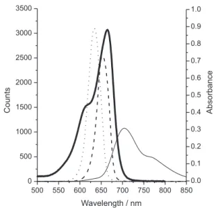
The emission spectrum of the equipment studied, superimposed with the photosensitizer absorption used is shown in Figure 2.
LED600 presents a wide emission area: 360 cm2,
suficient homogeneity (seen with spectrometer) and low heating. For this equipment we kept the distance of 11 cm for a better homogeneity/intensity relation, which was used in the microbiological experiments and in the degradation of DPBF. The systems based on LED arrays should present an ideal working distance seeing that, in a LED, the polymeric pack acts as a lens: a greater proximity means that the sample is very near to the focus, and, therefore, the irradiation is not homogeneous; the increase in the distance
Figure 2. Absorption spectrum for methylene blue (—); emission
tends to improve the distribution of the radiation, on the other hand great distances of the light source compromise the intensity of the light received.
AMS-II, although having intensity very close to LED600, has the peak of emission at 652 nm, being still closer to the maximum absorption of the drug (663 nm). Emission of AMS-II is not only more appropriate for this drug, as it is also very homogeneous, as seen in additional tests with the spectrometer. The emission area of 95 cm2
(Table 1) could be increased by the simple addition of more LED in the array. This system presented irradiance values near 43.35 mW cm-2. The information on the irradiation
area with homogeneous intensity, optical power, irradiance, and a fraction of excited molecules obtained from each equipment is summarized in Table 1.
The useful fraction of light that excites the fenotiazinic coloring is different for each equipment. It is possible to observe that the emission region of LED is suficiently adjusted for the region of better drug electronic excitement, since PHLS presents low magnitude in the excitement of the photosensitizer (R = 6.33), basically in regard to the reduced photonic emission in the region of the band of absorption of methylene blue. The PHLS region emitted is suficiently appropriate for other drugs, as for example, the class of phthalocyanines, since the absorption peak of the Q band of these drugs is located near the region between 675-700 nm.35
These emissions are normally dificult to be obtained using conventional LED, because commercially available LED emission values above 650-660 nm are not usual.
Even though the LED600 and AMS-II are close, it is expected that AMS-II would have greater eficiency in generating reactive species due to its irradiation being capable of exciting a greater fraction of molecules of the photosensitizer (R = 31.36) compared to the rest of the equipment. This phenomenon can be explained because of the emission of this equipment found in the band of greater light absorption by the photosensitizer.
Conirmation of singlet oxygen presence
Solutions with sodium azide (NaN3) were used in order to give evidence of the participation of singlet oxygen in the process (Figure 3). Sodium azide is known as a strong
singlet oxygen quencher (108 dm3 mol-1 s-1 in H 2O).
33,37,38
In all the equipments similar proiles of DPBF oxidation and suppression by azide were observed, Figure 3 shows the results observed with the LED600 equipment.
It is possible to observe in Figure 3B, that in the presence of NaN3, a reduction in the oxidation speed of DPBF occurs. The absorption peak in the DPBF region is found in the 429 nm region and shows the Figure that the mechanism of oxidation by participation of singlet oxygen is predominant.
Kinetic comparison of photo oxidation of DPBF
A kinetic study allows the quantiication ion of to express the eficiency in the generation of ROS by emitted light of equipment built in the photo degradation of DPBF. Equation 2 expresses the degradation of the oxidable substract by the action of singlet oxygen. The concentration of singlet oxygen can be admitted as constant because the solutions were keeping contact with air and under constant agitation. Thus, it is possible to incorporate [1O
2] into the
kinetic constant k, resulting in a pseudo-irst order constant, k’. In this way, the resultant kinetics is of pseudo-irst order (equation 3 and 4). After the integration the equation obtained for the process is express in equation 5.33,36
Table 1. Optical Power (P), Irradiation Area (A), Irradiance (Ir) and estimated excited fraction of photosensitizer (R), in the conditions used
Equipment P / mW Aa / cm2 Ir / (mW cm-2) Φ / (Einstein s-1) Rb
PHLS 2.90 132 1.80 1.7 × 10⁻⁸ 6.33
LED600 20.11 360 12.46 1.6 × 10⁻⁷ 18.58
AMS-II 19.69 95 12.21 1.07 × 10⁻⁷ 31.36
aHomogeneous irradiation area. Refers only to the possibility of irradiation of greater numbers of samples at the same time. Maximum variation in ±5% of intensity. bCalculated with respect to methylene blue 10-6 mol dm-3 in sodium chloride 0.9%.
Figure 3. Photo-induced oxidizing of DPBF in LED600, in the absence
(2)
(3)
(4)
(5)
A reduction in the concentration of DPBF in regard to time is shown in Figure 4, for all equipments.
Each spectrum was obtained after 5 sec of exposition to emitted light by the equipment. Thus, it is observed that for the AMS-II, only 20 sec were suficient for a reduction greater than 98% of intensity of the DPBF band.
These results suggest that AMS-II presented better performance. These tests are also in accordance with the values shown in Table 1. In these experimental conditions, it is still possible to say that AMS-II presents twice the eficiency of LED600 (k’AMS = 0.25532 s-1; k’
LED = 0.12668 s -1).
However, PHLS, in view of the small emission in the useful working band, presents an eficiency quite reduced when compared to LED-based equipments.
Photodynamic inactivation
Methylene blue, in the absence and in the presence of light, presents bactericide action in particular against gram-positive bacteria. These effects are described in the literature.39,40 The results of the bacterial inhibition by the
presence and absence of light, are arranged in Figure 5. The samples were irradiated for 20 min.
The bacterial inhibition in the dark, for the more diluted solution (5 × 10⁻⁷ mol dm-3), presents a reduction,
of approximately 50%, in the number of colony forming units. In the equipment of greater eficiency, AMS-II, methylene blue under irradiation photo inactivated 75% of the bacteria reducing by 50% of the colony forming units compared with the dark control.
In all the other concentrations, the AMS-II equipment presented photo inactivation superior to 99%. The other equipments, LED600 and HPLS also presented satisfactory performances of photo inactivation. At the concentration of 5 × 10⁻⁵ mol dm-3 and after light application, there
was no colony formation. Therefore, all the equipments described operate satisfactorily against growth inhibition of Staphylococcus aureus in vitro. The obtained results in photodynamic inactivation agree proportionally to the results obtained with the DPBF oxidation and with the respective R values of each equipament.
Conclusions
All the equipments built proved to be capable of generating oxygen singlet, as was observed in the discoloration of the DPBF, in the suppression by NaN3 and photo inactivity of Staphylococcus aureus.
The study enables one to conclude that high power LED equipment proved to be able to excite a more signiicant fraction of molecules of the photo synthesizer, and consequently showed a more signiicant generation of oxygen singlet, as was observed more signiicantly by the magnitude of the kinetics of discoloration of DPBF, and bacterial inhibition. The reasons for this are relatively
Figure 4. Kinetics of pseudo-irst order corresponding to the degradation
of DPBF induced by irradiation of various equipments developed. () PHLS; () LED600; () AMS-II.
Figure 5. Bacteria count before and after the exposure of light, under
simple: the high powered LED used emits in wavelengths near to the peak methylene blue absorption aside from having high luency. The system that uses a halogen bulb presents low photonic emissions, even though other optic ilters may be used.
High powered LED are more effective photochemically aside from showing cost advantages and set up practicality.
Acknowledgments
The authors are indebted to Prof. Dr. Antonio Eduardo da Hora Machado for his valuable contribution to this study. To Laboratório de Fotoquímica and Laboratório de Fotoquímica de Lignocelulósicos from Universidade Federal de Uberlândia, for the concession of the infra-structure and materials. To CNPq, FAPEMIG and Nanobrax (www.nanobrax.com) for research support.
References
1. Gad, F.; Zahra, T.; Hasan; T.; Hamblin, M. R.; Antimicrob. Agents Chemother. 2004, 48, 2173.
2. Wainwright, M.; J. Antimicrob. Chemother. 1998, 42, 13. 3. Perussi, J. R.; Quim. Nova2007, 30, 988.
4. Machado, A. E. H.; Quim. Nova 2000, 23, 237.
5. Dougherty, T. J.; Gomer, C. J.; Henderson, B.W.; Jori, G.; Kessel, D.; Korbelik, M.; Moan, J.; Peng, Q.; J. Natl. Cancer Inst.1998, 90, 889.
6. Weishaupt, K. R.; Gomer, C. J.; Dougherty, T. J.; Cancer Res.
1976, 36, 2326.
7. Komerik, N.; Nakanishi, H.; MacRobert, A. J.; Henderson, B; Speight, P.; Wilson, M.; Antimicrob. Agents Chemother.2003,
47, 932.
8. Wong, T-W.; Wang, Y-Y.; Sheu, H-M.; Chuang, Y-C; Antimicrob. Agents Chemother.2005, 49, 895.
9. Qin, Y.; Luan, X.; Bi, L.; He, G.; Bai, X; Zhou, C.; Zhang, Z.;
Lasers Med. Sci.2008, 23, 49.
10. Alaya, F.; Grimaldi, E.; Perfetto, B.; Donnarumma, M.; De Filippis, A.; Donnarumma, G.; Tufano, M. A.; Photodermatol. Photoimmunol. Photomed.2008, 24, 237
11. Donnelly, R. F.; McCarron, P. A.; Tunney, M.M.; Microbiol. Res.2008, 163, 1.
12 . Huang, Z.; Photodiagnosis Photodyn. Ther.2008, 5, 285. 13. Alves, E.; Costa, L.; Carvalho, C. M.; Tomé, J. P.; Faustino,
M. A.; Neves, M. G.; Tomé, A. C.; Cavaleiro, J. A.; Cunha, A.; Almeida, A.; BMC Microbiol. 2009, 9, 70.
14. Tirand, L.; Thomas, N.; Dodeller, M.; Dumas, D.; Frochot, C.; Maunit, B.; Guillemin, F.; Barberi-Heyob, M.; Drug Metab. Dispos.2007, 35, 806.
15. Stukavec, J.; Duchac, V.; Horak, L.; Pouckova, P.; Photomed. Laser Surg.2009, 27, 107.
16. Marotti, J.; Aranha, A. C.; Eduardo C. D.; Ribeiro, M. S.;
Photomed. Laser Surg.2009, 27, 357.
17. Almeida, J. M.; Theodoro, L. H.; Bonfante, S.; Luize, D. S.; Macarini, V. C.; Bosco, A. F.; Nagata, M. J. H.; Garcia, V. G.;
Periodontia2006, 16, 34.
18. Câmara, F. P.; Elias, C. A.; An. Bras. Dermatol. 1978, 4, 53. 19. Tardivo, J. P.; Petri, V.; Bonetti, T, I.; Oliveira, L. S.; Baptista,
M. S.; Jornal Brasileiro de Laser2007, 1, 28.
20. Tardivo, J. P.; Giglio, A. D.; Oliveira, C. S.; Gabrielli, D. S.; Junqueira, H. C.; Tada, D. B.; Severino, D.; Turchiello, R. F.; Baptista, M. S.; Photodiagnosis Photodynamic Ther.2005, 2, 175.
21. Farmacopéia Brasileira, 2a. ed., Atheneu São Paulo, 1959, p. 119.
22. Krol, J.; Cahiers D’Anesthésiologie1968, 17, 333. 23. Dahshan, A.; Donovan, G. K.; Pediatrics 2006, 117, e806. 24. Babilas, P.; Landthaler, M.; Szeimies, R-M.; Eur. J. Dermatol.
2006, 16, 340.
25. Demian, A. E.; Britto, J. R.; de Freitas, L. C.; Farias, V. J.; Coelho, E. A. A.; Vieira, J. B.; 12thEuropean Conference on
Power Electronics and Applications, Aalborg, Denmark, 2007. 26. Phillips Lumileds Lighting Company,Datasheet DS45, March
2006.
27. Patrice, T. In Comprehensive Series in Photochemistry and Photobiology; Hader, D. P.; Jori, G., eds.; The Royal Society of Chemistry: Cambridge, 1994, ch. 7.
28. Ferreira, I.; Rahal, S. C.; Rocha, N. S.; Gouveia, A. H.; Corrêa, T. P.; Carvalho, Y. K.; Bagnato, V. S.; Veterinary Dermatology
2009, 20, 174.
29. Szeimies, R. M.; Matheson, R. T.; Davis, S. A.; Bhatia, A. C.; Frambach, Y.; Klövekorn, W.; Fesq, H.; Berking, C.; Reifenberg, J.; Thaçi, D.; Dermatol. Surg.2009, 35, 586.
30. Barolet, D.; Semin. Cutan. Med. Surg.2008, 27, 227. 31. Bonacin, J. A.; Engelmann, F. M.; Severino, D.; Toma, H. E.;
Baptista, M. S.; J. Braz. Chem. Soc. 2009, 20, 31.
32. Kingbright Corporation, Datasheet WP7113PR51C/A, August 2007.
33. Bell, J. A.; MacGillivray, J. D.; J. Chem. Educ.1974, 51, 677. 34. Rubinstein, E.; Int. J. Antimicrob. Agents 2008, 32, S18. 35. Rosenthal, I.; Photochem. Photobiol.1991, 53, 859. 36. Young, R. H.; Brewer, D.; Keller, R. A.; J. Am. Chem. Soc.
1973, 95, 375.
37. Foote, C. S.; Org. Chem1979, 40, 139.
38. Harbour, J. R.; Issler, S. L.; J. Am. Chem. Soc.1982, 104, 903. 39. Li, W. Y.; Xu, J. G.; He, X. W.; Anal. Lett.2000, 33, 2453. 40. Rohs, R.; Sklenar, H.; Indian J. Biochem. Biophys.2001, 38, 1. 41. Zhao, G.-C.; Zhu, J.-J.; Zhang, J.-J.; Chen, H.-Y.; Anal. Chim.
Acta1999, 394, 337.
Received: June 6, 2009