Article
0103 - 5053 $6.00+0.00*e-mail: [email protected]
Evidences of Siderophores Synthesis by Grapevine Xylella fastidiosa,
Causal Agent of Pierce’s Disease, through Instrumental Approaches
Ana Valéria C. Simionato,*,a,c,d Maria Estela Silva-Stenico,b
Siu Mui Tsaib and Emanuel Carrilhoc,d
aGrupo de Toxicologia de Alimentos e Fármacos, Universidade Estadual de Campinas,
P.O.Box 6154, 13083-970 Campinas-SP, Brazil
bLaboratório de Biologia Celular e Molecular, Centro de Energia Nuclear na Agricultura,
Universidade de São Paulo, Av. Centenário, 303, CP 96, 13416-000 Piracicaba-SP, Brazil
cGrupo de Bioanalítica, Microfabricação e Separações, Instituto de Química de São Carlos,
Universidade de São Paulo, Av. Trab. São-Carlense, 400 CP 780, 13560-970 São Carlos-SP, Brazil
d Instituto Nacional de Ciência e Tecnologia em Bioanalítica, Universidade Estadual de Campinas,
CP 6154, 13083-970 Campinas-SP, Brazil
Os sideróforos provenientes de Xylella fastidiosa de videiras foram investigados. Tais metabólitos seqüestram ferro, um elemento essencial, do hospedeiro, o que os torna um potencial fator de patogenicidade. Em um meio de cultura em placa com limitação de ferro, tais sideróforos foram detectados pela reação com o complexo cromoazurol S (CAS). Diferentes métodos de análise instrumentais foram utilizados para caracterização dos sideróforos, como: cromatograia de ainidade por metal imobilizado (IMAC), cromatograia micelar eletrocinética capilar (MEKC) e espectrometria de massas com ionização por electrospray (ESI-MS). Os resultados obtidos conirmaram a produção de sideróforos. A extração do(s) composto(s) por IMAC com Fe3+
imobilizado foi uma etapa importante. O(s) sideróforo(s) não foi separado por eletroforese capilar de zona, indicando sua neutralidade sob os pHs investigados. As análises por MEKC apresentaram um pico diferente (quando comparadas à análise do controle), com caráter levemente hidrofóbico. A espectrometria de massas mostrou que os compostos alvos podem ter uma massa molecular relativa dentro da esperada para sideróforos, como: 875, 1004 e 1092 Da.
Siderophore molecules from grapevine Xylella fastidiosa were investigated. Such metabolites sequester iron, an essential element, from the host, making them a potential factor of pathogenicty. In an iron-limited medium, siderophores were detected in culture plates of X. fastidiosa containing the complex Chromeazurol S (CAS). A combination of different instrumental analyses was used for siderophore(s) characterization, such as: immobilized metal afinity chromatography (IMAC), micelar electrokinetic capillary chromatography (MEKC) and electrospray-mass spectrometry (ESI-MS). The results show that grapevine X. fastidiosa produced siderophore(s), as conirmed by the CAS plate assay. The extraction of the compound(s) using IMAC with immobilized Fe3+ was
important for analyte puriication. The chelator was not separated by capillary zone electrophoresis, indicating the possibility of a neutral compound under the investigated pHs. MEKC runs presented a different peak (when compared to the control analysis) which represented a slightly hydrophobic compound. Mass spectrometry showed that the compound(s) may have a relative molecular mass within the expected range for siderophore molecules, such as 875, 1004 and 1092 Da.
Keywords: IMAC, MEKC, ESI-MS, siderophores, iron sequestration
Introduction
Xylella fastidiosa is a gram-negative pathogenic bacterium in grapevine, resulting in Pierce’s disease, and
in a large variety of other hosts. Many diseases are caused by X. fastidiosa, citrus variegated chlorosis (CVC)3 and
Pierces’ disease of grapevine (PD)2,3 being economically
CVC has been an economical problem in Brazil towards production of orange, while Pierce’s disease has been the major concern in USA, therefore both countries have tried to control such diseases.8
The complete genome of Xylella fastidiosa (citrus strain
9a5c) has been sequenced by a Brazilian consortium.9 It
was the irst pathogenic phytobacterium with a sequenced
genome in the world. By X. fastidiosa genome inspection,
five outer membrane receptors - associated to iron transportation - have been recognized and about 67 genes are involved in iron metabolism.9
Siderophores are low molecular mass chelators that
guarantees iron uptake to several microorganisms.10
Besides iron, other metals may also complex with siderophores and most of them present the following afinities: Ca2+ < Ni2+ < Zn2+ < Cu2+ < Al3+ < Fe3+ (i.e.,
Fe3+ and Ca2+ have the strongest and the weakest complex
stability, respectively).11,12 Different molar ratios may be
observed according to the metal-siderophore complex,
while Fe3+ complexes present a higher formation constant
(Kf = 1013), than Fe2+ (K
f = 104) and Ni
2+ (data not presented
by the authors).13 Iron, as found in the environment, is
highly insoluble; therefore, siderophores are secreted by microorganisms to complex iron and transport it into the cell. Because siderophore-Fe3+ is found at very low
concentrations outside cells, diffusion through outer membrane would not sufice to supply the necessary uptake for this element. Therefore, a system of outer membrane high afinity siderophores receptors (probably similar to that recognized in X. fastidiosa genome) is required in order to transport the complex into the periplasma.14,15
In general, siderophores may be classified as:
cathecolates,16 hydroxamates,17 phenolics18 and
carboxylates,19 or a combination of them. Hydroxamates
and cathecolates siderophores present different mechanisms of iron release.20 The amino acid composition of such
molecules, which are synthesized non-ribossomicaly, may be predicted with the aid of molecular tools.21
Literature reports some of the most used siderophores bioassays,22 including Arnow assay for cathecolates,23
Csáky assay for hydroxamates,24 and Chrome Azurol
S (CAS),25 which is used as an universal assay, since it
does not classify the functional group of the detected siderophore. The CAS test is based on the reaction of a ligand added to the media containing siderophore, which shows a change of color from blue to orange in the presence of a siderophore. In solid plate cultures containing low concentration of Fe3+, in order to activate
the bacterium metabolism of siderophores synthesis, CAS assay results in orange halos around bacteria isolates (equation 1).
(1)
Capillary electrophoresis (CE) is a fast and eficient separation method, which has been extensively used in biomolecules analyses.26-28 Therefore, the application of
CE in siderophore analyses is a promising ield, since these analytes are found in very complex media, such as biological matrixes or microbial cultures. Within the several CE separation modes, micellar electrockinetic capillary chromatography (MEKC) is probably the most suitable one, as soon as siderophores with different hydrophobic properties are currently known. Besides, a simple preparation step is required before siderophore CE analyses. Mucha et al.29 have analyzed siderophores by
CE after isolation of the analytes by iltration followed by
adsorption on ionic exchange column.29
High performance liquid chromatography (HPLC) coupled to electrospray mass spectrometry (LC-ESI-MS and LC-ESI-MS/MS) is an eficient tool towards separation and identification of siderophores.30,31 Cox et al.32
extracted siderophores from culture supernatants by liquid extraction using ethyl acetate. The authors observed that cultivation time and iron deiciency conditions were critical factors in siderophores production, as observed in the HPLC chromatograms proiles. Electrospray ionization-mass spectrometry has also been used for the study of hydroxamate siderophores.33,34
Immobilized metal afinity chromatography (IMAC) is a sample preparation / separation technique based on
afinity of some proteins to immobilized metals.35 IMAC
is mainly used for puriication of proteins, but lately it has also been used for immobilized metal ion afinity capillary electrophoresis, among other applications.35,36
IMAC, first introduced by Porath,37 has gained wide
acceptance simultaneously to its development. Briely, A metal ion (Lewis acid) is immobilized to a support matrix. The incoming protein binds to the immobilized metal and separates from the rest of the sample. Elution of protein can be carried out by different mechanisms.38,39 Fe3+
-hydroxamate has already been used as IMAC adsorbent. Fe3+ exists in solution in the hexacoordinated form.
Therefore, when it is immobilized, some of its coordination sites are occupied by ixed ligands from the polymeric matrix.40 Thus, Fe3+ immobilization on IMAC columns
may be successfully used in extraction of siderophores from complex samples.
Van Sluys et al.41 compared the genome sequences from
that both strains present identical metabolic functions and most probably the same colonization and pathogenesis mechanisms. Bioassays and MS analyses strongly suggests that citrus X. fastidiosa secrets siderophores but their structures have not been elucidated yet.42,43 Some studies
have also shown that X. fastidiosa in vitro growth is aided by the presence of producing siderophores endophytic bacteria, such as Methylobacterium. Lacava et al.44 have concluded
that X. fastidiosa is able to use such siderophores as iron source. Therefore, the genome evidences of siderophore receptors and the ability of X. fastidiosa to use siderophores, suggests the potentiality of such metabolites in the plant pathogenicity process. Accordingly grapevine X. fastidiosa
must also secret siderophores. Therefore, investigations of such siderophores have been carried out herein by:
i) sample extraction from solid plate-CAS culture media;
ii) isolation of the analyte by IMAC; iii) and characterization by MEKC and electrospray MS.
Experimental
Materials
Acetonitrile, acetone, ammonium hydroxide, EDTA and sodium phosphate salts (analytical grade) were purchased from Mallinckrodt (Paris - USA or Xalostoc - Mexico). Sodium dodecyl sulfate was acquired from Polysciences (Warrington – USA). Formic acid and phosphoric acid (85%) was purchased from Merck (Rio de Janeiro – Brazil)
and NaOH was from Synth (Diadema – Brazil). Fe2(SO4)3
was purchased from Riedel-de Häen (Seelze – Germany) and Chrome azurol S was acquired from Aldrich (Steinheim – Germany) and its solution was prepared according to Schwyn & Neilands.25 All solutions were prepared with
puriied water obtained from a Milli-Q system (Millipore, Bedford – USA).
Bacterial strain and growth conditions
Grapevine X. fastidiosa (strain 6752) was obtained from INRA – Institut National de la Recherche Agronomique et Universite´ Victor Se´gale (Bordeaux – France). Bacteria were cultured three times in an iron-deicient agar medium
(MM9) containing the complex Chrome Azurol S plus Fe3+,
according to Silva-Stenico et al.42 Positive results were
recorded as a halo formation around the colonies.
Sample preparation
An agar block (ca. 1 cm2) excised from the halo region
was used for analysis and a corresponding agar block,
in which no bacterial growth was observed, was used as negative control. Compounds were extracted as follows: 500 µL of Milli-Q water was added and the agar was crushed and centrifuged for 5 min at 10000 × g. The supernatant was iltered on a 0.22 mm membrane ilter and analyzed by capillary zone electrophoresis (CZE) and MEKC.
Isolation of siderophores by IMAC
HiTrapTM Chelating HP Columns kit from GE
Healthcare – formerly Amersham (Uppsala, Sweden) was used for IMAC experiments, as follows. The IMAC column was conditioned by addition of Milli-Q water followed by 0.5 mL of Fe2(SO4)3 0.1 mol L-1 solution. The column was
rinsed with water twice and then 10 mL of starting buffer,
which contained 10 mmol L-1 of imidazol and 20 mmol L-1
of sodium phosphate (pH 7.4-7.6). The sample was loaded
(ca. 500 µL) and the column was lushed with 10 mL of
starting buffer. Fractions of 1 mL were collected. Elution
was carried out with 10 mL of EDTA 0.05 mol L-1, which
was also fractioned in 1 mL aliquots. All IMAC fractions were analyzed by MEKC and electrospray-MS.
Electrophoretic Analysis
The background electrolyte (BGE) consisted of
25 mmol L-1 phosphate buffer and the pH varied from 3
to 11.5, for method development. In MEKC experiments, sodium dodecyl sulfate was used as surfactant at 50 mmol L-1. Analyses were carried out in a HP3DCE
equipment with a diode array detector (Agilent, Waldbroon – Germany). Capillary conditioning was carried out daily with the following solutions: NaOH 1 mol L-1 (5 min);
NaOH 0.1 mol L-1 (5 min); Milli-Q water (5 min) and BGE
(10 min). After each run, the capillary was rinsed for 2 min with BGE solution. Applied potential varied from 15 to 25 kV and detection wavelength was set at 200 nm, while the full spectra data ranged from 200 to 600 nm. Capillary dimensions are described in the Figures. Electroosmotic low was determined with acetone - a neutral marker. The electropherograms were analyzed in the HP3DCE ChemStation System software.
Mass Spectrometry Analysis
Platform LC mass spectrometer (Manchester – United Kingdom), with a single quadrupole mass analyzer. Samples were directly injected with a microsyringe via an injection loop of 5 µL. A Phoenix 40 syringe pump (CE instruments, Rodano – Italy) introduced the sample and the make-up liquid into the system at 15 µL min-1; low
was changed to 100 µL min-1 after each analysis for faster
system clean up and to avoid carry over. Capillary and cone potential was 3.5 and 25.0 V, respectively. Nitrogen was used as nebulizer gas at 260 L h-1. Data was collected in scan
mode (from 200 to 1500 Da) at 1 Hz and data processing was carried out in the MassLynx software.
Results and Discussion
CE analysis
Separation of siderophores from samples not puriied by IMAC was not observed in CZE runs (Figure 1). In both evaluated BGEs (pH 7 and 10), peaks in the negative control and sample presented the same migration times, differing only in peak intensity. An acidic pH BGE was evaluated, although electroosmotic low (EOF) is extremely low under this condition, making the analysis time unnecessarily long. For this reason, this data was not presented herein. Indeed, as observed in Figure 1, peak migration time in pH 7 was longer than in pH 10, due to reduced ionization of the silanol groups in the capillary wall, which results in decreased EOF.
Since CZE does not separate neutral solutes, the investigated siderophores were either neutral or uncharged in both investigated pHs, as long as the detected peaks presented the same migration time as the EOF marker (data not shown). Thus MEKC seemed to be a better option to analyze these samples.
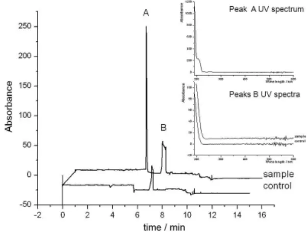
MEKC analysis showed a different pattern (Figure 2) compared to CZE. The sample and the control could be
distinguished by peak A. From peak A migration time it is concluded that this solute interacts less with micelle than peak B, thus it is more hydrophilic than the later, under the analysis conditions. Peak B UV-Vis spectra are practically identical in both sample and control, suggesting it is probably the same solute, which could be either a system peak or a compound from the culture medium. Therefore, as expected, these CE runs have proved the sample do contain a substance not present in the control, which probably is the searched siderophore(s).
CE analysis - IMAC samples
In order to selectively isolate the siderophores from the sample, afinity chromatography was applied. The puriication of samples extracted from solid plate culture medium was carried out by IMAC. A total of 24 fractions were collected and analyzed by MEKC. Fractions 1 to 16 (1 mL each) were obtained from the starting buffer elution and presented the same electrophoretic pattern (fraction 13 - Figure 3) – the other fractions electropherograms are not presented. This fact led us to conclude that no analytes were present in the washing elution fraction (starting buffer), thus reinforcing the idea of selective retention of the siderophores by the IMAC column. By UV-Vis spectra comparison, obtained ofline (in the spectrophotometer) and online (in the CE-UV equipment), peak C identity (Figure 3 and Figure 4a) was confirmed to be from imidazol, the main component of the starting buffer. Fractions 17 and 18 were obtained when the eluting buffer (EDTA) was passed through the column. They presented
the same color as Fe3+ solution, what strongly suggested
the presence of siderophore in these fractions. In fact, Figure 1. CZE analyses of samples and control for siderophore production,
not puriied by IMAC. BGE: 25 mmol L−1 phosphate buffer; injection time (ti): 5s (50 mBar); applied potential: 15 kV; λ = 200 nm. Capillary length (Lt) = 61 cm; effective length (Leff) = 52 cm; internal diameter (i.d.) = 50 µm.
Figure 2. MEKC analyses of control and sample for siderophore production, not puriied by IMAC. The insert shows UV-Vis spectra for peaks A and B. BGE: 25 mmol L-1 phosphate buffer at pH 10 plus 50 mmol L-1 SDS; t
electropherograms indicated a different peak pattern from the previous 16 fractions (see electropherograms in Figure 3).
It seems that at least three peaks were coeluting with peak D in Fraction 18 (Figure 3). This observation is corroborated by their slightly different UV-Vis spectra (Figure 4b). Fractions 17 and 19 showed the same pattern (data not shown). Peak C intensity decreased with eluting buffer passage and its migration time (ca. 2.5 min) was almost the same as the one observed in fractions 1 to 16. A
minor difference may be attributed to EOF variation due to sample-to-sample BGE ionic strength variation.
ESI-MS analysis - IMAC samples
The same fractions obtained by IMAC, and analyzed by CE (MEKC), were analyzed by ESI-MS. Both negative and positive ionization methods were tested and analyses were carried out by direct sample injection.
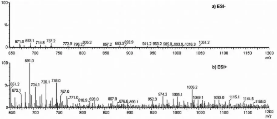
The fractions obtained with the starting buffer presented the same signals as blank samples (data not shown). However, mass spectra from fractions 17 to 24 demonstrated a different proile, both in negative and positive ionization modes. Nevertheless, negative ionization mode is less informative because signal intensity was low due to poor ion production (Figure 5), suggesting that these samples molecules present a low tendency to donate protons.
Comparison of fraction 18 mass spectra, obtained by negative and positive ionization modes (Figures 5a and 5b, respectively), shows some signals observed in both of them – with a difference of two mass units (corresponding to [M+H]- and [M-H]+, respectively), such as: m/z 671 and
673, m/z 658 and 660, m/z 737 and 739, m/z 857 and 859.
However, only peaks m/z 671 and 673 presented signiicant
abundance. The CAS dye (605 Da) and the corresponding Fe3+ complex (660.8 Da), which are present in the culture
medium, could have been observed in these spectra as the ions m/z 603 and 658.8 ([M-H]-) or m/z 607 and 663.8
([M+H]+), respectively. Nevertheless, such ions were not
present in these spectra, reinforcing the hypothesis that the observed ions are analytes present in the sample.
Focusing on the MS spectrum obtained by ESI+ (Figure 5b), some of the signals may indicate the presence of siderophore. The following ions are possible compounds of interest ([M+H]+) m/z 726, 876, 952, 1005, 1036 and 1093,
since the corresponding sodium adducts (m/z 748, 898, 974,
1027, 1058 and 1115) were observed as well ([M+23]+ or
[(M+H)+22]+). Besides the sodium adducts, the protonated
molecular ions m/z 876, 1005 and 1093 also presented possible
iron adduct signals m/z 930, 1059 and 1147, respectively
([M+55]+ or [(M+H)+54]+), what reinforces the probability
of their high afinity for this metal. Indeed, Simionato
et al.45 investigated Methylobacterium mesophilicum
samples by CE-ESI-MS and concluded they produce a siderophore with a relative molecular mass of 1004 Da. The Methylobacterium spp. is an endophytic bacterium which interacts with X. fastidiosa within the xylem of citrus trees or of grapevines. As a consequence, the endophyte siderophore may be used by X. fastidiosa for iron uptake, thus resulting in plant disease.44 The results observed herein show
that grapevine X. fastidiosa may also synthesize a similar Figure 3. Electrophoretic analyses of IMAC fractions obtained with
starting (Fraction 13) and eluting (Fractions 17, 18 and 19) buffers. Electrophoretic conditions are the same as in Figure 2, except for capillary dimensions (Lt = 50 cm; Leff = 42.5 cm). The insert shows time ampliication around peak D.
siderophore, since one of the protonated molecular ions observed in mass spectra presents the same molecular mass.
Conclusions
Grapevine strain Xylella fastidiosa plate cultures, containing Chrome azurol S solution, have indicated the production of a compound with high afinity by Fe3+ due to
halo formation around the bacterial colonies. Analyses of samples extracted from these halos by CZE have shown that this compound was neutral in both basic and neutral pH. In the other hand, MEKC analyses have shown a different proile for the sample and the control. A peak present in the sample, which was not observed in the control by MEKC analysis, refers to a neutral hydrophilic compound, conirming the secretion of a biomolecule from the bacterium isolate. IMAC puriication step has corroborated with the hypothesis that this compound has an extremely high afinity for Fe3+.
Electrophoretic and mass spectrometry analysis of IMAC fractions indicated the presence of different compounds in the same fractions. Indeed, Fraction 18 electropherogram showed the slight separation of three different species. Mass spectrometry experiments revealed possible siderophores molecular masses, which were identiied by blank spectra subtraction and sodium and iron adducts formation. Such biomolecules presented the molecular masses 875, 1004 and 1092 Da, which are within the expected range for siderophores molecules. Further investigation is underway for isolation and structural elucidation, to better characterize these compounds.
Acknowledgements
The authors would like to acknowledge the inancial support provided by Fundação de Amparo à Pesquisa do Estado de São Paulo (FAPESP 99/09177-1, 00/12207-9 and 08/57805-2), Coordenação de Aperfeiçoamento de Pessoal
de Nível Superior (CAPES) and Conselho Nacional de Desenvolvimento Cientíico e Tecnológico (573672/2008-3). We also would like to thank Professor Fernando Mauro Lanças by his laboratory facilities.
References
1. http://www.lbi.ic.unicamp.br/xf/, accessed in March 2009. 2. Liang, X.; Campopiano, D. J.; Sadler, P. J.; Chem. Soc. Rev.
2007, 36, 968.
3. Hartung, J. S.; Beretta, J.; Brlansky, R. H.; Spisso, J.; Lee, R.
F.; Phytopathology1994, 84, 591.
4. Hopkins, D. L.; Mollenhauer, H. H.; Science1973, 179, 298. 5. Davis, M. J.; Purcell, A. H.; Thomson, S. V.; Science1978, 199,
75.
6. Davis, M. J.; French, W. J.; Schaad, N. W.; Curr. Microbiol.
1981, 6, 309.
7. Lima, J. E. O.; Miranda, V. S.; Coutinho, A.; Roberto, S. R.; Carlos, E. F.; Fitopatol. Bras.1996, 21, 392.
8. Chatterjee, S.; Almeida, R. P. P.; Lindow, S.; Annu. Rev.
Phytopathol.2008, 46, 243.
9. Simpson, A. J. G.; Reinach, F.C.; Arruda, P.; Abreu, F. A.; Acencio, M.; Alvarenga, R.; Alves, L. M. C.; Araya, J. E.; Baia, G. S.; Baptista, C. S.; Barros, M. H.; Bonaccorsi, E. D.; Bordin, S.; Bove, J. M.; Briones, M. R. S.; Bueno, M. R. P.; Camargo, A. A.; Camargo, L. E. A.; Carraro, D. M.; Carrer, H.; Colauto, N. B.; Colombo, C.; Costa, F. F.; Costa1, M. C. R.; Costa-Neto, C. M.; Coutinho, L. L.; Cristofani, M.; Dias-Neto, E.; Docena, C.; El-Dorry, H.; Facincani, A. P.; Ferreira, A. J. S.; Ferreira1, V. C. A.; Ferro, J. A.; Fraga, J. S.; Franc¸a, S. C.; Franco, M. C.; Frohme, M.; Furlan, L. R.; Garnier, M.; Goldman, G. H.; Goldman, M. H. S.; Gomes, S. L.; Gruber, A.; Ho, P. L.; Hoheisel, J. D.; Junqueira, M. L.; Kemper, E. L.; Kitajima, J. P.; Krieger, J. E.; Kuramae, E. E.; Laigret, F.; Lambais, M. R.; Leite, L. C. C.; Lemos, E. G. M.; Lemos, M. V. F.; Lopes, S. A.; Lopes, C. R.; Machado, J. A.; Machado, M. A.; Madeira, A. M. B. N.; Madeira, H. M. F.; Marino, C. L.; Marques, M. V.; Martins, E.
Figure 5. ESI-MS of IMAC fraction 18. (a) Negative electrospray ionization mode. Cone potential: 25 V. ESI potential: −3.50 kV. Make-up liquid: CH3CN:H2O (1:1); low rate: 15 µL min-1. Nebulizer gas (N
2) low rate: 260 L h
A. L.; Martins, E. M. F.; Matsukuma, A. Y.; Menck, C. F. M.; Miracca, E. C.; Miyaki, C. Y.; Monteiro-Vitorello, C. B.; Moon, D. H.; Nagai, M. A.; Nascimento, A. L. T. O.; Netto, L. E. S.; Nhani Jr., A.; Nobrega, F. G.; Nunes, L. R.; Oliveira, M. A.; De Oliveira, M. C.; De Oliveira, R. C.; Palmieri, D. A.; Paris, A.; Peixoto, B. R.; Pereira, G. A. G.; Pereira Jr., H. A.; Pesquero, J. B.; Quaggio, R. B.; Roberto, P. G.; Rodrigues, V.; Rosa, A. J. de M.; De Rosa Jr., V. E.; De Sa´, R. G.; Santelli, R. V.; Sawasaki, H. E.; Da Silva, A. C. R.; Da Silva, A. M.; Da Silva, F. R.; Silva Jr., W. A.; Da Silveira, J. F.; Silvestri, M. L. Z.; Siqueira, W. J.; De Souza, A. A.; De Souza, A. P.; Terenzi, M. F.; Trufi1, D.; Tsai, S. M.; Tsuhako, M. H.; Vallada, H.; Van Sluys, M. A.; Verjovski-Almeida, S.; Vettore, A. L.; Zago, M. A.; Zatz, M.; Meidanis, J.; Setubal, J. C.; Nature 2000, 406, 151.
10. Neilands, J. B.; J. Biol. Chem.1995, 270, 26723.
11. Hernlem, B. J.; Vane, L. M.; Sayles, G. D.; Water Res.1999,
33, 951.
12. Nair, A.; Juwarkar, A. A.; Devotta, S.; J. Hazard. Mater.2008,
152, 545.
13. Simionato, A. V. C.; Cantú, M. D.; Carrilho, E.; Microchem. J.
2006, 82, 214.
14. Shanzer, A.; Libman, J.; Weizman, H.; Mester, B.; Hadar, Y.; Chen, Y.; Jurkevitch, E.; Ardon, O.; Pure Appl. Chem.1996,
68, 757.
15. Braun, V.; Killmann, H.; Trends Biochem. Sci.1999, 24, 104. 16. Miller, M. C.; Parkin, S.; Fetherson, J. D.; Perry, R. D.; DeMoll,
E.; J. Inorg. Biochem.2006, 100, 1495.
17. Essen, S. A.; Johnsson, A.; Bylund, D.; Pedersen, K.; Lundstrom, U. S.; Appl. Environ. Microbiol.2007, 73, 5857. 18. Goodell, B.; Jellison, J.; Liu, J.; Daniel, G.; Paszczynski, A.;
Fekete, F.; Krishnamurthy, S.; Jun, L.; Xu, G.; J. Biotechnol.
1997, 53, 133.
19. Drechsel, H.; Tschierske, M.; Thieken, A.; Jung, G.; Zahner, H.; Winkelmann, G.; J. Ind. Microbiol.1995, 14, 105. 20. Raymond, K. N.; Carrano, C. J.; Acc. Chem. Res.1979, 12, 183. 21. Etchegaray, A.; Silva-Stenico, M. E.; Moon, D. H.; Tsai, S. M.;
Microbiol. Res.2004, 159, 425.
22. Payne, S. M.; Methods Enzymol.1994, 235, 329. 23. Arnow, L. E.; J. Biol. Chem.1937, 118, 531. 24. Csáky, T. Z.; Acta Chem. Scand.1948, 2, 450.
25. Schwyn, B.; Neilands, J. B.; Anal. Biochem.1987, 160, 47. 26. Garcia-Campana, A. M.; Taverna, M.; Fabre, H.; Electrophoresis
2007, 28, 208.
27. Nunnally, B.; Park, S. S.; Patel, K.; Hong, M.; Zhang, X.; Wang, S. X.; Rener, B.; Reed-Bogan, A.; Salas-Solano, O.; Lau, W.; Girard, M.; Carnegie, H.; Garcia-Canas, V.; Cheng, K. C.; Zeng, M.; Ruesch, M.; Frazier, R.; Jochheim, C.; Natarajan, K.; Jessop, K.; Saeed, M.; Moffatt, F.; Madren, S.; Thiam, S.; Altria, K.; Chromatographia2005, 64, 359.
28. Hernandez-Borges J.; Neususs, C.; Cifuentes, A.; Pelzing, M.;
Electrophoresis2004, 25, 2257.
29. Mucha, P.; Rekowski, P.; Kosakowska, A.; Kupryszewski, G.;
J. Chromatogr., A1999, 830, 183.
30. Berner, I.; Greiner, M.; Metzger, J.; Jung, G.; Winkelmamm,
G.; Biol. Metals1991, 4, 113.
31. Essen, S. A.; Bylund, D.; Holmstrom, S. J. M.; Moberg, M.;
Biometals2006, 19, 269.
32. Cox, G. B.; Gibson, F.; Luke, R. K. J.; Newton, N. A.; O’Brien, I. G.; Rosenberg, H.; J. Bacteriol.1970, 104, 219.
33. Gledhill, M.; Analyst2001, 126, 1359.
34. Simionato, A. V. C.; de Souza, G. D.; Rodrigues-Filho, E.; Glick, J.; Vouros, P.; Carrilho, E.; Rapid Commun. Mass Spectrom.
2006, 20, 193.
35. Gaberc-Porekar, V.; Menart, V.; J. Biochem. Biophys. Methods
2001, 49, 335.
36. Hauput, K.; Roy, F.; Vijayalakshmi, M. A.; Anal. Biochem.
1996, 234, 149.
37. Porath, J.; Carlsson, J.; Olsson; I.; Belfrage, G.; Nature1975,
258, 598.
38. Wimalsena, R. L.; Wilson, G.S.; LC-GC1992, 10, 223. 39. Sulkowski, E.; Trends Biotechnol.1985, 3, 1.
40. Ramadan, N.; Porath, J.; J. Chromatogr.1985, 321, 93. 41. Van Sluys, M A.; De Oliveira, M. C.; Monteiro-Vitorello, C. B.;
Miyaki, C. Y.; Furlan, L. R.; Camargo, L. E. A.; da Silva, A. C. R.; Moon, D. H.; Takita, M. A.; Lemos, E. G. M.; Machado, M. A.; Ferro, M. I. T.; da Silva, F. R.; Goldman, M. H. S.; Goldman, G. H.; Lemos, M. V. F.; El-Dorry, H.; Tsai, S. M.; Carrer, H.; Carraro, D. M.; de Oliveira, R. C.; Nunes, L. R.; Siqueira, W. J.; Coutinho, L. L.; Kimura, E. T.; Ferro, E. S.; Harakava, R.; Kuramae, E. E.; Marino, C. L.; Giglioti, E.; Abreu, I. L.; Alves, L. M. C.; do Amaral, A. M.; Baia, G. S.; Blanco, S. R.; Brito, M. S.; Cannavan, F. S.; Celestino, A. V.; da Cunha, A. F.; Fenille, R. C.; Ferro, J. A.; Formighieri, E. F.; Kishi, L. T.; Leoni, S. G.; Oliveira, A. R.; Rosa, V. E.; Sassaki, F. T.; Sena, J. A. D.; de Souza, A. A.; Trufi, D.; Tsukumo, F.; Yanai, G. M.; Zaros, L. G.; Civerolo, E. L.; Simpson, A. J. G.; Almeida, N. F.; Setubal, J. C.; Kitajima, J. P.; J. Bacteriol.2003, 185, 1018.
42. Silva-Stenico, M. E.; Pacheco, F. T. H.; Rodrigues, J. L. M.; Carrilho, E.; Tsai, S. M.; Microbiol. Res.2005, 160, 429. 43. Pacheco, F. T. H.; Silva-Stenico, M. E.; Etchegaray, A.; Gomes,
J. E.; Carrilho, E.; Tsai, S. M.; Genet. Mol. Biol.2006, 29, 137. 44. Lacava, P. T.; Silva-Stenico, M. E.; Araújo, W. L.; Simionato, A.
V. C.; Carrilho, E.; Tsai, S. M.; Azevedo, J. L.; Pesqui. Agropec.
bras.2008, 43, 521.
45. Simionato, A. V. C.; Simó, C.; Cifuentes, A.; Lacava P. T.; Araújo, W. L.; Azevedo, J. L.; Carrilho, E.; Electrophoresis
2006, 27, 2567.
Received: April 1, 2009 Web Release Date: December 15, 2009