Article
J. Braz. Chem. Soc., Vol. 27, No. 6, 1032-1039, 2016. Printed in Brazil - ©2016 Sociedade Brasileira de Química 0103 - 5053 $6.00+0.00
*e-mail: [email protected], [email protected]
Antiprotozoal Activity of the Cyclopalladated Complexes Against
Leishmania
amazonensis
and
Trypanosoma cruzi
Angela M. A. Velásquez,a,b Rodrigo A. de Souza,b Thaís G. Passalacqua,a,b Aline R. Ribeiro,c Mateus Scontri,a Chung M. Chin,a Leticia de Almeida,a,b Mayara L.
Del Cistia,a João A. da Rosa,a Antonio E. Maurob and Marcia A. S. Graminha*,a
aFaculdade de Ciências Farmacêuticas and bInstituto de Química, Universidade Estadual Paulista,
UNESP, 14800-900 Araraquara-SP, Brazil
cInstituto de Ciências Biológicas, Universidade Estadual de Campinas,
13083-862 Campinas-SP, Brazil
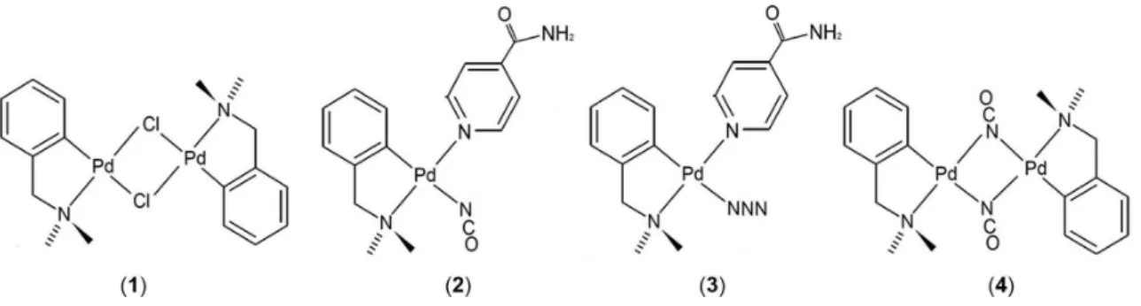
The present study describes the antiprotozoal activities of four cyclopalladated compounds, [Pd(dmba)(µ-Cl)]2, [Pd(dmba)(NCO)(isn)], [Pd(dmba)(N3)(isn)] and [Pd(dmba)(µ-NCO)]2,
(dmba: N,N’-dimethylbenzylamine and isn: isonicotinamide), against the diseases leishmaniasis (Leishmania amazonensis and Leishmania infantum), Chagas disease (Trypanosoma cruzi) and human African trypanosomiasis (Trypanosoma brucei). [Pd(dmba)(µ-NCO)]2 exhibited good
leishmanicidal and trypanocidal activities against L. amazonensis and T. cruzi intracellular amastigote forms, with a 50% inhibitory concentration (IC50) value of less than 9 µM and selectivity
indexes of 14.47 and 28.42, respectively. Stability essays were conducted in phosphate buffer saline (PBS) pH 7.0 and showed that [Pd(dmba)(µ-NCO)]2 is the most stable molecule. These findings
indicate that this compound presented higher selectivity for these parasites than the other tested compounds. The data presented here suggest that this compound should be considered in the development of new and more potent drugs for the treatment of leishmaniasis and Chagas disease.
Keywords: cyclopalladated, leishmaniasis, Chagas disease, Leishmania amazonensis, Trypanosoma cruzi, trypanosomiasis
Introduction
Leishmaniasis, Chagas disease (American trypanosomiasis, AT) and sleeping sickness (human African trypanosomiasis, HAT) are parasitic diseases caused by flagellated protozoa related to the family Trypanosomatidae. According to the World Health Organization (WHO), 1.3 million new cases of leishmaniasis and 20,000 to 30,000 deaths occur annually in all continents,1 7 to
8 million people are infected worldwide with Chagas disease2 and the estimated number of actual cases of HAT
is currently 30,000.3
More than 20 species of Leishmania4 are responsible
for different clinical manifestations, including cutaneous (CL), mucocutaneous (MCL) and visceral forms (VL).5
In the Old World, Leishmania major, Leishmania tropica, Leishmania aethiopica and some zymodemes from Leishmania infantum are the causative agents of CL.
In the New World, CL is mainly caused by Leishmania amazonensis, Leishmania guyanensis, Leishmania panamensis and Leishmania braziliensis.6 MCL can be
caused by L. braziliensis, L. panamensis, L. guyanensis
and L. amazonensis in the New World and by L. major
and L. infantum in the Old World.7 Pentavalent antimonial
drugs are the most frequently prescribed treatments for leishmaniasis.8 The main adverse effects that occur in
systemic administration of these antimonials are arthralgias, myalgias, anorexia, and nausea, and other serious side effects include pancreatitis, liver-enzyme abnormalities, cardiac malfunctions and severe renal toxic effects.9
Other drugs, such as amphotericin B, pentamidine and miltefosine, are second choice drugs but they also produce side effects that can endanger the patient’s life.
Trypanosoma cruzi is the causative agent of Chagas
disease, which is present mainly in Latin America but also in North America.10-12 Nifurtimox and benznidazole
effective against the chronic phase of the disease, and they present multiple side effects, from dermatitis to bone marrow depression.15 HAT is caused by Trypanosoma
brucei gambiense and Trypanosoma brucei rhodesiense.16
Treatment of the hemolymphatic stage of HAT relies on suramin and pentamidine. In the meningoencephalitis stage, melarsoprol, a highly toxic arsenic-based drug that is effective against both T. b. gambiense and T. b. rhodesiense
is used. Eflornithine is useful only against T. b. gambiense.
An eflornithine/nifurtimox combined therapy has been used, but this also causes several side effects.17 In summary,
the conventional drugs that have been employed to treat these diseases are antiquated, have high toxicity and are ineffective due to drug resistance. Therefore, is critical to develop new drugs for the treatment of these trypanosomiases.
The use of transition metal-based drugs is increasing significantly in the therapy of cancer and tropical diseases; in fact, many organometallic compounds have been designed to specifically bind to a well-defined target site in different biomolecules.18-20 The emergence of drug resistance in
tropical diseases has led to alternatives to chloroquine and its analogues, such as metallocene derivatives for the treatment of malaria.19,20 Several scaffolds of metal
complexes containing palladium, platinum, gold, iridium, rhodium, osmium and iron have shown leishmanicidal18-21
and trypanocidal22,23 activity. For palladium compounds,
it was recently verified that a cyclopalladated complex showed a high selectivity index with trypanocidal activity in the treatment of Chagas disease.24 Literature has reported
a cyclopalladated compound with leishmanicidal activity25
against L. amazonensis promastigote and amastigote forms;
and the activity of some palladium(II) cyclometalated complexes26 against T. cruzi and Leishmania, which
indicate on preliminary data that the compounds inhibited the growth of intracellular amastigote forms.
Cyclopalladated complexes have high thermodynamic and kinetic stability compared to others palladium(II) compounds due to the formation of a stable chelate ring.27
Based on the potential activity of the cyclopalladated
complexes reported in the literature,28-34 in this study,
we describe the evaluation of the leishmanicidal and trypanocidal activity of [Pd(dmba)(µ-Cl)]2 (1), [Pd(dmba)
(NCO)(isn)] (2), [Pd(dmba)(N3)(isn)] (3) and [Pd(dmba)
(µ-NCO)]2 (4) (Figure 1), dmba: N,N’-dimethylbenzylamine
and isn: isonicotinamide. All complexes have been previously described35-38 but their protozoal activities have
not been reported yet.
Herein we tested the above mentioned cyclopalladated complexes against L. amazonensis, L. infantum, T. cruzi
and T. brucei.
Experimental
Infrared spectra (IR) were recorded on a Spectrum 2000, PerkinElmer, in the spectral range 4000-370 cm-1.
Samples were prepared in KBr pellets. 1H and 13C{1H}
nuclear magnetic resonance (NMR) spectra were referred to the high field SiMe4 signal, on a INOVA 500 spectrometer;
the magnetic field applied was 11.7 T and the resonance of 1H and 13C{1H} nucleus were at 500 and 125 MHz,
respectively. Full spectroscopic data are presented in the Supplementary Information section.
The elemental analysis was performed with an Elemental Analyzer 2400 CHN, PerkinElmer, at Central Analítica, Instituto de Química, USP, São Paulo, Brazil.
Synthesis of cyclopalladated complexes
All synthesis was carried out at room temperature. All reagents were obtained from commercial suppliers and used without further purification.
Compound (1), [Pd(dmba)(µ-Cl)]2 was prepared
as previously described in the literature,36,37 with some
modifications. In summary, 22.6 mmol of LiCl (Carlo Erba) was added to a solution of 11.3 mmol of PdCl2
(Degussa S. A.) in methanol. The mixture was stirred at 60 °C followed by the addition of 11.3 mmol of dmba (N,N’-dimethylbenzylamine, Sigma-Aldrich) and
14.4 mmol of triethylamine (Carlo Erba). After 6 h, the
yellow solid formed was filtered off, washed thoroughly with methanol and diethyl ether, and dried in vacuo.
The yield was 90%; mp 185 °C (dec.); anal. calcd. for C18H24N2Cl2Pd2: C 39.1, H 4.3, N 5.1; found: C 39.4, H 4.2,
N 5.0; MW = 552.14.
Compound [Pd(dmba)(µ-NCO)]2 (4) and starting
species, [Pd(dmba)(µ-N3)]2.H2O, for the synthesis of
(2) and (3), were prepared based on Almeida et al.35 by
replacement of chlorido ligands in (1) by the corresponding
pseudohalides. The reactions were carried out in acetone and the obtained yellow solids were filtered off, washed with water and pentane, and dried in vacuo. The yield
was 79% for compound (4) and 87% for the precursor,
mp 177 °C (dec.), mp 187 °C (dec.), respectively.
[Pd(dmba)(µ-NCO)]2: anal. calcd. for C20H24N4O2Pd2:
C 42.5, H 4.3, N 9.9; found: C 42.3, H 4.3, N 9.4; MW = 552.14.
[Pd(dmba)(µ-N3)]2.H2O: anal. calcd. for C18H24N8Pd2.H2O:
C 37.1, H 4.5, N 19.2; found: C 37.0, H 4.2, N 18.5; MW = 565.28.
Compounds of the type [Pd(dmba)X(isn)]; X = NCO (2),
N3 (3); isn: isonicotinamide, were prepared according to the
literature,35,38 with some modifications.
In summary, compounds (2) and (3) were obtained in
acetone by reacting the suitable [Pd(dmba)(µ-X)]2 precursor
with isn (Sigma-Aldrich). The obtained white solids were washed thoroughly with acetone and pentane. The yield was 90% for compound (2) and 94% for (3), mp 206 °C
(dec.), mp 196 °C (dec.), respectively.
[Pd(dmba)(NCO)(isn)] anal. calcd. for C16H18N4O2Pd:
C 47.5, H 4.5, N 13.8; found: C 47.3, H 4.3, N 13.0; MW = 404.76.
[Pd(dmba)(N3)(isn)] anal. calcd. for C15H18N6OPd:
C 44.5, H 4.5, N 20.8; found: C 44.0, H 4.3, N 20.5; MW = 404.76.
Stability assay
The samples 1- 4were dissolved in acetonitrile (minimum
amount to solubilize) and the solutions then prepared in phosphate buffer saline (PBS) pH 7.0 to give concentrations of 317, 312, 220 and 143 µmol L-1. The solutions were
prepared and the samples were left with occasional stirring at room temperature being analyzed aliquots of these solutions in the days immediately after the preparation (time 0) and after 12, 24, 48 and 72 hours. The experiments were performed in HPLC Shimazu LC-20AT CNM-20A UV detector SPD-20A using ODS (C-18) column, particle size 5 µm, 4.6 × 250 mm, mobile phase: methanol:water (70:30, v/v/v), flow 0.8 mL min-1 and λ = 254 nm. For more
details see Supplementary Information section.
Biological assays
Compounds
Cyclopalladated complexes 1, 2, 3, 4 and controls
(pentamidine, amphotericin B and benznidazole (Sigma-Aldrich)) were dissolved in DMSO, dimethyl sulfoxide, (the highest concentration was 1.4%, which was not hazardous to the parasites, as previously determined), added to the parasite suspension to final concentrations between 0.5 and 100.0 µmol L-1.
Parasites: L. amazonensis, L. infantum, T. cruzi and T. brucei
Promastigotes of L. amazonensis MPRO/BR/1972/
M1841-LV-79 strain and epimastigote forms of T. cruzi
Y strain39 were maintained at 28 ± 2 ºC in liver-infusion
tryptose medium (LIT)40 supplemented with 10%
heat-inactivated fetal bovine serum (FBS) (Gibco). Promastigotes of L. infantum (MHOM/BR/72) were freshly
isolated from golden hamsters (Mesocricetus auratus)
and maintained at 28 °C in M199 medium (Cultilab) without phenol red supplemented with 10% FBS (Gibco), penicillin Aldrich) and streptomycin (Sigma-Aldrich). Promastigote and epimastigote forms cultures were carried out until to obtain in the exponential growth phase (1.107 parasites mL-1).
Procyclic forms of T. brucei 427 strains41 were cultured
at 28 ± 2 °C in SDM-79 medium42 containing 10% FBS,
penicillin (Sigma) and streptomycin (Sigma). Procyclic forms cultures were carried out until to obtain the exponential growth phase (1.106 parasites mL-1).
Cytotoxicity using murine macrophages
To determine the cytotoxicity in murine macrophages we used a method previously described.43 Adult male Swiss
albino mice (20 to 35 g) were used in the experiments. They were housed in single-sex cages under a 12 h light/12 h dark cycle in a controlled-temperature room (22 ± 2 °C). The mice had free access to food and water. Groups of three animals were used in each test group. The experiments were performed in concordance to protocol approved by the Institutional Ethics Committee-CEUA (Comissão de Ética no Uso de Animais), protocol CEUA/FCF/CAr No. 20/2013. The mice were stimulated with thioglycolate to collect peritoneal macrophages. Murine peritoneal macrophages were seeded in 96-well flat-bottom plates (TPP) at a density of 1 × 10⁵ cells per well
in RPMI 1640 medium44 supplemented with 10%
heat-inactivated FBS, 25 mM HEPES, and 2 mM L glutamine and incubated for 4 h at 37 ± 2 °C in a 5% CO2-air mixture.
concentrations of compounds and controls. Cells without drugs were used as a negative control. After that, plates were incubated for 24 h at 37 ± 2 °C in a 5% CO2-air mixture.
Subsequently, the MTT (3-4(4,5-dimethyl-2-thiazolyl)-2,5-diphenyl-2H-tetrazolium bromide) colorimetric assay
was carried out as described further on. Absorbance was read in a 96-well plate reader (Robonik) at 595 nm. The drug concentration corresponding to 50% of cell growth inhibition was expressed as the inhibitory concentration (CC50, µM).45
MTT colorimetric assay
L. amazonensis and L. infantum promastigote, T. brucei
procyclic and T. cruzi epimastigote forms were treated with
the cyclopalladated complexes and their respective controls to calculate the half maximal inhibitory concentration (IC50)
by MTT colorimetric assay.46,47 The MTT assay is based
on the determination of the ability of living cells to reduce 3-4(4,5-dimethyl-2-thiazolyl)-2,5-diphenyl-2H-tetrazolium
bromide (MTT). To avoid interaction of compounds with the MTT, the cells were gently washed with PBS before the addition of MTT solution on the cells. The compounds were tested in different concentrations and incubated at 28 ± 2 °C for 72 h for L. amazonensis, L. infantum and T. cruzi and 24 h for T. brucei.23,43 The assays were carried
out in triplicates. Absorbance was read in a 96-well plate reader (Robonik) at 490 nm for Leishmania species and
595 nm for T. cruzi and T. brucei. The drug concentration
corresponding to 50% parasite growth inhibition was expressed as the IC50 in µM.
The safety index (SI) was calculated and SI around 10 means that a compound can be better evaluate for further studies. Data obtained were processed with the software Origin 7.0.48
Leishmanicidal and trypanocidal activities against intracellular amastigote forms
To determine the leishmanicidal and trypanocidal activities against intracellular amastigote forms we used a method previously described.43 Murine peritoneal
macrophages were plated at 3 × 10⁵ cells per well on
coverslips (13 mm diameter) previously arranged in a 24 well plate in RPMI 1640 medium44 supplemented
with 10% inactivated FBS and allowed to adhere for 4 h at 37 ± 2 °C in 5% CO2. Adherent macrophages
were infected with Leishmania promastigotes in the
stationary growth phase or T. cruzi trypomastigote using
a ratio of 5/10:1 parasites per cell (L. amazonensis or L. infantum/T. cruzi: macrophages) at 37 °C in 5% CO2
for 4 h for L. amazonensis, 18 h for L. infantum and 24 h
for T. cruzi. After that time, the non-internalized parasites
were removed by washing and infected cultures were incubated in RPMI 1640 medium for 24 h at 37 ± 2 °C in 5% CO2 to allow parasite multiplication. Then, infected cells
were treated with different concentrations of compounds, amphotericin B Aldrich), pentamidine (Sigma-Aldrich) or benznidazole (Sigma-(Sigma-Aldrich) for 24 h. The cells were then fixed in a methanol solution and stained with Giemsa. The number of amastigotes/100 macrophage cells and the percent of infected cells were determined. The concentration that caused a 50% decrease of growth inhibition compared to the control was determined by regression analysis and expressed as IC50 in µM.45
The experiments were performed in accordance with the protocol approved by the Institutional Ethics Committee-CEUA (Comissão de Ética no Uso de Animais), protocol CEUA/FCF/CAr No. 20/2013.
Differentiation of T. cruzi epimastigote to trypomastigote
forms
In vitro differentiation of T. cruzi was made as
previously described in literature.49,50 The epimastigote
forms were collected from the LIT culture on the 7th day
when the plateau phase of the growth curve was reached and then, re-suspended in artificial triatomine urine (TAU) (190 mmol L-1 NaCI, 8 mmol L-1 phosphate buffer, pH 6.0,
17 mmol L-1 KCl, 2 mmol L-1 CaCI
2, 2 mmol L-1 MgCI2), and
incubated for 2 h at room temperature. The parasites were diluted to a final concentration of 5 × 10⁶ parasites mL-1 in
TAU supplemented with 2.5% (v/v) sodium bicarbonate 1.4%, 500 units penicillin mL-1, 10 mmol L-1 proline (TAUP
medium) and incubated at 27 °C for 10 days.
Results and Discussion
Compounds 1-4 (Figure 1) were obtained as stable
solids (yields ranging from 79% to 94%), and their structures are fully supported by infrared spectroscopy(IR) and elemental analysis.
The cyclopalladated complexes were tested against
L. amazonensis, L. infantum (promastigote and amastigote), T. cruzi (epimastigote and amastigote) and T. brucei
(procyclic forms) using the MTT colorimetric assay44-46
and an in vitro intracellular amastigote assay43,49,50 to
evaluate the antiprotozoal activity. Amphotericin B and pentamidine were included in the assays as positive controls for Leishmania species, benznidazole for T. cruzi
and pentamidine for T. brucei. The MTT assays were
carried out after 72 h (L. amazonensis, L. infantum and T. cruzi) and 24 h (T. brucei) of incubation. In general, all
The antiprotozoal effect of the cyclopalladated complexes on intracellular amastigotes was evaluated in murine peritoneal macrophages obtained from adult male Swiss albino mice infected with Leishmania species
promastigote forms and T. cruzi trypomastigote forms.
We observed a decrease in the amount of intracellular amastigote forms after treatment with the cyclopalladated complexes. A cytotoxicity assay was also carried out to evaluate the toxicity of the compounds in the peritoneal macrophages, showing lower toxic activity on mammalian cells. All data are presented in Table 1.
The ligands dmba and isn by themselves did not exhibit antiprotozoal activities (Table 1), suggesting that the observed biological activity was due to the cyclopalladated complexes. In the present study, we observed that compound 2 showed in vitro activity against T. cruzi amastigotes (IC50 = 4.45 ± 0.45 µM), while
compound 4 showed activity against both L. amazonensis
(IC50 = 9.29 ± 2.08 µM) and T. cruzi amastigotes
(IC50 = 4.73 ± 1.19 µM). It is interesting to note that for
L. amazonensis amastigote, compound 4 showed almost 3
times higher leishmanicidal activity compared to compounds
2 (IC50 = 22.45 ± 1.17 µM) and 3 (IC50 = 28.82 ± 2.15 µM)
(Table 1), suggesting that the isn group complexed to Pd decreases the anti-Leishmania activity but does not
influence the anti-T. cruzi activity.
The replacement of chlorido by the cyanato ligand (1→4) did not result in any increase in leishmanicidal
activity against the promastigote and amastigote forms; however compound 1 was 2 times more active than
compound 4 against the T. cruzi epimastigote. On the
other hand, different efficacy was observed in the T. cruzi
amastigote form, where compound 4 was approximately
3 times more active than compound 1. The presence of
Table 1. Antiparasitic activities (IC50, half-maximal inhibitory concentration, µM), mammalian cell toxicity (CC50, half-maximal cytotoxicity concentration,
µM) and safety index (SI = CC50/IC50) of the cyclopalladated complexes. The SI values ≥ 10 in bold indicate promising compounds, i.e., those with low
mammalian toxicity and high toxicity to parasites. Each value is the mean of three experiments performed in triplicate ± standard error
Compound
IC50 (SI) / µM CC50 / µM
L. infantum L. amazonensis T. cruzi Y T. brucei 427
Macrophage Promastigote Amastigote Promastigote Amastigote Epimastigote Amastigote Procyclic
1 [Pd(dmba)(µ-Cl)]2 24.72 ± 1.40
(2.39)
42.00 ± 1.00 (1.41)
13.53 ± 1.76
(4.37)
9.57 ± 2.60
(6.18)
7.06 ± 1.76
(8.38)
11.82 ± 1.17
(5.01)
7.14 ± 1.54
(8.29)
59.18 ± 5.29
2 [Pd(dmba)(NCO)(isn)] 17.54 ± 0.30 (5.60)
82.30 ± 2.30 (1.19)
32.12 ± 3.09
(3.06)
22.45 ± 1.17
(4.37)
7.72 ± 0.62
(12.72)
4.45 ± 0.45
(22.07)
41.56 ± 1.68
(2.36)
98.22 ± 11.70
3 [Pd(dmba)(N3)(isn)] 12.35 ± 0.30
(9.56)
59.50 ± 0.80 (1.98)
25.79 ± 2.20
(4.58)
28.82 ± 2.15
(4.10)
14.82 ± 1.09
(7.97)
3.40 ± 0.26
(34.73)
46.39 ± 1.17
(2.55)
118.09 ± 14.67
4 [Pd(dmba)(µ-NCO)]2 16.57 ± 2.10
(8.11)
53.20 ± 0.10 (2.53)
13.67 ± 1.54
(9.83)
9.29 ± 2.08
(14.47)
11.06 ± 1.76
(12.16)
4.73 ± 1.19
(28.42)
16.38 ± 0.70
(8.21)
134.44 ± 24.53
5 Dmba − − NA NA NA NA NA NA
6 Isn − − NA NA NA NA NA NA
7 Pentamidine 67.71 ± 8.10 (0.53)
19.77 ± 0.50 (1.81)
7.62 ± 0.10 (4.68)
5.07 ± 1.14 (7.04)
− − 6.44 ± 0.01 (5.54)
35.69 ± 6.84
8 Benznidazole − − − − 4.07 ± 0.33
(242.86)
5.28 ± 1.38
(187.20)
− 988.43 ± 38.10
9 Amphotericin B 0.92 ± 0.01 (25.11)
2.98 ± 0.40 (7.75)
3.22 ± 0.03
(7.17)
4.92 ± 0.14
(4.70)
− − − 23.10 ± 2.52
NA: not active in the maximal inhibitory concentration tested (100 µmol L-1).
Figure 2. Percentage of viable (a) L. amazonensis promastigote; (b) T. cruzi
NCO instead of Cl in compound 4 increased its chemical
stability after 72 hours and decrease in its contentwhich suggests a profile of Pd release/time without structure change (Figure 3). The advantage of compound 4 is its
effectiveness on amastigotes forms that’s present in human host while epimastigotes is present in the bugs.
All cyclopalladated complexes analyzed showed low mammalian toxicity, mainly for compounds 2, 3 and 4 (CC50 = 98.22, 118.09 and 134.44 µM, respectively)
compared to pentamidine and amphotericin B (CC50 = 35.69
and 23.10 µM, respectively). The decrease in cytotoxicity of these compounds varies according to their ligands in the following order: Cl < NCO (for dimeric compounds), NCO < N3 (for mononuclear complexes).
Among the tested complexes, compound 4 showed
good activity against the selected pathogens as well as low toxicity, with SI values of approximately 10 and 30, except for the L. infantum amastigote. In particular,
for L. amazonensis promastigote and amastigote forms,
compound 4 presented SI values of 9.83 and 14.47,
respectively, which are safer than those of amphotericin B (7.17 and 4.70) and pentamidine (4.68 and 7.04).
For T. brucei procyclic forms, we did not observe
any compound with higher trypanocidal activity than pentamidine. However, the compounds 1 and 4 were
more selective (SI = 8.29 and 8.21, respectively) than pentamidine (SI = 5.54).
Cyclopalladated 2, 3 and 4 evaluated against intracellular
amastigote forms of T. cruzi showed the best SI values
compared to those obtained for the intracellular amastigote forms of the Leishmania species (Table 1). This data
showed that compounds 2, 3 and 4 were more selective
for T. cruzi when compared to other parasites. Further
studies on 3 and 4 are required in order to gain a better
understanding about their mechanism of action.
NMR analysis of complexes 2 and 3 were recorded
in DMSO which indicated that isn is free of its dimeric precursors, [Pd(dmba)(µ-N3)]2 and [Pd(dmba)(µ-NCO)]2;
besides we do not observe species containing DMSO coordinated to PdII. According to the literature,51
cyclopalladated complexes containing pyridine generate its corresponding binuclear precursors in solution at room temperature. Herein, the mononuclear derivatives of cyclopalladated with isn and pseudohalides do not maintain their structure completely intact in solution acting possibly as a prodrug since we observed anti-T. cruzi activity,
therefore, the activity of compounds 2 and 3 might be the
result of fractionation of these species present in solution and their precursors. Probably the observed dissociation in solution would explain the biological activity of the compound and the formation of pro-active species. This could contribute to the bioactivity displayed by compound 4.
This hypothesis can be supported by cisplatin mode of action since it undergoes successive hydrolysis reactions resulting in active species that react more rapidly with the target.52 Further pharmacokinects studies are needed to
prove the pro-action model of the cyclopalladed compound. Some authors24 observed that cyclopalladated complex
against T. cruzi reduced the parasitemia and mortality in vivo with very low and nontoxic doses, as well as
inhibited amastigote intracellular proliferation, similar to observed for us in vitro in T. cruzi and L. amazonensis
intracellular amastigote.
The mechanism of actions proposed by Matsuo et al.24
suggests that cyclopalladated complex interacts with the mitochondria of T. cruzi, causing a collapse in the cell
extrusion of protons, followed by deoxyribonucleic acid (DNA) fragmentation and exposure of phosphatidylserine on the surface, a process resembling to apoptosis in mammalian cells. Already the same authors mentioned that the cyclopalladated complexes are “much more stable and less toxic than other derivatives of palladium(II)53 and
were able to inhibit cathepsin B (CpB)”.54 Other authors55-57
reported that cathepsin B and L are involved in the growth of Leishmania and their virulence in vitro and in vivo. Other
authors25 observed that a palladacycle compound showed
inhibition of the cysteine protease activity expressed in
L. amazonensis amastigotes, being significant the inhibition
of CpB activity. It was also reported that cyclometalled palladium(II) complexes26 inhibited cathepsin B in other
Leishmania species. However, the compounds did not
affect the CpB activity of macrophages.25 Probably, for this
latter reason, our compounds were relatively innocuous on
Figure 3. Stability study of compound 4. Chromatogram profile of
compound [Pd(dmba)(µ-NCO)]2 (4)at 0, 12, 24, 48 and 72 hours in
peritoneal macrophages which is very interesting for new drugs evaluation.
Other mechanism of action by palladiumcomplexes58
described that “the production of oxidative stress as a result of their bioreduction and extensive redox cycling”, as well as the interaction with DNA.21,56
Studies on the mechanism of action of cyclopalladated complexes 1-4 against both L. amazonensis and T. cruzi are
underway, and so are the tests with other T. cruzi strains,
due to genetic variability, to determine if our compounds may be following the similar mechanisms of action reported in the literature.
Conclusions
The results presented in this work suggest that the cyclopalladated compounds 1-4 (in special 4 showed to
be the most active compound against L. amazonensis
and T. cruzi intracellular amastigote forms whit low
cytotoxicity) should be further considered as potential new hit in the search for new drugs for the chemotherapy of Chagas disease and leishmaniasis. In addition, the cyclopalladated 2 and 3 showed a promising anti-T. cruzi
activity. Our biological results demonstrated that all cyclopalladated complexes presented low cytotoxicity towards mammalian cells.
Supplementary Information
Supplementary data (IR spectra, 1H NMR spectra, 13C
spectra and chromatogram profile) are available free of charge at http://jbcs.sbq.org.br as PDF file.
Acknowledgments
The authors thank FAPESP (Fundação de Amparo à Pesquisa do Estado de São Paulo), process number 201308248-1, for financial support and CAPES (Coordenação de Aperfeiçoamento de Pessoal de Nível Superior), for research fellowship to AMAV, TGP, MLDC and ARR. We are also grateful for Regina M. B. Cicarelli for donate the T. brucei 427 strain to our lab.
References
1. http://www.who.int/mediacentre/factsheets/fs375/en/, accessed in December 2015.
2. http://www.who.int/mediacentre/factsheets/fs340/en/, accessed in December 2015.
3. http://www.who.int/trypanosomiasis_african/en/, accessed in December 2015.
4. Pinto, M. C.; Barbieri, K.; Silva, M. C. E.; Graminha, M. A. S.; Casanova, C.; Andrade, A. J.; Eiras, A. E. J.; J. Med. Entomol.
2011, 48, 39.
5. Magill, A. J.; Dermatol. Clin. 1995, 13, 505.
6. Torres, F. A. E.; Passalacqua, T. G.; Velásquez, A. M. A.; Souza, R. A.; Colepicolo, P.; Graminha, M. A. S.; Rev. Bras. Farmacogn.2014, 24, 265.
7. Goto, H.; Lindoso, J. A.; Expert Rev. Anti-Infect. Ther. 2010,
8, 419.
8. Hepburn, N. C.; Curr. Opin. Infect. Dis.2001, 14, 151.
9. González, U.; Pinart, M.; Rengifo-Pardo, M.; Macaya, A.; Alvar, J.; Tweed, J. A.; Cochrane Database Syst. Rev. 2009, 2,
CD004834.
10. Jannin, J.; Villa, L.; Mem. Inst. Oswaldo Cruz 2007, 102 S1,
95.
11. Schmunis, G. A.; Mem. Inst. Oswaldo Cruz 2007, 102 S1, 75.
12. Teixeira, A. R. L.; Nitz, N.; Guimaro, M. C.; Gomes, C.; Santos-Buch, C. A.; Postgrad. Med. J. 2006, 82, 788.
13. Coura, J. R.; Mem. Inst. Oswaldo Cruz 2009, 104, 549. 14. Wilkinson, S. R.; Kelly, J. M.; Expert Rev. Mol. Med. 2009, 11,
e31.
15. Coura, J. R.; de Castro, S. L.; Mem. Inst. Oswaldo Cruz 2002,
97, 3.
16. Cox, F. E. G.; Infect. Dis. Clin. North Am. 2004, 18, 231.
17. Babokhov, P.; Sanyaolu, A. O.; Oyibo, W. A.; Fagbenro-Beyioku, A. F.; Iriemenam, N. C.; Pathog. GlobalHealth 2013,
107, 242.
18. Jaouen, G.; Vessières, A.; Butler, I. S.; Acc. Chem. Res. 1993,
26, 361.
19. Schatzschneider, U.; Metzler-Nolte, N.; Angew. Chem., Int. Ed.
2006, 45, 1504.
20. Sharma, V.; Piwnica-Worms, D.; Chem. Rev. 1999, 99, 2545.
21. Navarro, M.; Betancourt, A.;Hernández, C.; Marchán, E.;
J.Braz. Chem. Soc. 2008, 19, 1355.
22. Farrell, N. P.; Williamsom, J.; Mclaren, D. J. M.; Biochem. Pharmacol. 1984, 33, 961.
23. Velásquez, A. M. A.; Francisco, A. I.; Kohatsu, A. A.; Silva, F. A.; Rodrigues, D. F.; Teixeira, R. G.; Chiari, B. G.; de Almeida, M. G.; Isaac, V. L.; Vargas, M. D.; Cicarelli, R. M.;
Bioorg. Med. Chem. Lett.2014, 24, 1707.
24. Matsuo, A. L.; Silva, L. S.; Torrecilhas, A. C.; Pascoalino, B. S.; Ramos, T. C.; Rodrigues, E. G.; Schenkman, S.; Caires, A. C.; Travassos, L. R.; Antimicrob. Agents Chemother. 2010,
54, 3318.
25. Paladi, C. S.; Pimentel, I. A. S.; Katz, S.; Cunha, R. L. O. R.; Judice, W. A. S.; Caires, A. C. F.; Barbiéri, C. L.; PLoS Negl. Trop. Dis. 2012, 6, e1626.
27. Caires, A. C. F.; Mauro, A. E.; Quim. Nova 1996, 19, 59. 28. Caires, A. C. F.; Almeida, E. T.; Mauro, A. E.; Hemerly, J. P.;
Valentini, S. R.; Quim. Nova 1999, 22, 329.
29. Moro, A. C.; Mauro, A. E.; Netto, A. V. G.; Ananias, S. R.; Quilles, M. B.; Carlos, I. Z.; Pavan, F. R.; Leite, C. Q. F.; Hörner, M.; Eur. J. Med. Chem. 2009, 44, 4611.
30. Moro, A. C.; Urbaczek, A. C.; de Almeida, E. T.; Pavan, F. R.; Leite, C. Q. F.; Netto, A. V. G.; Mauro, A. E.; J. Coord. Chem.
2012, 65, 1434.
31. Rocha, M. C.; Santana, A. M.; Ananias, S. R.; de Almeida, E. T.; Mauro, A. E.; Placeres, M. C. P.; Carlos, I. Z.; J. Braz. Chem. Soc. 2007, 18, 1473.
32. Rodrigues, E. G.; Silva, L. S.; Fausto, D. M.; Hayashi, M. S.; Dreher, S.; Santos, E. L.; Pesquero, J. B.; Travassos, L. R.; Caires, A. C.; Int. J. Cancer 2003, 107, 498.
33. Souza, R.; Stevanato, A.; Treu-Filho, O.; Netto, A. V. G.; Mauro, A. E.; Castellano, E. E.; Carlos, I. Z.; Pavan, F. R.; Leite, C. Q. F.; Eur. J. Med. Chem. 2010, 45, 4863.
34. Spencer, J.; Rathnam, R. P.; Motukuri, M.; Kotha, A. K.; Richardson, S. C.; Hazrati, A.; Hartley, J. A.; Male, L.; Hursthouse, M. B.; Dalton Trans. 2009, 14, 4299.
35. Almeida, E. T.; Mauro, A. E.; Santana, A. M.; Ananias, S. R.; Netto, A. V. G.; Ferreira, J. G.; Santos, R. H. A.; Inorg. Chem. Commun.2007, 10, 1394.
36. Cope, A. C.; Friedrich, E. C.; J. Am. Chem. Soc. 1968, 90, 909. 37. Lucca Neto, V. A.; Mauro, A. E.; Caires, A. C. F.; Ananias,
S. R.; de Almeida, E. T.; Polyhedron1999, 18, 413.
38. Stevanato, A.; Mauro, A. E.; Netto, A. V. G.; J.Therm. Anal. Calorim. 2009, 97, 149.
39. Brun, R.; Schönenberger, M.; Acta Trop.1979, 36, 289.
40. Silva, L. H. P.; Nussenzweig, V.; Fol. Clín. Biol. 1953, 20, 191. 41. Croos, G. A.; Manning, J. C.; Parasitology1973, 67, 315.
42. Wirtz, E.; Clayton, C.; Science 1995, 268, 1179.
43. Dutra, L. A.; de Almeida, L.; Passalacqua, T. G.; Reis, J. S.; Torres, F. A.; Martinez, I.; Peccinini, R. G.; Chin, C. M.; Chegaev, K.; Guglielmo, S.; Fruttero, R.; Graminha, M. A.; dos Santos, J. L.; Antimicrob. Agents Chemother. 2014, 58, 4837. 44. Moore, G. E.; Woods, L. K.; TCA Man. 1976, 3, 503.
45. dos Santos, V. A.; Leite, K. M.; Siqueira, M. C.; Regasini, L. O.; Martinez, I.; Nogueira, C. T.; Galuppo, M. K.; Stolf, B. S.; Pereira, A. M.; Cicarelli, R. M.; Furlan, M.; Graminha, M. A.;
Molecules 2013, 18, 1053.
46. Mosmann, T.; J. Immunol. Methods 1983, 65, 55.
47. Cotinguiba, F.; Regasini, L. O.; Bolzani, V. S.; Debonsi, H. M.; Passerini, G. D.; Cicarelli, R. M. B.; Kato, M. J.; Furlan, M.;
Med. Chem. Res. 2009, 18, 703.
48. Wass, J. A.; Biotechnol. Softw. I. J. 2002, 3, 130.
49. Contreras, V. T.; Morel, C. M.; Goldenberg, S.; Mol. Biochem. Parasitol. 1985, 14, 83.
50. Rimoldi, A.; Alves, R. T.; Ambrósio, D. L.; Fernandes, M. Z. T.; Martinez, I.; de Araújo, R. F.; Cicarelli, R. M. B.; da Rosa, J. A.;
Parasitology 2012, 139, 37.
51. Black, D. St. C.; Deacon, G. B.; Edwards, G. L.; Aust. J. Chem.
1994, 47, 217.
52. Neves, A. P.; Vargas, M. D.; Rev. Virtual Quim. 2011, 3, 196. 53. Navarro-Ranninger, C.; Lopez-Solera, I.; Perez, J. M.;
Rodriguez, J.; Garcia-Ruano, J. L.; Raithby, P. R.; Masaguer, J. R.; Alonso, C.; J. Med. Chem. 1993, 36, 3795.
54. Bincoletto, C.; Tersariol, I. L.; Oliveira, C. R.; Dreher, S.; Fausto, D. M.; Soufen, M. A.; Nascimento, F. D.; Caires, A. C.;
Bioorg. Med. Chem. 2005, 13, 3047.
55. Bart, G.; Frame, M. J.; Carter, R.; Coombs, G. H.; Mottram, J. C.; Mol. Biochem. Parasitol.1997, 88, 53.
56. Mottram, J. C.; Souza, A. E.; Hutchison, J. E.; Carter, R.; Frame, M. J.; Coombs, G. H.; Proc. Natl. Acad. Sci. U.S.A. 1996, 93, 6008.
57. Mottram, J. C.; Brooks, D. R.; Coombs, G. H.; Curr. Opin. Microbiol. 1998, 1, 455.
58. Otero, L.; Vieites, M.; Boiani, L.; Denicola, A.; Rigol, C.; Opazo, L.; Olea-Azar, C.; Maya, J. D.; Morello, A.; Krauth-Siegel, L. R.; Piro, O. E.; Castellano, E.; González, M.; Gambino, D.; Cerecetto, H.; J. Med. Chem. 2006, 49, 3322.
Submitted: August 24, 2015 Published online: December 21, 2015
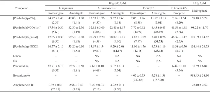
![Figure 3. Stability study of compound 4. Chromatogram profile of compound [Pd(dmba)(µ-NCO)] 2 (4) at 0, 12, 24, 48 and 72 hours in PBS buffer, pH 7, room temperature, C-18 column, particle size 5 µm, 4.6 × 250 mm, mobile phase: methanol:water (70:30,](https://thumb-eu.123doks.com/thumbv2/123dok_br/19000030.463447/6.892.97.436.246.492/figure-stability-compound-chromatogram-compound-temperature-particle-methanol.webp)
