Rev Bras Med Esporte vol.19 número1
Texto
Imagem
Documentos relacionados
The two points considered at the alternate sides, of the tangents through the diameter of the circle, and then the line joining these points divides the circle
At the first stage of the measurements results analysis, the gear wheel cast surface image was compared with the casting mould 3D-CAD model (fig.. Next, the measurements results
The structure of the remelting zone of the steel C90 steel be- fore conventional tempering consitute cells, dendritic cells, sur- rounded with the cementite, inside of
social assistance. The protection of jobs within some enterprises, cooperatives, forms of economical associations, constitute an efficient social policy, totally different from
The eight main recommendations are: all European countries that have not signed the Bern Convention should do so urgently; 1995 will be the Second European Year for
The probability of attending school four our group of interest in this region increased by 6.5 percentage points after the expansion of the Bolsa Família program in 2007 and
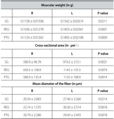
Measurement results of the masseter muscle thickness found in this study are compatible with ultrasound measurements obtained by the area of the transversal section parameter,
analyzed (body weight, volumes of the tibia-fibula and talocrural joints, muscle weight, muscle length and cross- sectional area of the muscle fibers and connective tissue)
