Universidade Nova de Lisboa
Instituto de Higiene e Medicina Tropical
Propagation and Molecular Characterization of
Chlamydia trachomatis strains isolated in Portugal
Ana Rita Serra Valente Caldeira
Dissertação para a obtenção do grau de Mestre em Ciências Biomédicas especialidade de Biologia Molecular em Medicina Tropical e Internacional
Universidade Nova de Lisboa
Instituto de Higiene e Medicina Tropical
Propagation and Molecular Characterization of Chlamydia trachomatis
strains isolated in Portugal
Autor: Ana Rita Serra Valente Caldeira
Título do Candidato: Licenciada em Anatomia Patológica, Citológica e Tanatológica
Orientadora: Doutora Maria José Borrego, Investigadora Auxiliar no Instituto Nacional
de Saúde Doutor Ricardo Jorge
Coorientadora: Doutora Rita Castro, Professora Auxiliar no Instituto de Higiene e
Medicina Tropical
Dissertação apresentada para cumprimento dos requisitos necessários à obtenção do grau de Mestre em Ciências Biomédicas
Agradecimentos
Gostaria de agradecer:
À minha orientadora Dra. Maria José Borrego, Responsável pelo Laboratório Nacional de Referência das Infeções Sexualmente Transmissíveis do INSA, por me ter aceite no seu laboratório, por todos os conhecimentos transmitidos, por toda a paciência, pela permanente disponibilidade, amizade e apreço.
À minha coorientadora Dra. Rita Castro, Professora Auxiliar do IHMT, por ter aceite ser minha orientadora pela disponibilidade e simpatia.
Ao Dr. Celso Cunha, Coordenador do Mestrado de Ciências Biomédicas do IHMT, pela simpatia e disponibilidade.
Ao Dr. Vítor Borges, Investigador na Unidade de Bioinformática do Departamento de Doenças Infeciosas do INSA, pelo importante contributo dado à realização da presente tese, pelos conhecimentos científicos, simpatia e amizade.
Ao Dr. João Paulo Gomes, Responsável da Unidade de Investigação do Departamento de Doenças Infeciosas do INSA.
À Dora Cordeira, Técnica no Laboratório do Infeções Sexualmente Transmissíveis, pelos conhecimentos transmitidos e amizade desde o primeiro dia.
Aos demais colegas, nomeadamente: Miguel Pinto pela amizade, companheirismo e entreajuda; e, Alexandra Nunes, pela simpatia e entreajuda.
Agradeço também aos meus pais por todo o suporte para a realização do mestrado e por me dar a possibilidade de concluir mais uma etapa.
Por fim, agradeço ao Hugo Barreto por tudo o suporte, compreensão, paciência, disponibilidade e entreajuda. O principal pilar para a conclusão deste trabalho.
Resumo
Chlamydia trachomatis é uma bactéria intracelular que infeta exclusivamente o Homem. Constitui o principal agente etiológico bacteriano de infeções sexualmente transmissíveis e, na ausência de tratamento, pode evoluir para complicações clínicas graves, como a infertilidade tubária. C. trachomatis foi classificada em 15 genótipos, os quais causam preferencialmente infeções oculares (A-C), infeções genitais (D-K) ou linfogranuloma venéreo (LGV) (L1-L3). A tipagem das estirpes de clamídia baseia-se na principal proteína da membrana externa (MOMP) codificada pelo gene ompA. O estudo do genoma de C. trachomatis tem permitido identificar diferenças genéticas entre estirpes; no entanto, os mecanismos de adaptação e virulência não estão ainda bem definidos mas, tal tem sido observado noutras bactérias, podendo decorrer de mecanismos de variação de fase.
No presente estudo foi realizada a tipagem de 278 amostras clínicas com base na sequência do gene ompA, através de técnicas de nested-PCR, de sequenciação pelo método de Sanger e de análise de similaridades das sequências obtidas relativamente a sequências de estirpes protótipo de C. trachomatis; tal processo possibilitou identificar variantes genotípicas previamente descritas assim como novas variantes, sugerindo a existência de um contínuo processo de adaptação na bactéria. Os genótipos mais frequentes foram o E, D e F, tal como descrito na maioria dos estudos em populações heterossexuais femininas; contudo um número apreciável (26) de estirpes do genótipo L2 foi igualmente detetado, em particular em amostras anorretais masculinas, o que está igualmente de acordo com o descrito em populações de homens que têm sexo com homens. Tal constatação revela a necessidade de um método de identificação expedita dos genótipos LGV, pelo que contribuímos para a implementação de uma metodologia mais rápida que a tradicional genotipagem-ompA. Pretendemos ainda contribuir para a avaliação da virulência das estirpes de C. trachomatis e para tal, realizamos um estudo preliminar de identificação de potenciais alvos genéticos sujeitos a variação de fase, com recurso à sequenciação de nova geração (NGS).
Este estudo constitui um contributo para a vigilância epidemiológica das infeções por C. trachomatis em Portugal, uma situação pouco conhecida, um contributo para a celeridade da identificação de casos de LGV e um contributo para a avaliação da virulência das estirpes de C. trachomatis, pela identificação de alterações em genes potencialmente relacionados com a variação de fase.
Palavras-chave: Chlamydia trachomatis; genotipagem ompA; variação de fase.
Abstract
Chlamydia trachomatis is an intracellular bacterium that exclusively infects humans. It is the main bacterial etiological agent of sexually transmitted infections and, in the absence of treatment, may lead to serious clinical complications such as tubal infertility. C. trachomatis was classified into 15 genotypes, which preferentially cause ocular infections (A-C), genital infections (D-K) or lymphogranuloma venereum (LGV) (L1-L3). Typing of chlamydial strains is based on the major outer membrane protein (MOMP), which is encoded by the ompA gene. The study of the genome of C. trachomatis allowed identifying genetic differences between strains, and although the mechanisms of adaptation and virulence still require proper definition, these mechanisms may derive from mechanisms of phase variation, as has been observed in other bacteria. In the present study, 278 clinical samples were typed based on their ompA gene sequence, determined by using a nested-PCR technique, Sanger method sequencing and gene sequence similarity analysis regarding the ompA-sequence of C. trachomatis prototype strains. This procedure allowed to identify genotypic variants previously described, as well as new variants, suggesting the existence of a continuous process of adaptation in the bacterium. The most frequent genotypes in our study were E, D and F; this finding is in accordance to the described in most of the studies held in female heterosexual populations. However, an appreciable number (26) of L2 strains was also detected, in particular in male anorectal samples, which is an in agreement with the described for men who have sex with men. The number of L2 strains reveals the need for an expeditious LGV-genotype identification method. Thus, we contributed to the implementation of a methodology that should provide faster identification of LGV strains in comparison to the traditional ompA-genotyping. We also intended to contribute to the evaluation of the virulence of C. trachomatis strains and for this, we participated in a preliminary study of identification of potential genetic targets subjected to phase variation, using a new generation sequencing (NGS) approach
This study contributes to the epidemiological surveillance of C. trachomatis infections in Portugal, a poorly known situation, contributes to a faster identification of LGV cases, and contributes to the evaluation of virulence of C. trachomatis strains by identifying changes in genes potentially related to phase variation.
Keywords: Chlamydia trachomatis; ompA-genotyping; phase variation.
Table of contents
Agradecimentos ... i Resumo ... ii Abstract ... iii Table of contents ... iv Figure index ... vi
Table index... vii
List of abbreviations ... viii
I. Introduction ... 1
1. Sexually Transmitted Infections ... 1
2. Chlamydia ... 1
2.1. The genus Chlamydia ... 1
2.2 Chlamydia trachomatis ... 2
2.2.1 Biology: The life cycle of C. trachomatis ... 2
2.2.2 Epidemiology and Human diseases ... 6
2.2.3 Genomics features ... 11
3. Objectives ... 13
II. Material and Methods ... 15
1. Cell culture and inoculation of C. trachomatis ... 15
2. Typing C. trachomatis and bioinformatic analysis ... 19
3. Strategies for quick subtyping LGV strains ... 23
3.1 Subtyping LGV: Using sets of primers discriminatory of L1, L2 and L3 ... 23
3.2 Subtyping LGV: real time-PCR for C. trachomatis ompA and pmpH ... 24
4. Homopolymeric tracts counts and bioinformatics analyses ... 26
4.1 Study design: selection of homopolymeric tracts and C. trachomatis positive DNA samples ... 26
4.2 Amplicon-based NGS and bioinformatics analyses ... 27
III. Results and Discussion ... 31
1. Cell culture and C. trachomatis typing ... 31
1.1 Cell culture, the old gold standard ... 31
1.2 Characterization of C. trachomatis clinical strains ... 33
2. Strategies for quick subtyping LGV strains ... 44
3. Phase variation in C. trachomatis – Preliminary study ... 47
3.2 Allelic variation among strains ... 51 IV. Conclusion ... 59 V. References ... 61 VI. Annexes ... 75
Figure index
Figure 1 - Schematic representation of the development cycle of C. trachomatis 3
Figure 2 – Schematic representation of C. trachomatis outer membrane 5
Figure 3 – Phylogenetic tree of C. trachomatis 7
Figure 4 – Schematic representation of inoculation in a 24 well plate 18
Figure 5 – Schematic representation of homopolymeric traits counts 29
Figure 6 – Results from propagating C. trachomatis strains in cell culture 31
Figure 7 – Rate of C. trachomatis ompA-genotyped strains 33
Figure 8 – C. trachomatis strains by gender 34
Figure 9 – C. trachomatis strains by age 35
Figure 10 – Frequency of biological samples positive for C. trachomatis 36 Figure 11 – Frequency of the different ompA-genotypes among the studied C.
trachomatis strains
37
Figure 12 – Number of ompA variants among detected C. trachomatis genotypes 39 Figure 13 – C. trachomatis ompA-genotype distribution according to anatomical site of infection
42
Figure 14 – C. trachomatis ompA-genotype frequency per age group 43
Figure 15 – Conventional PCR approach for detecting L1, L2 and L3 45
Figure 16 – Results for real time PCR assay 46
Figure 17 – Comparison between WGS and amplicon-based NGS techniques 49
Figure 18 – Comparison between 35 and 45 cycles PCR 50
Figure 19 – Profile of the CT166 homopolymeric tract in several C. trachomatis strains
52
Figure 20 – Profile of the homopolymeric tract affecting the regulatory region of CT533 for several strains
53
Figure 21 – Analysis of tropism-specific homopolymeric lengths 55
Figure 22 – Intra-strain homopolymeric tracts variability 57
Table index
Table 1 – Formula for complete medium preparation 16
Table 2 – Formula for enriched medium 17
Table 3 – Nested-PCR mix 20
Table 4 – C. trachomatis ompA Nested-PCR amplification profile 20
Table 5 – Sequencing mix 21
Table 6 – Sequencing profile 22
Table 7 – Master mix for LGV subtyping real time – PCR 24
Table 8 – Amplification profile for LGV subtyping real time – PCR 25
Table 9 – PCR mix for homopolymeric targets 27
Table 10– Amplification profile for homopolymeric regions 28
Table 11 - Nucleotide sequence variation within ompA in relation with the respective prototype strain
40
List of abbreviations
µg – Microgram µl - Microliter µm – Micrometer 2SP – 2 sucrose phosphateATCC – American Type Culture Collection bp – Base pair
CD – Conserved domain CO2 – Carbon dioxide
COMC – Chlamydia Outer Membrane Complex ddNTPs – Dideoxynucleotide
DFA – Direct Fluorescence Antibody DMSO – Dimethyl Sulfoxide
DNA – Deoxyribonucleic acid dNTP – Deoxynucleotide
DPBS – Dulbecco’s Phosphate Buffer Saline EB – Elementary Body
EDTA – Ethylenediaminetetraacid EIA – Enzyme Immunoassays
ELISA – Enzyme-Linked Immunosorbent Assay FBS – Fetal Bovine Serum
HBV – Hepatitis B Virus HCV – Hepatitis C Virus
HIV – Human Immunodeficiency Virus HPV – Human Papilloma Virus
HSV – Herpes Simplex Virus IFN-γ – Interferon-γ
IgG – Immunoglobulin G IgM – Immunoglobulin M
INSA – National Institute of Health (from the Portuguese Instituto Nacional de Saúde) kb – Kilobase (= 1 000 bp)
LGV – Lymphogranuloma venereum LPS – Lipopolysaccharide
MACPF – Membrane Attack Complex/ Perforin Mb – Megabase (= 1 000 000 bp)
MEM – Minimum Essential Medium mg – Milligrams
MgCl2 – Magnesium chloride ml – Milliliter
mM – Millimolar
MOMP – Major Outer Membrane Protein MSM – Men who have Sex with Men NAATs – Nucleic Acid Amplification Tests NGS – New Generation Sequencing
ºC – Degrees Celsius ORF – Open Reading Frame PCR – Polymerase Chain Reaction PID – Pelvic Inflammatory Disease PLD – Phospholipase D
pmol – Picomolar
PMP – Polymorphic Membrane Proteins POC – Point-Of-Care
RB – Reticulate Body
RFLP – Restriction Fragment Length Polymorphism STI – Sexually Transmitted Diseases
TAE – Tris-Acetate-EDTA
TARP – Translocated Actin Recruiting Phosphoprotein UTI – Unit of Technology and Information
v - Volume V – Volts
VD – Variable Domain w – Weight
WGS – Whole Genome Sequencing WHO – World Health Organization
I. Introduction
1. Sexually Transmitted Infections
Sexually transmitted infections (STI) can be caused by different microorganisms, such as: a) bacteria (like Chlamydia trachomatis, Neisseria gonorrhoeae, Treponema
pallidum, Haemophilus ducreyi, Mycoplasma genitalium and Ureaplasma urealyticum),
b) virus (Human Immunodeficiency Virus [HIV], Herpes Simplex Virus [HSV] type 1 and 2, Human Papilloma Virus [HPV], Hepatitis B Virus [HBV] and Hepatitis C Virus [HCV]), c) parasites, either protozoa (Trichomonas vaginalis) or metazoa (Phthirus pubis and Sarcoptes scabiei) (1).
In 2016, WHO (2), estimated in 357 million the number of new cases of the four curable STI, being C. trachomatis the second most frequent with 131 million after T.
vaginallis, next N. gonorrhoeae and T. pallidum. There are several factors associated to
mortality and morbidity of STI, such as fetal and neonatal death, cervical cancer, infertility and higher risk of HIV acquisition.
2. Chlamydia
2.1. The genus Chlamydia
C. trachomatis is an obligate intracellular bacterium and one of the eleven species
within the family Chlamideaceae, order Chlamydiales, phylum Chlamydiae and genus
Chlamydia. Beyond C. trachomatis, the Chlamydia genus also includes C. pneumoniae, C. pecorum, C. felis, C. psittaci, C. abortus, C. caviae, C. avium, C. gallinacea, C. suis
Introduction
human) and tissue tropism (3–5). The most relevant chlamydial species infecting humans are C. trachomatis, that only infects humans and is associated with ocular and anogenital infections, the later, sexually transmitted (6) and C. pneumoniae (7), related to a wide range of diseases including respiratory illnesses, arthritis (8) and some chronic pathologies involving the cardiovascular (9), or the central nervous systems (10). C.
psittaci can also infect humans, who usually are accidental hosts, among who some
genotypes are more prone for causing disease, called psittacosis, a pathology that affects the respiratory system (11).
2.2 Chlamydia trachomatis
2.2.1 Biology: The life cycle of C. trachomatis
The family Chlamideaceae is constitute by obligate intracellular bacteria that evolved by acquiring the ability to adapt and colonize its host. Therefore, C. trachomatis presents a unique and complex biphasic development cycle along which it alternates between two morphologically distinct forms: the elementary body (EB) capable of extracellular survival for a short period, infectious and metabolically inactive and the intracellular reticulate body (RB), non-infectious and metabolically active (12).
The EB’s are small gram-negative cocci (~ 0,3µm) presenting a polarized architecture while RB’s are larger (~ 1µm) (13).
The life cycle comprehends four major events: it initiate with binding and invasion of the host cell (14), that is followed by differentiation of EB into RB that undergo several cell division cycles before re-differentiate into EB that are liberated to infect new cells by the end of the cycle upon eclosion of the inclusion/ host cell (13). Until 2016, chlamydial cell division was believed to occur through binary fission, despite the absence of FtsZ protein in these bacteria; however, this year, it has been shown that Chlamydia divides by a polarized cell division process through which major outer membrane protein (MOMP) first restricts to one pole of the bacterial cell, where the chlamydial cell division
Introduction
machinery also accumulates, and it is from there that the bacterial cell daughter emerges, a so called budding process (15). A complete development cycle takes 48 to 72 hours and can be interrupted by environmental stress factors, like nutrient deprivation or exposure to antibiotics (12,16). (Figure 1).
Figure 1 - Schematic representation of the development cycle of C. trachomatis. [Available in (17)]
C. trachomatis, as any gram-negative bacteria, is provided of a double membrane;
the outer membrane exhibits a type III secretion system (T3SS), the chlamydia outer membrane complex (COMC), polymorphic membrane proteins (Pmp), porins, lipids and polysaccharides (18,19) (Figure 2).
Introduction
The T3SS has been identified in C. trachomatis in 1997 (20), firstly described as rosette structure present in the outer membrane. A set of T3SS is located in one of the EB hemisphere (14) and composes a macromolecular structure called “injectisomes” constitute by several proteins (21). The T3SS is a widely used secretion system among gram-negative bacteria and is essential for virulence of human pathogens (22). For chlamydial species it is essential for bacterial protein delivery across the eukaryotic cell membrane, contributing to chlamydial pathogenesis (21,23).
The COMC is constitute by the MOMP and by two other major proteins, the OmcA and the OmcB (24–26). MOMP represents 60% of membrane dry weight (25), and has been associated to pathogenesis given its variability and antigenic properties (27). MOMP also acts as a cytoadhesin (28), interacting with the host, and has been characterized also as a porin (29). Beyond MOMP another porin has been identified in the outer membrane, the PorB, a conserved porin that seems to function exclusively for transport of nutrients for chlamydial survival (30).
Also present in the outer membrane are the pmp, which were characterized as autotransporters and adhesins, being coded by a highly heterogenic gene family presenting a different number of pmp genes amongst the different chlamydial species (31,32).
Well represented in the surface of C. trachomatis is the lipopolysaccharide (LPS) being the main lipid present in the outer membrane (33). LPS functions as a barrier to permeability (19) enabling cell viability, and beyond that, it has been related to interactions with host cells, and thus associated with infectivity (34).
In 2013 (35) it was first demonstrate the presence of peptidoglycan between the double membrane of the Chlamydia cell wall; its functions were related to cellular integrity and it should also play a role during cell division. The goal of detecting peptidoglycan in the chlamydial cell wall was not an easy task; however, recent labeling and proteomics methodologies made that possible (35,36).
Introduction
Figure 2 – Schematic representation of C. trachomatis outer membrane [Adapted from (37)]
Since EBs start interact with the host cell, bacterial proteins entry in the cytoplasm of the host cell mediated by the T3SS (22). Next, several effectors are recruited to entry location; one of them is the translocated actin recruiting phosphoprotein (TARP) that will be involved in the inclusion formation. The chlamydial inclusion is a cytoplasmic vacuole surrounded by host cell membrane, where specific proteins called Inc, are identifiable. Incs interact with the host cell during infection, becoming important factors for the maintenance and growth of C. trachomatis (38,39).
Gene transcription starts when EB differentiate into RB. Genes have been categorized according to three temporal transcription classes (in relation to the chlamydial life cycle) in: early, midcycle and late (13,40). Early genes are related to inclusion remodeling and host-pathogen interaction. Midcycle genes, which are the largest group, are associated with nutrient acquisition, growth and replication. Finally, late genes start to be transcribed after the second differentiation (RB to EB) and are related to DNA condensation and the outer membrane complex formation (13,18). To note, the second differentiation occurs in an asynchronous manner, which means that the differentiation of RBs into EBs do not occur simultaneously in all particles (12,13).
There are two critical phases during chlamydial infections, the entry in the host cell to perform the infections and the exit of the host cell to keep the infection. The exit from the host cell usually happens by a lytic mechanism, through which a new progeny
Introduction
of EBs is released (13), after inclusion and host cell membranes rupture. The lytic mechanism has been associated to LGV infections and it is thought to be regulated by the plasmid (41). A non-lytic exit is characterized by the extrusion of the whole inclusion and involves host cell actin and myosin allowing chlamydial survival inside macrophages and infection at further anatomic sites (42–44).
2.2.2 Epidemiology and Human diseases
C. trachomatis is the most common bacterial STI, with an estimated incidence of
131 million new cases per year, affecting in particular individuals aged 15 to 49 years (2). Moreover, C. trachomatis infections potentiate HIV transmission due to epithelial damage (45).
Three biovars can be identified in C. trachomatis, related to tissue tropism. These biovars are named trachoma, genital tract and lymphogranuloma venereum (LGV) (13). Each biovar includes several bacterial types that were first identified through the use of monoclonal antibodies developed against C. trachomatis MOMP (46). The nucleotide differences of the MOMP encoding gene, ompA, reflect the differences found in the amino acid sequence and for this reason the 15 genotypes are defined reflecting the 15 serovars (A, B, Ba, C, D, E, F, G, H, I, J, K, L1, L2 and L3) (47). The majority of the amino acid sequence of MOMP is conserved, except within four variable domains (VD). These VD are responsible for the major antigenic differences among the 15 identified ompA-genotypes (48,49) (Figure 3).
Introduction
Figure 3 – Phylogenetic tree of C. trachomatis. The neighbor joining phylogenetic tree (50) is based on
the nucleotide differences among ompA sequences, computed using the Kimura 2-parameter method (51). Bootstraps values (1000 replicates) are shown next to the branch nodes. Evolutionary analyses were conducted in MEGA7 (52).
Based on ompA gene diversity, three phylogenetic groups can be defined: the B-complex (includes ompA-genotypes B, Ba, D, E, L1 and L2), the C-B-complex (includes
genotypes A, C, H, I, J, K and L3) and the intermediate complex (includes
ompA-genotypes F and G) (48,53). These three phylogenetic do not reflect the three biovars, indicating that chlamydial ompA genotyping does not reflect the whole genome in terms of pathobiology (53).
In terms of associated pathology, ompA-genotypes A to C cause trachoma while
ompA-genotypes D to K constitute the major bacterial cause of STI and ompA-genotypes
Introduction
Trachoma affects mainly the conjunctival epithelia, being the most common infectious cause of blindness, yet preventable, related to poverty and poor hygiene conditions (54). Trachoma affects 40 million people, being endemic in 50 countries and includes the list of 17 neglected tropical diseases (55).
C. trachomatis D-K ompA-genotypes are the major cause of bacterial STI
worldwide (6). C. trachomatis genital infection is asymptomatic in about 70% of women and 50% of men (56). When existing, the clinical manifestations in males include urethritis, epididymitis, epididymo-orchitis, prostatitis and infertility (57,58). In women
C. trachomatis genital infections, besides the clinical manifestations as cervicitis and
urethritis, may also evolve to complications such as salpingitis, endometritis, ectopic pregnancy, pelvic inflammatory disease (PID) and tubal infertility (6,59). Associated to the genital infections, C. trachomatis can also be transmitted to newborns during delivery. These neonatal infections appear as conjunctival or nasopharyngeal infection that can progress to pneumonia (6,60).
LGV ompA-genotypes beyond the capacity of infecting epithelial cells (like other
ompA-genotypes), can also infect mononuclear phagocytes, an invasive characteristic, as
migration through immune cells allow LGV genotypes to infect regional lymph nodes or lymphatic tissue of the intestine (61). The clinical manifestation of LGV are diverse; the classic clinical picture included the appearance of a genital ulcer, followed by an inguinal stage during which some patients presented the “groove sign” that was characterized by the swallow of the inguinal nodes, finally a third stage often called “anogenitorectal syndrome”, described as a chronic inflammatory lesion that leads to lymphatic obstruction (61–64). Endemic in several tropical countries such in some parts of Africa, Asia, South America and the Caribbean, LGV has been spreading since 2004 to industrialized countries (61,65). Since then, several outbreaks have been reported (66), such as in France (67), Great Britain (68), The Netherlands (69), Canada (70), Australia (63) and Portugal (71,72). L2 became the most frequent ompA-genotype associated to the recent LGV epidemics (65,66), during which new variants have been discovered, namely one L2b subtype associated to the Sweden outbreak (73). Curiously, this new Swedish L2b variant presented a deletion of 377 base pairs within the plasmid target region of the most popular molecular diagnostic method in Sweden, as discovered through gene sequencing (73); consequently, false negative cases escaped from treatment and were
Introduction
allowed to propagate the infection. The diagnostic of C. trachomatis through acid nucleic amplification had become the gold standard in terms of diagnostic in the 2000s, and the omnipresent 4 – 8 copies C. trachomatis plasmid appeared as an attractive target (74,75). However, as a consequence of the Swedish outbreak, commercial nucleic acid amplification tests implemented multi-gene target systems, for avoiding false negatives caused by mutation, insertion or deletion phenomena that can affect the performance of single gene target based tests. It also contributed to show the importance of epidemiological data and external quality assessment programs (74).
Worldwide several molecular epidemiology studies have been done, evidencing that the most common ompA-genotype among heterosexuals are E, D and F, while G, D, J and L2 are most frequent among MSM (76).
In terms of public health, the major problem related to C. trachomatis STI is the high number of asymptomatic cases that remain untreated and continue propagating, resulting in millions of new infections per year. When left untreated, C. trachomatis infections may progress to severe reproductive tract complications and sequelae, like PID, ectopic pregnancy and tubal infertility (6,77). At present, the recommendations for treating C. trachomatis are azithromycin and doxycycline (77,78). Azithromycin is recommended for uncomplicated genital chlamydial infections in a single dose of 1 gram, while 100 milligram of doxycycline twice a day during 7 or 21 days is prescribed for anorectal chlamydial infections and LGV cases, respectively (78).
Due to the high number of asymptomatic cases, the guidelines for the management of chlamydial infections include the screening for STI in high risk populations (such as sexworkers), partner notification and management of pregnant woman in order to prevent adverse clinical outcomes for the newborn (1,79,80).
The serological techniques were among the earliest diagnostic methods for C.
trachomatis, determining the antibody response to chlamydial antigens (81,82).
Microimmunofluorescence is the recommended serological technique, although several enzyme-linked immunosorbent assay (ELISA) tests (6) are commercially available for detecting specific IgM, IgG an IgA. However cross reactivity to different Chlamydia species is common and antibody responses can be scarce; thus, serological testing should
Introduction
be restricted to diagnose newborn infections, and to be used as a marker in case of LGV suspicion or tubal infertility (81–83).
To achieve a better management of chlamydial infections, detection techniques have greatly evolved since the isolation in cell culture, being the first isolation performed in 1957 (84). Ever since, the isolation of chlamydial strains through cell culture has been considered as a gold standard; however, time consumption and stringent transport (Sucrose Phosphate buffer – 2SP) and storage conditions (4ºC up to 24 hours, -80ºC for longer periods), together with the delay to obtain a result, have limited its applicability (82,85). For these reasons, chlamydial culture is nowadays restricted to reference laboratories and research (82).
Antigen detection based diagnostic techniques have also been developed, namely enzyme immunoassays (EIA) and the direct fluorescence antibody (DFA) (6). The EIA techniques were also implemented for use as point-of-care (POC) tests to allow diagnosis during a clinical visit; however, all EIA tests reveal low sensitivity and poor specificity, and for this reason their use is no longer recommended (82). DFA performance relies on monoclonal antibody quality, on fluorescence microscopy availability and most of all, on personal skills for recognizing EBs, a process that is time consuming; furthermore, antigen detection techniques are restricted to exudates (82).
The major advance of chlamydial infections diagnosis was the introduction of nucleic acid amplification tests (NAATs), which are nowadays the recommended due to their superior performance characteristics in terms of specificity and sensitivity, close to 100% (81,82). Moreover, these tests allow the use of first void urine, a non-invasive biological sample that is a well-accepted and recommended sample for male urethra evaluation. Several NAATs methodologies have been developed, but real-time polymerase chain reaction (PCR), is the most used. In addition, recent NAATs allow the simultaneous amplification and detection of various STI agents, in particular the two most common bacterial STI, C. trachomatis and N. gonorrhoeae (82). Some NAATs include several targets for each microorganism to avoid false negatives caused by target gene changing’s (as explained above); as an example, COBAS 4800® has two targets for C.
trachomatis, one in the chromosome and the other in plasmid (86).
Introduction
2.2.3 Genomics features
The genomes of intracellular parasites are relatively small when compared to genomes of free-living microorganisms, due to the adaptation to their lifestyle (4,87). In fact, intracellular bacteria suffered a genome reduction with genetic material loss while becoming metabolic parasites, through which the bacteria benefits from living within the host cell cytoplasm (87,88).
Accordingly, the Chlamydiaceae family presents a small genome (87), specifically, C. trachomatis has a single circular chromosome with approximately 1Mb, where around 935 genes were identified, 887 of them are coding genes (89–91) and a 7,5 kb plasmid (only a few plasmidless strains were described), comprehending 8 coding genes (89,90,92). Excluding the plasmid, there are no evidence of other extrachromosomal or mobile genetic elements (90,91).
Genomic analysis of C. trachomatis shows a highly conserved genome with the exception of a 50 kb region of high variability, named plasticity zone, dissimilar among chlamydial strains (90). This hypervariable region contains some genes related to pathogenesis and to interactions with the host. Some of the genes present in the plasticity zone are the tryptophan synthase (trp) operon, the cytotoxin, the membrane attack complex/ perforin (MACPF) and phospholipase D (PLD) (91). The trp operon is considered a virulence factor, as it provides the ability to synthesize tryptophan in the presence of interferon (IFN)-γ from the host immune response (93). However this trp only exists in genital ompA-genotypes, which are the only having a functional tryptophan synthase. The chlamydial cytotoxin, a highly polymorphic gene, appears differently among strains, but only genital ompA-genotypes encode both functional domains of the cytotoxin ORF (CT166), the glycosyltransferase and UDP-glucose binding domains. Ocular ompA-genotypes encode only one domain, the UDP-glucose binding domain, and LGV ompA-genotypes lack the CT166 ORF. The chlamydial cytotoxin has a significant homology with the clostridial cytotoxin, and leads to a cytopathic effect since it is involved in disabling the actin filaments of the host cell cytoskeleton (94–96). Other genes, like the MACPF and the PDL that were also related to pathogenesis, are present in all ompA-genotypes; their functions have been further related to acquisition of
Introduction
metabolites from the host, a function that is essential for the chlamydial survival and successful development cycle (97).
Outside the plasticity zone, other genes have been related to virulence and pathogenicity such as Pmp genes, TARP genes, Inc genes and the ompA gene (98). Genes encoding the Pmp family, unique of the order Chlamydiales, constituted by 9 members (Pmp A to I) localized in the outer membrane, involve 3,2% of the genome coding capacity. Pmps are also described as autotransporters, pointed as virulence factors (because they promote antigenic polymorphisms), adhesion and differential tissue tropism (31). The type III secretion system effector TARP is involved in actin remodeling during invasion; it exhibits a high degree of variability and it is known to be involved in niche adaptation (99). The analysis of the TARP of different ompA-genotypes showed variation in the number of tyrosine-rich repeat regions and in the actin binding domain. In respect to tyrosine-rich repeat regions the ocular strains have the lowest number, while LGV strains present the highest; related to acting binding domains, the ocular strains have a higher number than LGV strains; genital strains are in the middle for both (100). Inc’s are also effectors of the type III secretion system family and are proteins inserted in the inclusion membrane and thus exposed to the host cytosol. Inc proteins are species-specific, and very conserved among ompA-genotypes, although evidencing some divergence between strains with different tissue tropism (39). MOMP, encoded by the
ompA gene, is a porin implicated in attachment (functioning as an adhesin), in
pathogenesis, due to its variability and antigenic properties (includes genus, species, and type epitopes and induces humoral and cellular immune response in the host, justifying being considered a potential candidate for the development of a vaccine (27)) and, recently, it was further implicated in cell division (15).
Besides the knowledge of genetic variation between strains, phase variation mechanisms have been shown to play a role in adaptation and pathogenesis in C.
pneumoniae (101,102) and it could be expected that the same functions occur in C. trachomatis (32,103–105). The phase variation mechanism relies on the ability of bacteria
to quickly adapt in response to a stimuli, being usually associated with a reversible switching between an ON and OFF state of specific proteins, such as: surface-exposed proteins or proteins involved in the biosynthesis/ expression of the bacterial capsule and the LPS (a major component of gram-negative bacteria surface); the switching occurs due
Introduction
to changes in DNA that lead to different phenotypes (106). The phase variation mechanism in many cases occur due to expansion and contraction of homopolymeric tracts, described as repetitive regions of a DNA single base [poly(A), poly (T), poly (G) and poly (C)] (107,108).
3. Objectives
Propagation of C. trachomatis clinical strains in cell culture
ompA-genotyping of C. trachomatis clinical strains
Implementation of molecular biology techniques to specifically and rapidly detect L1, L2 and L3 ompA-genotypes
II. Material and Methods
1. Cell culture and inoculation of C. trachomatis
During our work we used the HeLa 299 immortal cell line (American Type Culture Collection® [ATCC] CCL – 2.1TM), which derives from cervical cancer cells,
and is commonly used for the propagation of C. trachomatis.
The specimens inoculated in cell culture were cervical, urethral and anorectal exudates kept in 2SP buffer at -80ºC upon arrival to the laboratory. These specimens had tested positive for C. trachomatis in the host laboratory during its routine diagnosis service, through COBAS 4800® (Roche). INSA provides routine C. trachomatis diagnosis to attendees of STI, family planning, general practice, urology and gynecology clinics, among others, and also participates in C. trachomatis screening studies in specific populations (i.e. sex workers). C. trachomatis prototype strains were also inoculated,
namely G-UW57 (ATCC® VR-878TM), H-UW43 (ATCC® VR-879DTM), I-UW12
(ATCC® VR-880TM), F-IcCal3 (ATCC® VR-346TM), and L3-404 (ATCC® VR-903TM).
For culturing HeLa 299 cells we used the Minimum Essential Medium (MEM) (41090 Gibco™) supplemented with fetal bovine serum (FBS) (16000 Gibco™), antibiotic and antifungal agents, hereby designated by “complete medium” (Formula in Table 1). In a first step and using a water bath at 37ºC, we defrosted an aliquot of cells stored at -195,79ºC in liquid nitrogen. After defrosting, cells were put in a T25 flask in which 8 ml of complete medium were added together with an additionally 10% of FBS. Cells were left to incubate at 37ºC, 5% CO2 for 24 hours to obtain a cell monolayer that
could be observed with an inverted microscope (objective x20). The maintenance of the cell culture is performed by passaging, a process that involves two times washing with 10 ml of Dulbecco’s Phosphate-Buffered Saline (DPBS) (14190 Gibco™), for medium and dead cells removal, cell detaching by adding 4 ml of 1x trypsin solution (15090 Gibco™), suspension of cells with fresh complete medium (10 ml) and transfer of a variable volume
Material and Methods
(depending on time until next passage and cell culture surface area ) to a new tissue culture flask containing fresh complete media.
Table 1 – Formula for complete medium preparation
Supplements (in %) to complete MEM (41090 Gibco™)
0,3% Gentamicin (10 mg/ml)*
0,3% Amphotericin B (250 µg/ml)**
11% Fetal Bovine Serum (FBS)***
*15710 **15290 ***16000 Gibco™
We also prepared HeLa 299 cell stock, by adding 1 ml of dimethyl sulfoxide (DMSO, Amresco®) to a 10 ml cell-suspension (10% v/v) from a T175-flask confluent monolayer, distributing by 10 cryotubes (1 ml/cryotube) and allowing a slow drop of the temperature until -80ºC by using a “Cryo 1ºC freezing container” (Nalgene™). Frozen aliquots of HeLa 229 cells were then stored in a liquid nitrogen container (or at -80ºC for a shorter usage).
For inoculation of C. trachomatis positive specimens or prototype strains, we transferred HeLa 299 cells into a 24 well plate at a final concentration of 2,3x10⁵
cells/ml/well (see Annex 1) and let to incubate for 24 hours at 37ºC, 5% CO2. For ATCC
strains we removed 0,8 ml of medium from each well and added 0,2 ml of prototype strain suspension. For clinical samples, we removed 0,5 ml of medium from each one of four wells and added 0,2 ml of the sample to each one of four wells. The inoculated plates were centrifuged 1 hour at 32ºC, 2126 g in order to promote adhesion of C. trachomatis to the cell monolayer and let to incubate for another hour at 37ºC, 5% CO2. The
supernatant was replaced by “enriched medium” (Formula in Table 2), followed by incubation at 37ºC, 5% CO2 for 48 hours.
Material and Methods
Table 2 – Formula for enriched medium
Supplements (in %) to prepare enriched medium
1% Vitamins (100x)*
1% Non-essential aminoacids (100x)**
5,4% Glucose (10x)ª
0,5% Cycloheximide (100 µg/ml)ᵇ
Until perform the total volume Complete medium
* 11120 ** 11140 Gibco™ ª INSA ᵇ Sigma-Aldrich
C. trachomatis characteristic inclusions can be observed under an inverted
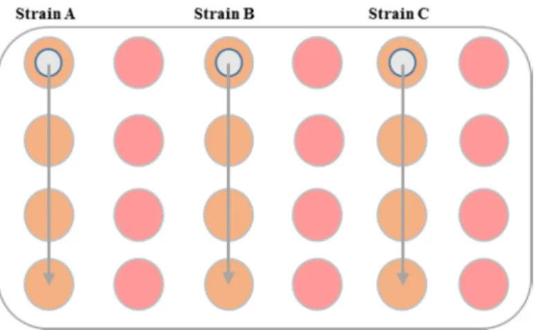
microscope (objectives x10 and x40) after 48 hours of incubation. However, inclusions are better observed and counted by fluorescence microscopy (objective x20 and x40). For this, when preparing the cell culture in the 24 well – plate, a 12 mm diameter glass coverslip was placed at the bottom of one of the selected four wells for each specimen (Figure 4). After the 48 hours incubation, media from coverslip containing wells is withdrawn, 1 ml of methanol is added for fixing and coverslips are detached for further staining with a monoclonal antibody specific for C. trachomatis (Pathfinder Chlamydia
trachomatis Direct Specimen Monoclonal Antibody kit) (Bio-Rad®), according to the
manufacturer’s instructions.
Material and Methods
Figure 4 – Schematic representation of inoculation in a 24 well plate. Schematic representation of a 24
well-plate, where in pink are represented non-inoculated wells and in orange are represent inoculated wells; in the latter, glass coverslips have been prior added to the first well of each column.
For negative and low efficiency inoculations (1 – 4 inclusions per microscope field) we scraped the cell monolayer, collected the whole content of the well, performed cell disruption through sonication (Bioblock scientific, vibracell®) for 7 minutes, 80% Cycle Actif, power 9 and briefly centrifuged at room temperature for 7 minutes at 99 g, to eliminate cell debris, prior repeating inoculation procedure (keeping a 500 µl aliquot + 500 µl 2SP at -80ºC as a stock). When a high inoculation efficiency (≥ 5 inclusions per microscope field) is observed, cultures were treated as described above but they did not require further inoculation; thus, supernatant was divided into two cryotubes and added with an equal volume of 2SP, one to be kept at -80ºC and another to be stored in liquid nitrogen.
We also prepared stocks of chlamydial prototype strains. The preparation of these stocks involved the inoculation of aliquots of prototype strains (stored at -80ºC or liquid nitrogen) on HeLa 229 monolayers prepared in 24 well plates. Chlamydial cultures treated as described above and when culture efficiency reached ≥ 5 inclusions per microscope filed, supernatants were inoculated into HeLa 229 monolayer prepared in T25 flasks. Prototype chlamydial culture was controlled by inverted microscopy (objective x10 and x40), and when culture efficiency again reached ≥ 5 inclusions per microscope field and just prior inclusion rupture by the end of the chlamydial life cycle, cell monolayer was detached with glass beads and 4 ml of complete medium. This chlamydial suspension was sonicated, further centrifuged (as described above), and distributed by cryotubes to which
Material and Methods
an equal volume of 2SP buffer is added. We performed a slow mixing of the cryotubes prior to freezing in “Cryo 1 ºC freezing container” (Nalgene™) and further storing at -80ºC or liquid nitrogen.
2. Typing C. trachomatis and bioinformatic analysis
C. trachomatis typing was performed over samples that: a) arrived at INSA for
diagnosis and revealed positive; b) in specimens that were determined positive in other laboratories and sent to INSA for national collection purposes. The specimens were exudates (cervical, urethral, anorectal and conjunctival) and urines. For exudates, 200µl of the original sample were taken for DNA extraction, while for urines, DNA was extracted from 1 ml. DNAs were kept at -20ºC until further investigation.
C. trachomatis DNA was extracted using the NucliSENS®easyMag® of
bioMérieux, according to manufacturer’s instructions and using the specific EasyMag equipment.
A nested-PCR technique was used for ompA typing, through which two sequential PCR amplifications are performed in order to increase sensitivity. Primers used for typing are listed in Annex 2. In the first amplification (outer amplification) we used the primers NLO and NRO, and for the second amplification (inner amplification) we used the primers PCTM3 and SERO2A (109). We prepared PCR mixes (Table 3) for a total volume of 15 µl that were added with 10 µl of each DNA sample for the first PCR; for the second PCR, we prepared a total mix volume of 23 µl that were added with 2 µl of PCR product from the first round of amplification. The two pairs of primers are specific of the C. trachomatis ompA gene and promote an amplicon of about 1000 bp.
Material and Methods
Table 3 – Nested-PCR mix
Volume per sample Reagent Name
5 µl dNTPs (1 mM)*
2,5 µl Buffer (10x)ª
1,4 µl Magnesium chloride (MgCl2) (50 mM)ª
0,37 µl Bio-x-act short polymerase (4 U/ml)ª
2 µl Primers pair (25 pmol/µl)ᵇ
3, 73 µl / 11, 73 µl Water DNase, RNase, Protease-free ͨ
* Applied Biosystems ª Bioline ᵇ Invitrogen ͨ 5Prime
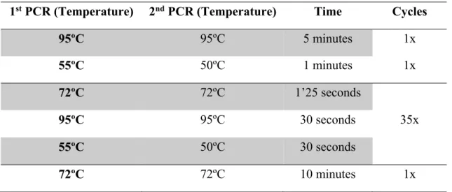
The amplification profile of the nested-PCR reaction comprehended a first step of denaturation and annealing, followed by 35 cycles of extension, denaturing and annealing, and a final extension step, according to the conditions described in Table 4.
Table 4 – C. trachomatis ompA Nested-PCR amplification profile
1st PCR (Temperature) 2nd PCR (Temperature) Time Cycles
95ºC 95ºC 5 minutes 1x 55ºC 50ºC 1 minutes 1x 72ºC 72ºC 1’25 seconds 35x 95ºC 95ºC 30 seconds 55ºC 50ºC 30 seconds 72ºC 72ºC 10 minutes 1x
After the nested amplification, we performed amplicon detection through gel electrophoresis in TAE buffer [1% (w/v) agarose in TAE buffer, SYBR® safe DNA gel stain (10.000x in DMSO; Invitrogen)], for 1 hour at 90 V.
Material and Methods
According to the ompA nested-PCR results, positive samples were further sequenced. When negatives were from urine samples no further action was done. If the sample was an exudate nested-PCR was repeated and if they remained negative they were excluded.
For sequencing, PCR products were purified using ExoSAP® (Affymetrix) according to the manufacturer’s instructions. Briefly, they were subjected to an enzymatic PCR cleanup that consists in two cycles of 15 minutes each (one at 37ºC followed by another at 80ºC) the first with the aim of activating the enzymes responsible for removing contaminants (i.e. excess of primers and dNTPs or single strand DNA) and the second for inactivating enzymes used in the previous step.
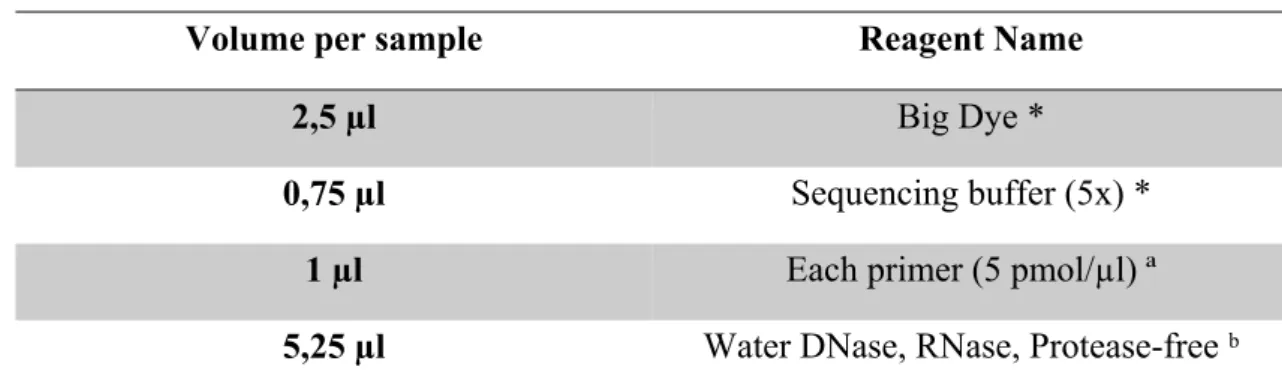
After purification, the reaction mix for the sequencing reaction was performed as described in Table 5, using the set of primers described in Annex 3, for a final volume of 9,5 µl, to which 0,5 µl of purified PCR product was added. We used Big Dye technology that is a fluorescent molecule attached to the ddNTPs allowing each one to carry a different color of dye (110).
Table 5 – Sequencing mix
Volume per sample Reagent Name
2,5 µl Big Dye *
0,75 µl Sequencing buffer (5x) *
1 µl Each primer (5 pmol/µl) ª
5,25 µl Water DNase, RNase, Protease-free ᵇ
* Applied Biosystems ª Invitrogen ᵇ 5Prime
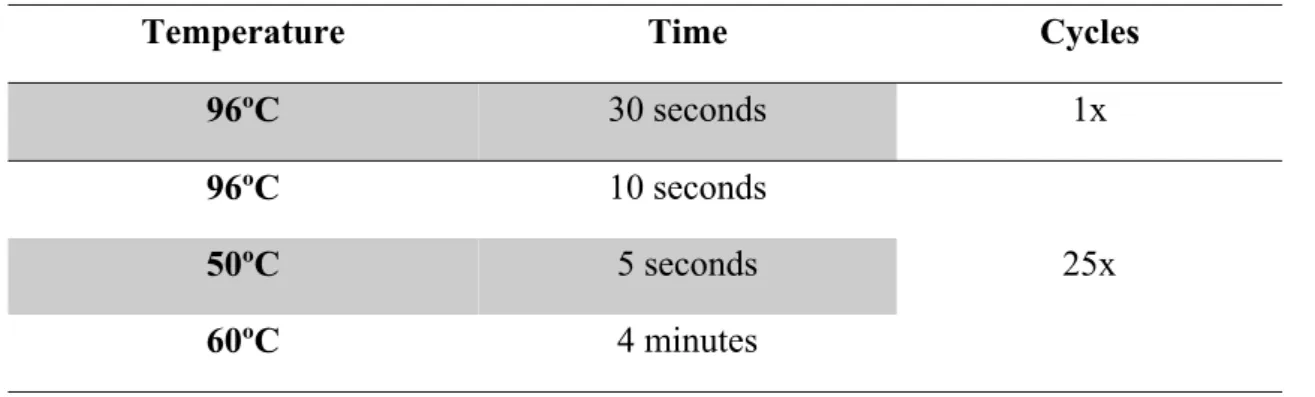
The amplification profile for the sequencing reaction is described in Table 6.
Material and Methods
Table 6 – Sequencing profile
Temperature Time Cycles
96ºC 30 seconds 1x 96ºC 10 seconds 25x 50ºC 5 seconds 60ºC 4 minutes
Sanger sequencing was performed by the Unit of Technology and Information (UTI) of INSA. Sequences were further evaluated by us using Chromas Lite program (Technelysium Pty Ltd). For each ompA sequence of each strain, we generated a nucleotide alignment with ompA sequences from C. trachomatis prototype strains representing the various ompA genotypes (and variants previously identified in the laboratory). This was done by applying the MegAlign software (DNASTAR) that, besides the alignment, also performs phylogenetic analyses and pairwise comparisons. When new variants were determined, the whole ompA typing procedure (nested-PCR plus sequencing) was repeated to confirm the observed mutations.
Statistical analysis was performed in order to determine the odds ratio (a statistical measure of association) between factors; the hypothesis was rejected if the P value for Fisher’s exact test was less than 0.05 (111). This analysis was done using GraphPad Prism 6 software (https://www.graphpad.com/scientific-software/prism/).
Material and Methods
3. Strategies for quick subtyping LGV strains
The Portuguese legislation regarding the obligatory notification of transmissible diseases (Despacho nº 5681 – A/2014 of 29 April 2014, plus Desclaração de Rectificação nº 609 – A/2014 16 June 2014, plus Despacho nº 15385-A/2016 21 December 2016), establishes that the identification of serovars L1, L2 and L3 is necessary to confirm a case of lymphogranuloma venereum. This can be done by using the routine ompA genotyping methodology described in the previously section but this technique takes about a week before getting to a result, and for this reason, whenever a clinical suspicion exists together with the detection of C. trachomatis, a quicker methodology should be applied. However, such a test is not yet commercially available. Thus, we intended to implement a methodology that would quickly identify C. trachomatis L1, L2 and L3 genotypes. For this, we developed a PCR targeting the ompA gene, using sets of primers for amplify a potential region that would differentiate between L serovars. We also implemented a real time-PCR with ompA and pmpH as target genes, freely available from the web (112), where the pmpH gene is used as it clearly separates LGV strains when a phylogenetic tree is constructed based on this gene (31). For both approaches, we used DNA samples from prototype chlamydial strains and from clinical specimens previously ompA-genotyped.
3.1 Subtyping LGV: Using sets of primers discriminatory of
L1, L2 and L3
Oligonucleotides were designed by the Bioinformatics Unit of the Department of Infectious Diseases of INSA (Annex 4). The PCR mix was prepared as described in the previously section (Table 3) and the PCR profile was identical to the described (Table 4) for the first round of amplification.
After the amplification, we performed amplicon detection through gel electrophoresis in TAE buffer [1,5% (w/v) agarose in TAE buffer, SYBR® safe DNA gel stain (10.000x in DMSO; Invitrogen)], for 30 minutes at 90 V.
Material and Methods
To assure the quality of amplicons, PCR products were further purified and sequenced; sequences were analyzed as mentioned in section 2.
3.2 Subtyping LGV: real time-PCR for C. trachomatis ompA
and pmpH
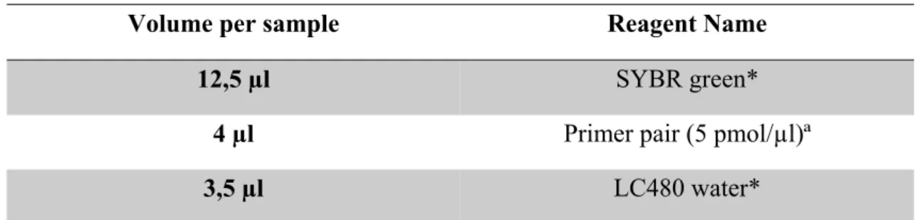
Subtyping of LGV through a real time-PCR approach, was based on a protocol available from the web (112) that implicates C. trachomatis ompA and pmpH genes. This procedure was done using the oligonucleotides described by authors (2) (Annex 5), and the detection was done by using the SYBR Green technology (LightCycler 480® Sybr green Master Mix I, Roche) according to the manufacturer’s instructions. Each set of primers was tested in a COBAS® z 480 Analyzer (Roche). The master mix for each real time-PCR was performed for a total volume of 20 µl as shown in Table 7, to which 5µl of DNA sample was added. The amplification profile is described in Table 8.
Table 7 – Master mix for LGV subtyping real time – PCR
Volume per sample Reagent Name
12,5 µl SYBR green*
4 µl Primer pair (5 pmol/µl)ª
3,5 µl LC480 water*
*LightCycler 480® Sybr green Master Mix I (Roche) ª Eurofins Genomics
Material and Methods
Table 8 – Amplification profile for LGV subtyping real time – PCR
Temperature Time Cycles
Pre-incubation 95ºC 10 minutes 1x PCR 95ºC 15 seconds 40x 60ºC 1 minute Melting 95ºC 15 seconds 1x 60ºC 20 seconds 1x 95ºC Continuous 1x Cooling 40ºC 30 seconds 1x
Material and Methods
4. Homopolymeric tracts counts and bioinformatics analyses
4.1 Study design: selection of homopolymeric tracts and C.
trachomatis positive DNA samples
For the study of potential targets of phase variation in C. trachomatis, we were supported by the Bioinformatics Unit of the Department of Infectious Diseases of INSA, which has been developing a strategy focused on sequencing, through a high throughput next-generation sequencing (NGS) technology with amplification products (amplicons) obtained with PCR schemes targeting homopolymeric tracts. Using whole-genome sequences available on Genbank, we firstly looked for regions in the C. trachomatis genome displaying homopolymeric tracts [poly (Ns)] with base counts equal or above to eight. Additionally, whole-genome sequence data from the host laboratory previously studied C. trachomatis population (104) was systematically analyzed for the presence of intra-strain heterogeneous and non-heterogeneous homopolymeric tracts. Altogether, this allowed us to select 20 genome-dispersed homopolymeric tracts identified to be likely prone to allelic variation (targets) and 4 homopolymeric tracts highly conserved within
C. trachomatis intra-populations (potential future controls for each base type). The poly
(N) selection have also taken into account the poly (N) location in relation to the potentially affected gene (upstream or coding region) and also on the function of the gene (Annex 6).
Although it is our goal to perform a future large-scale study involving multiple DNA samples obtained from C. trachomatis positive clinical specimens associated with distinct anatomical sites (genital versus anorectal mucosa) and distinct C. trachomatis
ompA-genotypes, in the present study, we performed a preliminary assay of
validation/optimization of the novel approach by evaluating the selected 24 poly (N)-targeting amplicons for 10 clinical samples (Annex 7). In particular, we specifically aimed at: i) implementing PCR schemes targeting each one of the 24 selected poly (N) s; ii) comparing the results obtained through the amplicon-based approach with previous
Material and Methods
results generated through WGS; iii) evaluating the impact of extending the PCR run on the poly (N) counts profile; iv) getting insight on both the profile of intra-patient variation of putative phase-variable loci and the existence of the tropism-specific homopolymeric lengths.
4.2 Amplicon-based NGS and bioinformatics analyses
PCR primers targeting each one of the 24 selected poly (Ns) were designed using Primer Select of DNASTAR (Lasergene 9 Core Suite) (see Annex 8) and oligonucleotides were ordered complemented with Illumina adapter sequences at their 5’ ends (see Annex 9).
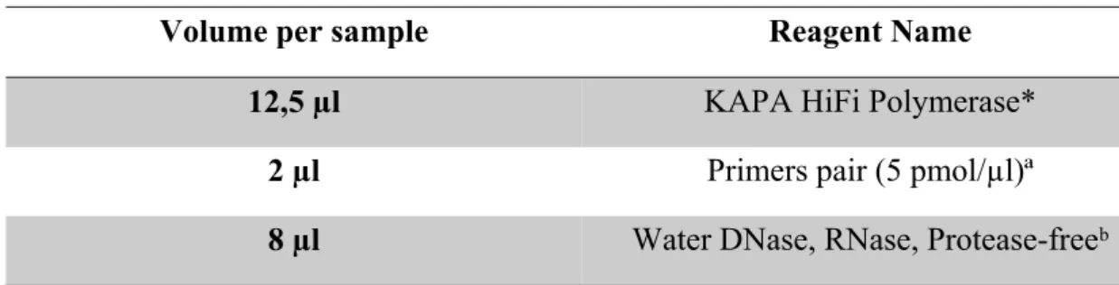
A PCR technique was used to amplify each homopolymeric region, using the DNA polymerase KAPA HiFi HotStart ReadyMix PCR Kit from KAPA Biosystems, an enzyme recognized by minimizing polymerase induced errors, according to the manufactures’ instructions. We prepared PCR mixes (Table 9) for a total volume of 22,5 µl to which 2,5 µl of each DNA sample were added. Each PCR promotes an amplicon of about 250 bp.
Table 9 – PCR mix for homopolymeric targets
Volume per sample Reagent Name
12,5 µl KAPA HiFi Polymerase*
2 µl Primers pair (5 pmol/µl)ª
8 µl Water DNase, RNase, Protease-freeᵇ
* KAPA Biosystems ª Invitrogen ᵇ 5Prime
The amplification profile comprehended a first step of denaturation, followed by 35 or 45 cycles of denaturing, annealing and extension, ending with a final extension step, according to the conditions described in Table 10. Annealing temperatures were optimized for each PCR scheme.
Material and Methods
Table 10– Amplification profile for homopolymeric regions
Temperature Time Cycle
95ºC 3 minutes 1x
95ºC 30 seconds
35 or 45x
Annealing temperature a 30 seconds
72ºC 30 seconds
72ºC 5 minutes 1x
a This temperature was adjusted for each PCR scheme
Amplicons generated for all gene targets for each clinical isolate were pooled in a single tube and sent to the UTI – INSA, where they were further cleaned-up, independently indexed and subjected to cluster generation and paired-end sequencing (2 x 150 bp) in an Illumina MiSeq equipment (Illumina Inc., San Diego, CA, USA), according to the manufacturer’s instructions. In order to evaluate the variation within DNA homopolymeric tracts, the Bioinformatics Unit of the Department of Infectious Diseases of INSA applied an in-house Python script [described in (113)] that enables the extraction and counting (directly from raw reads, both forward and reverse) of DNA sequences that are contiguously flanked by two conserved, user-defined, small DNA strings, as represented in Figure 5 and previously described by the host laboratory (113). As such, after defining conserved regions flanking each one of the 25 homopolymeric tracts under study, we have applied the script to determine the precise relative frequency of clones carrying specific base counts within a given sample.
Material and Methods
Figure 5 – Schematic representation of homopolymeric traits counts
III. Results and Discussion
1. Cell culture and C. trachomatis typing
1.1 Cell culture, the old gold standard
As previously mentioned, the cell culture used to be the gold standard technique for the diagnosis of C. trachomatis (81). However the requirements of the cell culture technique and for maintaining the bacteria alive until inoculation, led to the implementation of new techniques more affordable for diagnosis. These facts become obvious from our results; in fact for a total of 29 strains submitted to propagation in cell culture (7 ATCC prototype strains and 22 clinical isolates), only 4/7 (57%) ATCC and 6/22 (27%) clinical isolates were able to grow (Figure 6A and Figure 6B, respectively).
Figure 6 – Results from propagating C. trachomatis strains in cell culture. Panel A represents ATCC
prototypes strains and panel B characterizes clinical samples.
73% 27%
B
43% 57%A
Positive NegativeResults and Discussion
The ATCC strains were cultured with the aim of maintenance of prototype strains in the laboratory, providing an opportunity for learning cell culture and chlamydial propagation. ATCC samples were expected to have high bacterial loads; however, from our experience, almost half provided negative cultures. This could be related with deficient storage conditions, due to deficient freezing (above -80ºC) or broad temperature variations of the ultra-freezer; thus, the storage (even for short periods of time) under those non-optimal conditions might have contributed to the decrease of the infectivity. Clinical strains were cultured as a contribution for the Portuguese collection of C.
trachomatis. The success of chlamydial culture relies on the accomplishment of several
factors that can easily fail, from which transport in appropriate media, temperature and storage under -80ºC, stand out. Furthermore, samples that are selected for culture have prior been considered positive through commercial nucleic acids amplification techniques, which are recognizably more sensitive. Even the nested-PCR technique used for ompA-genotyping provides more positive results than culture, as 12/16 (75%) specimens that were negative in culture revealed positive upon nested-PCR. The four negative specimens, both in culture and ompA-nested-PCR, cannot be fully excluded from being true negatives; however, the lack of sensitivity of these methodologies (86,114,115) is the most probable explanation.
Live C. trachomatis prototype and clinical strains are considered of great interest for research studies, and the prior have been long used for such purposes; however, recent (104) whole genome sequencing studies upon which the genome of clinical strains was sequenced before and after several cycles of propagation in cell culture, revealed that C.
trachomatis genome rapidly evidences several changings. This phenomenon has been
considered (104) as an adaptation to in vitro life conditions, whereby some genes can be lost because they would no longer be needed (i.e. to provide nutrients that are made available through culture media), or because of their involvement in the in vivo infectious process and would no longer be required when the microorganism has no host immune system to fight against. Despite these considerations, much of chlamydial research will still depend on live strains and for this reason, the collection of clinical strains that are infecting humans’ remains obligatory to a national reference laboratory.
Results and Discussion
1.2 Characterization of C. trachomatis clinical strains
We got a collection of 278 C. trachomatis positive samples to perform ompA – genotyping, and we were successful for 245 (88,1%) of them (Figure 7).
Figure 7 – Rate of C. trachomatis ompA-genotyped strains
From the 278 C. trachomatis strains, 2 were obtained from the conjunctiva of newborns, due to mother to child transmission during delivery upon passage through an infected birth canal. These 2 samples will not be considered for analysis regarding to gender and age. Detected from women were 112 (41%) specimens, whereas 164 (59%) were detected from men samples (Figure 8).
12% 88% Positive Negative
Results and Discussion
Figure 8 – C. trachomatis strains by gender
Unlike most chlamydial studies, that comprehend a majority of women, a gender considered more prone to search for medical care, namely gynecological routine examination, our population involved more men. This relates to the attendees of the STI clinic from which most of the samples are taken for C. trachomatis diagnosis at INSA, mostly constituted by MSM, obviously leading to a bigger frequency of the male gender. In addition, laboratories that are currently sending positive chlamydial specimens for
ompA-typing at INSA, also provide C. trachomatis screening to MSM, contributing to a
collection where strains detected in men are more represented.
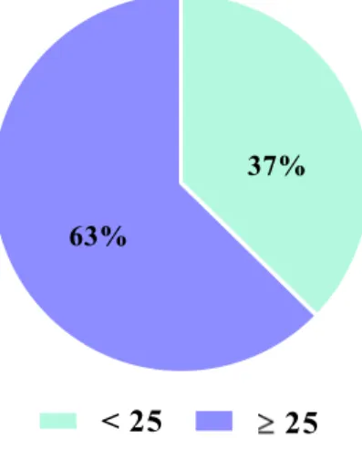
We evaluated 276 strains according to two age groups, one including strains from people with less than 25 years old and the other including people having 25 years old or more. People with 25 years old or more provide 173 (63%) strains and people with less than 25 years old, 103 (37%) strains (Figure 9).
59% 41%
Results and Discussion
Figure 9 – C. trachomatis strains by age
Strains were ompA-genotyped from infections detected in persons from 14 to 61 years old, median 27 years old and media 29 years old.
Most of our strains were detected in persons aged 25 years old or more, most probably due to the age of the attendees of STI clinics to which INSA provides routine
C. trachomatis diagnosis. The fact that C. trachomatis infects people of older ages shows
to the evidence that, despite the development of some immunity through several episodes of infection (116), it does not provide enough protection to avoid the occurrence of new episodes of chlamydial infections. However, in terms of public health, the infection is more important for people with less than 25 years old due to the clinical sequelae related to infertility.
Figure 10, represents the distribution of positive C. trachomatis samples per anatomical site. Urine was the most common biological sample providing positives, with a total of 124/278 (44,6%), followed by cervical swabs with 76/278 (27,3%), and anorectal swabs with 56/278 (20,1%). Less usual were urethral swabs (nowadays replaced by urine, particularly in men, due to its non-invasive character) with 13/278 (4,7%) samples, and two sorts of specimens where chlamydial diagnosis is rarely asked at INSA, the oropharynx, 7/278 (2,5%) and the conjunctiva 2/278 (0,7%).
63%
37%
![Figure 2 – Schematic representation of C. trachomatis outer membrane [Adapted from (37)]](https://thumb-eu.123doks.com/thumbv2/123dok_br/15599220.1051789/17.892.233.636.123.395/figure-schematic-representation-c-trachomatis-outer-membrane-adapted.webp)