Upper arm measurements of healthy neonates
comparing ultrasonography and anthropometric
methods
a ,
*
b aL. Pereira-da-Silva
, J. Veiga Gomes , A. Clington , J.M.
Videira-a c
Amaral , S.A. Bustamante
aˆ
Department of Pediatrics, Neonatal Intensive Care Unit, Dona Estef ania Hospital, Faculty of Medical
Sciences, Universidade Nova de Lisboa, Rua Jacinta Marto, 1169-045 Lisbon, Portugal b
ˆ
Department of Radiology, Dona Estef ania Hospital, Lisbon, Portugal c
Department of Pediatrics, University of Medicine and Dentistry of New Jersey, Robert Wood Johnson
Medical School, Camden, The Children’s Regional Hospital at Cooper Hospital, Camden, NJ, USA
Received 16 February 1998; received in revised form 5 July 1998; accepted 22 September 1998
Abstract
Objective: To compare measurements of the upper arm cross-sectional areas (total arm area,
arm muscle area, and arm fat area of healthy neonates) as calculated using anthropometry with the values obtained by ultrasonography. Materials and methods: This study was performed on 60 consecutively born healthy neonates: gestational age (mean6SD) 39.661.2 weeks, birth weight 3287.16307.7 g, 27 males (45%) and 33 females (55%). Mid-arm circumference and tricipital skinfold thickness measurements were taken on the left upper mid-arm according to the conventional anthropometric method to calculate total arm area, arm muscle area and arm fat area. The ultrasound evaluation was performed at the same arm location using a Toshiba sonolayer SSA-250A, which allows the calculation of the total arm area, arm muscle area and arm fat area by the number of pixels enclosed in the plotted areas. Statistical analysis: whenever appropriate, parametric and non-parametric tests were used in order to compare measurements of paired samples and of groups of samples. Results: No significant differences between males and females were found in any evaluated measurements, estimated either by anthropometry or by ultrasound. Also the median of total arm area did not differ significantly with either method (P 5 0.337). Although there is evidence of concordance of the total arm area measurements (r 5 0.68, 95% CI: 0.55–0.77) the two methods of measurement differed for arm muscle area and arm fat area. The estimated median of measurements by ultrasound for
*Corresponding author. Tel.: 1 351-1-3126613; fax: 1 351-1-7167203; e-mail: l.pereira.silva@mail.telepac.pt
0378-3782 / 99 / $ – see front matter 1999 Elsevier Science Ireland Ltd All rights reserved. P I I : S 0 3 7 8 - 3 7 8 2 ( 9 8 ) 0 0 0 8 5 - 1
arm muscle area were significantly lower than those estimated by the anthropometric method, which differed by as much as 111% (P , 0.001). The estimated median ultrasound measure-ment of the arm fat was higher than the anthropometric arm fat area by as much as 31% (P , 0.001). Conclusion: Compared with ultrasound measurements using skinfold measure-ments and mid-arm circumference without further correction may lead to overestimation of the cross-sectional area of muscle and underestimation of the cross-sectional fat area. The correlation between the two methods could be interpreted as an indication for further search of correction factors in the equations. 1999 Elsevier Science Ireland Ltd All rights reserved.
Keywords: Anthropometry; Skinfold thickness; Ultrasound; Upper arm areas
1. Introduction
In recent years, upper arm cross-sectional areas have been widely used as a simple, non-invasive and inexpensive method for assessment of the nutritional status of adults and children [1,2]. Standards for full-term and preterm neonates have also been reported recently [3,4]. These values are extrapolated from equations derived from mid-arm circumference (MAC) and tricipital skinfold thickness (TSF) [1,3,5]. It is not clear, however, how the conventional assumptions made in these calculations may stand compared to real time images of the arm cross-section. For the calculation of the arm muscle area, the conventional assumptions are that: (1) the mid-arm is cylindrical; (2) the subcutaneous fat is a concentric ring evenly distributed around the muscle; (3) that fat thickness is half the TSF; (4) that the muscle compartment is circular; and (5) that the muscle includes the humeral diameter. Any variation in bone diameter is not taken account [1,3,5]. Possible source of error using conventional assumptions is the variable tissue compression relative to differences in the skin tension when measuring TSF [6].
This study was designed to compare the upper arm cross-sectional areas of healthy neonates as measured by anthropometry with the values obtained by ultrasonography, a potential real time imaging method for assessment of arm areas in this age group.
2. Materials and methods
ˆ This study was performed on 60 neonates consecutively born at Dona Estefania Maternity Hospital, after informed parental consent was obtained. Only full-term newborn infants with a birth weight of more than 2500 g, Apgar score more than 6 at 5 min, and with no detectable malformation were included in the study. Gestational age, birth weight and sex were recorded. To avoid inter-observer variability [7], all anthropometric measurements were carried out by a single trained observer (LPS). Measurements were taken within 24 h of birth on the left arm mid distance between the tip of acromion and the olecraneon process. The arm was always held extended. MAC was measured to the nearest millimeter using a plastic flexible non-extendible
tape. The measured site was marked with a pen. TSF was evaluated using a Harpenden calliper at the same marked level of the extended arm. Measurements to the nearest 0.1 mm were made 60 s after application of the calliper, exerting a
2
pressure of 10 g / mm of contact surface area [8]. All anthropometric measurements were made in triplicate, and the results averaged. Cross-sectional arm area (AA), cross-sectional arm muscle area (AMA) and cross-sectional arm fat area (AFA) were calculated from TSF and MAC using conventional assumptions [1].
The ultrasound study was performed in all neonates using a Toshiba sonolayer SSA-250A, using a mechanical SM-708A sectorial probe of 7.5 MHz with an incorporated water cushion. The ultrasound examination was performed within 6 h of anthropometric assessment by a single operator experienced in pediatric examinations (JVG). The infants were placed in the supine position and the left arm was kept in an extended position slightly away from the body, and secured in such a way that no external pressure was exerted.
Close attention was given to the study protocol for obtaining a single transverse ultrasound view of the mid arm. To prevent any appreciable deformation of the arm image, it was established that the probe should never be applied to the posterior part of the arm. In the newborn, compression of subcutaneous tissues may deform the image, potentially changing the measurements. This deformation was minimized by applying the probe with the least possible pressure on the anterior part of the arm, thus reducing the probability of error. In order to achieve a near optimal image of the newborn’s arm, a water cushion was adapted to the probe. It was found to be an essential modification of the technique to achieve the best transversal, well defined view of the limits of the tissues involved.
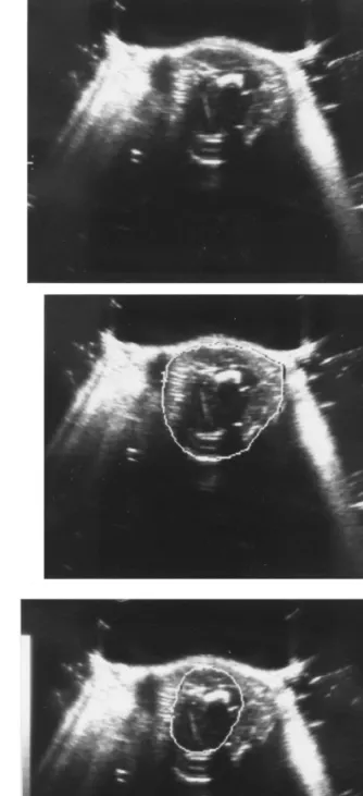
The probe was applied on the anterior part of the left upper arm at the same level of the anthropometric measurements, in the location previously marked. The probe was positioned perpendicular to the long axis of the upper arm to obtain a transversal plane. Using electronic callipers built in the ultrasound equipment, measurements were taken of both the total area of that section of the arm and of the area which included the muscular tissue and the humerus. In this set of newborn infants, the authors established that the cone of posterior acoustic shadow created by the transversally cut humerus of the neonate was not sufficiently intense to prevent the ultrasound imaging of the posterior structures. A single plane transversal cut view of the arm allowed us to perform the various measurements to be obtained (Fig. 1). In the Toshiba sonolayer SSA-250A the calculation of the areas was made by the number of pixels enclosed in the plotted areas (Fig. 1A–C). The reproducibility of the ultrasound measurements was tested in a separate sample of 16 healthy newborn infants. Statistical analysis of four consecutive measurements was used as the standard. The coefficients of variation of the measurements obtained for each variable were analyzed. Based on the Toshiba sonolayer SSA-250Atechnical information, the systematic error was established at 6%. The maximum measurement error was estimated to be as much as twice the systematic error. In fact, the mean of the coefficients of variation obtained ranged from 1.4% (SEM 5 0.2%) for the mid arm circumference to 3.4% (SEM 5 0.6%) for the arm muscle area, clearly acceptable for the purpose of these measurements (data not shown).
Fig. 1. Mid-arm cross-sectional ultrasonic view. (A) Plot of the mid-arm circumference obtained in the ultrasonic view regarding the calculation of the arm area. (B) Plot of the exterior limits of the muscular tissue obtained in the ultrasonic view regarding the calculation of the arm muscle area. (C) Diagram of A) and (B): UMAC, mid-arm circumference; UAA, arm area; UMMC, mid-muscle circumference; UAMA, arm muscle area (includes bone); arm fat area, UAA minus UAMA.
Fig. 1. (continued )
Statistical analysis was performed using the Stata 4.0 software (Stata Press, TX, USA 1995). Each measurement was analyzed by central tendency measures (means and medians), and dispersion measures (standard deviation, minimum, maximum and coefficient of variation). The central tendency results were compared according to the method of measurement (anthropometric or by ultrasound) and sex. Normality distribution was tested by Shapiro–Wilk test in order to apply the most appropriate test as follows: (1) in the case of data being normally distributed, t-test for paired samples was used when measurement methods were compared, and t-test for independent samples was used when sex was compared. (2) For data non-normally distributed we used the Wilcoxon test for paired samples and the Mann–Whitney test when measurements by sex were compared.
Concordance correlation coefficients [9] and its 95% confidence intervals (95% CI) were determined for each parameter, and the Bland and Altman approach [10] was followed in order to better define the limits of concordance.
A P 5 0.05 or less was chosen as the level of statistical significance.
3. Results
The 60 healthy neonates studied had a gestational age of (mean6SD) 39.661.2 weeks and birth weight of 3287.16307.7 g. The sample included 27 male (45%) and 33 female (55%) infants.
No significant differences between males and females were found in relation to all measurements studied: TSF, and MAC, AA, AMA and AFA assessed either by
Table 1
Tricipital skinfold thickness (mm)
Total Males Females
Median 3.4 3.4 3.4 Mean 3.5 3.4 3.6 SD 0.6 0.5 0.8 CV (%) 17.1 14.7 22.2 Minimum 2.4 2.6 2.4 Maximum 5.4 4.4 5.4
anthropometry or by ultrasound. As an example, the distribution of TSF is represented in Table 1.
In contrast to the conventional assumptions of the anthropometric method, cross-sectional ultrasound shows that the mid-arm is not circular, but elliptical, and that the subcutaneous layer of adipose tissue is markedly asymmetric. The fat thickness over the triceps is considerably greater than over the biceps, as one could have expected. The muscle compartment does not form a circle and is variable in shape, resembling a ‘clover leaf’ in the majority of subjects.
An attempt was made to determine the agreement limits according to the approach of Bland and Altman. However, the distribution of anthropometric and ultrasound sample differences for MAC, AA and AFA, but not AMA, did not pass normality tests, and the logarithmic transformation did not produce reliable results. In this sense, the agreement between the two methods of measurement was evaluated through scatterplots and its concordance by correlation coefficients.
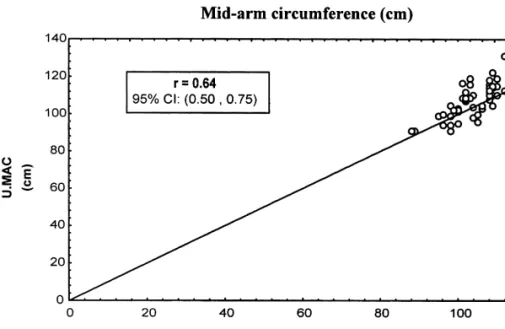
Both MAC assessed by anthropometry (AMAC) and MAC assessed by ultrasound (UMAC) (Table 2) follow a normal distribution. Values of AMAC are significantly lower than UMAC (P , 0.001). A minimal but definable compression of tissues may account for smaller arm circumference measured by tape. Intuitively the tape measure of the arm circumference should be the most accurate. Concordance correlation between the two measurement methods for MAC (r 5 0.64, 95% CI: 0.50–0.75) is depicted in Fig. 2.
The Wilcoxon test was used to evaluate the differences between the two methods for assessment of AA, AMA and AFA, because the results of the ultrasound
Table 2
Mid-arm circumference (mm)
Anthropometry Ultrasound
Total Males Females Total Males Females
Median 104 104 103 109 110 105 Mean 104.4 104.8 104.0 108.5 108.8 108.2 SD 6.5 4.9 7.6 10.3 6.9 12.5 CV (%) 6.2 4.7 7.3 9.5 6.3 11.6 Minimum 88 95 88 91 94 91 Maximum 118 118 116 136 119 136
Fig. 2. Concordance correlation between mid-arm circumference (MAC) measured by anthropometry (AMAC) and measured by ultrasound (UMAC).
measurement of AA (UAA), AMA (UAMA), and AFA (UAFA) were not normally distributed.
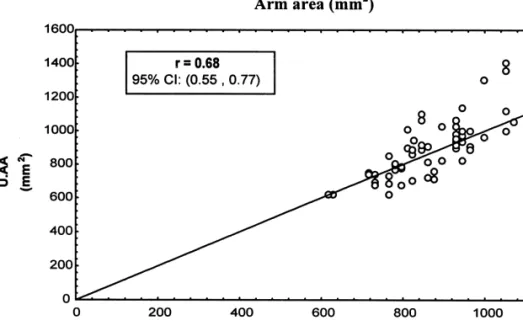
The median of AA does not differ significantly (P 5 0.337) if assessed by ultrasound or by anthropometry (AAA) (Table 3). The concordance correlation between the two measurement methods for AA (r 5 0.68, 95% CI: 0.55–0.77) is represented in Fig. 3.
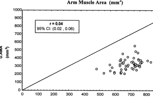
The median of AMA measured by ultrasound differs significantly from that measured by anthropometry (AAMA) (P , 0.001) (Table 4). AAMA overestimates muscle area when compared to the UAMA by as much as 111%. The concordance correlation between the two measurement methods for AMA is (r 5 0.04, 95% CI: 0.02–0.06) and is represented in Fig. 4.
Table 3 2 Arm area (mm )
Anthropometry Ultrasound
Total Males Females Total Males Females
Median 861 861 844 896.5 904 849 Mean 869.4 875.5 864.6 892.6 888.0 896.3 SD 107.2 82.4 124.9 170.2 110.6 208.5 CV (%) 12.3 9.4 14.4 19.1 12.5 23.3 Minimum 616 718 616 617 672 617 Maximum 1108 1108 1071 1410 1060 1410
Fig. 3. Concordance correlation between arm area (AA) measured by anthropometry (AAA) and measured by ultrasound (UAA).
In contrast to the measurements of muscle areas, the median of AFA is significantly less (P , 0.001) if it is assessed by ultrasound compared to the estimated areas using anthropometry (AAFA) (Table 5). The AAFA underestimates UAFA by 31%. The concordance correlation between the two measurement methods for AFA (r 5 0.04, 95% CI: 0.03–0.05) is represented in Fig. 5.
4. Discussion
Some authors have argued that estimates of muscle and fat content of the arm based on MAC and TSF are not accurate. Real time image technology is likely to
Table 4
2 Arm muscle area (mm )
Anthropometry Ultrasound
Total Males Females Total Males Females
Median 708 721 683 321.5 320 323 Mean 696.4 706.3 688.3 330.2 336.9 324.7 SD 80.5 66.5 90.6 77.4 70.0 83.7 CV (%) 11.6 9.4 13.2 23.4 20.8 25.8 Minimum 480 583 480 210 227 210 Maximum 906 906 888 598 553 598
Fig. 4. Concordance correlation between arm muscle area (AMA) measured by anthropometry (AAMA) and measured by ultrasound (UAMA).
provide very accurate estimates of such measurements, but this technology may not be as readily available as a tape and calliper. It is important to recognize the sources of inaccuracy and correct them in the assessment of tissue contents of the arm based on simple measurements like MAC and TSF. It has been assumed that larger arms have disproportionately more adipose tissue than smaller arms [1,5]. Using these criteria, cross-sectional arm areas have been preferred by some authors [2,11,12], as they represent better estimators of the relative contribution of fat and muscle to the total arm area than MAC and TSF used alone.
Some studies have demonstrated that conventional assumptions in the calculations of cross-sectional areas of the mid-arm are not accurate enough to appropriately estimate tissue size. Heymsfield et al. [13] identified errors inherent in the
anth-Table 5
2 Arm fat area (mm )
Anthropometry Ultrasound
Total Males Females Total Males Females
Median 167.5 169 165 556.5 564 554 Mean 173.1 169.2 176.3 560.8 551.1 568.7 SD 38.6 26.8 46.2 124.1 95.3 144.4 CV (%) 22.3 15.8 26.2 22.1 17.3 25.4 Minimum 102 122 102 352 387 352 Maximum 287 224 287 1059 704 1059
Fig. 5. Concordance correlation between arm fat area (AFA) measured by anthropometry (AAFA) and measured by ultrasound (UAFA).
ropometric method, using mid-arm computerized tomography scan performed in adults. In their study they found that the arm is elliptical and that in the majority of subjects the muscle compartment is rarely circular, rather resembling a ‘clover leaf’ as in our study. Furthermore, they concluded that the fat area model assuming an annulus of fat is not accurate enough. Instead, fat is asymmetrically distributed around the arm, with greater fat thickness behind the triceps than in front of the biceps. These results confirmed the findings of previous studies [14].
Another convention in the anthropometric method is to assume that the skinfold thickness is a measure of subcutaneous fat, assuming that the skinfold represents a double thickness of subcutaneous fat plus skin. The degree to which fat tissue and other tissues can be compressed during the TSF measurement may lead to an error [6]. The error may be predicted if every compression is done with the same pressure and time relationship [8]. The correction factor for better estimate of the cross-sectional areas of tissues has not been determined in newborn infants. It is apparent from the results of our study that, in order to make the conventional measurements based on MAC and TSF more accurate, there needs to be a constant factor to correct for tissue compression in the newborn, otherwise skinfolds will always underestimate actual thickness [6]. Therefore, according to our study, when uncorrected skinfold measurements and MAC are used to estimate cross-sectional mid-arm tissue areas, the assumptions lead to overestimation of the cross-sectional area of the muscle and underestimation of the cross-sectional fat area [6]. In neonates this effect may be more relevant, since differences in skinfold compressibility are influenced by many
factors, such as differences in skin turgor related to nutritional status and hydration [6,8,15].
In the present study a possible source of error using ultrasound could be the cone of posterior acoustic shadow created by the transversally cut humerus. In the neonate this shadow was not sufficiently intense to prevent the ultrasound view of the posterior structures, allowing us to use the images for measuring the cross-sectional areas of the mid-arm in this age group. For ethical and economic reasons, the comparison of the measurements made by ultrasound with other real time imaging methods at the cutting edge of technology such as CT scan or MRI was not done in this study. Nevertheless, we were able to prove the reproducibility of the ultrasound method, which provides internal validity to our data. The differences between values derived from ultrasonic method compared with anthropometric method require re-definition of the variables involved in the calculations of cross-sectional areas based on MAC and TSF. Further studies are needed to define the factors (constants) that can be used to improve the estimate of cross-sectional areas of muscle and fat in the arm of neonates, a key element of nutritional assessment. Due to the inherent limitations of the ultrasonic technique we would recommend using the most accurate of imaging technologies currently available, magnetic resonance imaging, to de-termine the gold standard for these measurements [16].
Acknowledgements
The authors are grateful to Daniel Virella, MD, ME, and to Sonia Doria Nobrega, statistician from Bioestat Portugal, for the assistance with the statistical analysis, to Eng. Pedro Ferreira of Toshiba Medical Systems Portugal for the technical collabora-tion, and to Wyeth-Lederle Portugal for the provided assistance.
References
[1] Frisancho AR. New norms of upper limb fat and muscle areas for assessment of nutritional status. Am J Clin Nutr 1981;34:2540–5.
[2] Georgieff MK, Mills MM, Zempel CE, Chang P-N. Catch-up growth, muscle and fat accretion, and body proportionality of infants one year after newborn intensive care. J Pediatr 1989;114:288–92. [3] Sann L, Durand M, Picard J, Lasne Y, Bethenod M. Arm fat and muscle areas in infancy. Arch Dis
Child 1988;63:256–60.
[4] Armanath UM, Weaver N, Armanath RP. Arm muscle and fat area in appropriate for gestational age, large for gestational age and small for gestational age neonates (Abstract). Clin Res 1988;36:894. [5] Gurney JM, Jelliffe DB. Arm anthropometry in nutritional assessment: nomogram for rapid
calculation of muscle circumference and cross-sectional muscle and fat areas. Am J Clin Nutr 1973;26:912–5.
[6] Himes JH, Roche AF, Siervogel RM. Compressibility of skinfolds and the measurement of subcutaneous fatness. Am J Clin Nutr 1979;32:1734–40.
[7] Branson RS, Vaucher YE, Harrison GG, Vargas M, Thies C. Inter- and intra-observer reliability of skinfold thickness measurements in newborn infants. Hum Biol 1982;54:137–43.
[8] Bustamante SA, Jacobs P, Gaines JA. Body weight, static and dynamic skinfold thickness in small premature infants during the first months of life. Early Hum Dev 1983;8:217–24.
[9] Lin LI-K. A concordance correlation coefficient to evaluate reproducibility. Biometrics 1989;45:255– 68.
[10] Bland JM, Altman DG. Statistical methods for assessing agreement between two methods of clinical measurement. Lancet 1986;i:307–10.
[11] Pereira-da-Silva L, Berdeja A, Ventosa L, Leal F, Clington A, Videira-Amaral JM. Upper arm areas in low birth weight (LBW) infants—Which usefulness for routine nutritional assessment? (Abstract). Biol Neonate 1994;66:160.
[12] Pereira-da-Silva L, Guerreiro N, Leal F, Videira-Amaral JM. Better nutritional support for low birth weight infants—‘better’ body composition? (Abstract). Prenat Neonat Med 1996;1:368.
[13] Heymsfield SB, McManus C, Smith J, Stevens V, Nixon DW. Anthropometric measurement of muscle mass: revised equations for calculating bone-free arm muscle area. Am J Clin Nutr 1982;36:680–90. [14] Malina RM, Habicht JP, Yarbrough C, Martorell R, Klein RE. Skinfold thicknesses at seven sites in
rural Guatemalan Ladino children birth through seven years of age. Hum Biol 1974;46:453–7. [15] Brozek J, Kinzey W. Age changes in skinfold compressibility. J Gerontol 1960;15:45–51. [16] Seidell JC, Bakker CJG, van der Kooy K. Imaging techniques for measuring adipose-tissue
distribution—a comparison between computed tomography and 1.5-T magnetic resonance. Am J Clin Nutr 1990;51:953–7.


