Toxic, antimicrobial and
hemagglutinating activities of the
purple fluid of the sea hare
Aplysia
dactylomela
Rang, 1828
Departamentos de 1Biologia and 2Bioquímica e Biologia Molecular,
Universidade Federal do Ceará, Fortaleza, CE, Brasil V.M.M. Melo1,
A.M. Fonseca1,
I.M. Vasconcelos2 and
A.F.F.U. Carvalho1
Abstract
The antimicrobial, hemagglutinating and toxic activities of the purple fluid of the sea hare Aplysia dactylomela are described. Intact or dialyzed purple fluid inhibited the growth of species of Gram-positive and Gram-negative bacteria and the action was not bactericidal but bacteriostatic. The active factor or factors were heat labile and sensi-tive to extreme pH values. The fluid preferentially agglutinated rabbit erythrocytes and, to a lesser extent, human blood cells, and this activity was inhibited by the glycoprotein fetuin, a fact suggesting the presence of a lectin. The fluid was also toxic to brine shrimp nauplii (LD50
141.25 µg protein/ml) and to mice injected intraperitoneally (LD50
201.8 ± 8.6 mg protein/kg), in a dose-dependent fashion. These toxic activities were abolished when the fluid was heated. Taken together, the data suggest that the activities of the purple fluid are due primarily to substance(s) of a protein nature which may be involved in the chemical defense mechanism of this sea hare.
Correspondence
V.M.M. Melo
Departamento de Biologia Universidade Federal do Ceará 60455-760 Fortaleza, CE Brasil
Fax: 55 (085) 287-2768
Received July 11, 1997 Accepted March 26, 1998
Key words
•Aplysia
•Antimicrobial
•Purple fluid
•Toxicity
•Hemagglutinating activity
Introduction
Many sea hares, which are opistobranch molluscs, discharge a fluid from the purple gland when disturbed. This reaction sug-gests that this fluid contains bioactive fac-tors which may act against potential enemies since the defense mechanisms of the sea hare differ from those of highly developed vertebrates (1). Sea hare species have at-tracted the interest of many workers investi-gating the chemical compounds secreted by the purple gland or present in different tis-sues, possibly involved in the defense of these invertebrates. Thus, some sea hare spe-cies have been shown to contain low
molec-ular mass substances with antimicrobial (2-6) and antitumor activities (7-10) and also high molecular mass compounds such as those from Aplysia kurodai, Aplysia juliana
and Dolabella auricularia with similar ac-tivities, and which were named aplysianins (11,12), julianins (13,14) and dolabellanins (15,16), respectively. This study describes some biological properties (antibacterial, antifungal, hemagglutinating and toxic ac-tivities) of the purple fluid of the sea hare
Material and Methods
Collection of the purple fluid
Specimens ofAplysia dactylomela Rang, 1828 were collected at Pacheco Beach, Caucaia, State of Ceará, Brazil, in June and July. The purple fluid was obtained by irri-tating the hare and squeezing it gently for a few minutes outside the water. The secretion was collected into a sterile bottle and subse-quently frozen at -10oC until used.
Protein determination
A manual colorimetric procedure for measuring ammonium nitrogen in Kjeldahl digests (18) was used for the determination of total nitrogen and protein content, which was calculated using a nitrogen conversion factor of 6.25.
Antibacterial assays
Inhibition of bacterial growth by the purple fluid samples was determined as de-scribed by Bauer et al. (19). Briefly, bacte-rial cultures were maintained in Müller-Hinton broth (Difco Laboratories, Detroit, MI). Sterile swabs were immersed in the microbial suspensions (108 cells/ml) and
evenly applied to Petri dishes containing Müller-Hinton agar. Sterile Whatman AA filter paper disks (6 mm in diameter) were fully imbibed with 30 µl of the purple fluid samples and placed over the agar in the plates. Tobramicin disks (10 µg; Cecon, São Paulo, SP) were used as positive control. The plates were incubated overnight at 35oC
and then examined for zones of growth inhi-bition around each disk.The bacteria used were Serratia marcescens, Citrobacter fre-undii, Vibrio cholerae, Salmonella thyphi-murium and Proteus vulgaris (all from the collection of Universidade Federal do Ceará),
Bacillus subtillis (ATCC 6633), Escherichia
coli (ATCC 13863), Staphylococcus aureus
(ATCC 6538), and Pseudomonas aerugino-sa (ATCC 25619). To investigate whether the antibacterial action was bacteriostatic or bactericidal, fluid samples were serially di-luted in 1% peptone broth and incubated with cells of Staphylococcus aureus and
Pseudomonas aeruginosa for 18 h at 35oC.
After this period, the minimum inhibitory concentration (MIC) was determined (20) and the mechanism of growth inhibition evaluated by subculturing the cells in media without purple fluid.
Antifungal assays
Growth inhibition of Candida albicans
and Saccharomyces cerevisiae (all from the UFC collection) by the purple fluid was determined as described by Roberts and Selitrennikoff (21). Briefly, agar assay plates were prepared by autoclaving agar Sabouraud medium (Difco Laboratories). After cooling to 45oC, the yeasts were added to a final
concentration of 107 cells/ml.
Fifteen-milli-liter aliquots of the suspension were dis-pensed into 100-mm diameter Petri dish and allowed to solidify before placing 6-mm di-ameter sterile paper disks on the surface of the agar. Thirty microliters of the purple fluid was added to each disk, and plates were incubated overnight at 35oC. Plates were
Effects of dialysis, heat treatment and pH on antibacterial activity
Aliquots of the fluid were dialyzed (cut-off 12,000) thoroughly against water, at 4ºC, and subsequently tested for antibacterial ac-tivity (Pseudomonas aeruginosa and Staph-ylococcus aureus), as described before. Fluid samples were heated at 80oC for 2, 5 and 15
min. After heating, the samples were cooled and centrifuged and the supernatants tested for activity. For the pH stability test, aliquots were adjusted to pH 2.0 (HCl) and 12.0 (NaOH) and kept in a refrigerator for up to 30 min. After this period, the fluid had its pH adjusted back to its original value of 6.4 and was tested for antibacterial activity.
Erythrocyte agglutination and inhibition assays
The hemagglutinating activity was as-sayed according to Vasconcelos et al. (23). Serial 1:2 dilutions of the fluid dialyzed against 25 mM Tris-HCl, pH 7.5, were mixed in small glass tubes with 0.25 ml of a 2% suspension of untreated or trypsin-treated erythrocytes (horse, chicken, pig, cow, rab-bit or human). The enzyme-treated cells were obtained by incubation of trypsin (0.1 mg; Type I, Sigma Chemical Co., St. Louis, MO) with 25 ml of a 2% suspension of cells in 150 mM NaCl for 60 min at 4oC. After washing
six times, a 2% suspension was prepared in 150 mM NaCl. The extent of agglutination was monitored visually after the tubes had been left at 37oC for 30 min and
subse-quently at room temperature for a further 30 min. The results are reported as the number of hemagglutination units (HU) per mg of fluid protein able to induce visible erythro-cyte agglutination. One HU was defined as the minimum protein concentration required to produce visible agglutination. The carbo-hydrate-binding specificity of the protein was assessed by the ability of sugars or glycoproteins in 150 mM NaCl to inhibit
agglutination of rabbit erythrocytes. The fluid was added to each tube at a concentration of 0.4 µg protein/ml, the minimum concentra-tion required to produce visible agglutina-tion. The lowest glycoprotein or sugar con-centration giving full inhibition of agglutina-tion was determined by two-fold serial dilu-tion of soludilu-tions at 1 mg/ml initial concentra-tion.
Toxicity bioassay against brine shrimp nauplii
A method using brine shrimp (Artemia
sp), proposed as a simple bioassay for re-search on natural products, was employed (24). Brine shrimp eggs (5 mg) were hatched in a rectangular dish (32 x 22 x 10 cm) filled with 5 l of sea water. A plastic sieve was clamped to the dish to form two unequal compartments. The eggs were sprinkled into the larger one which was darkened, while the smaller one was illuminated. After 48 h, the phototrophic larvae (nauplii) were col-lected with a pipette from the lighted side and transferred (10 shrimps) to vials filled with sea water (5 ml) containing 1 drop of casein peptone solution (3 mg/5 ml) as food. Dialyzed fluid was added to the vials to final concentrations of 50, 500 and 5000 µg pro-tein/ml. As control, a group of vials was filled with sea water containing the casein peptone solution. The vials were kept illumi-nated during 24 h of contact with the sub-stances, and survivals were counted with the aid of a magnifying glass. This assay was carried out three times with five replicates for each fluid concentration tested. To calcu-late the LC50 (mean lethal concentration) the
results were plotted as logit % mortality vs
log concentration. Logit is defined as ln (% mortality/% survival) (25).
Mouse toxicity assay
intraper-itoneal injections of the fluid exhaustively dialyzed against 25 mM Tris-HCl, pH 7.5. One LD50 unit (26) was taken as the amount
of protein (in mg protein/kg body weight) producing death of 50% of the tested ani-mals (six doses; six mice per dose).
SDS-polyacrylamide gel electrophoresis
Electrophoresis of the crude and dialyzed purple fluid was carried out by the method of Laemmli (27) on a 2-mm vertical slab gel consisting of stacking gel mix, 3.95% total acrylamide, and main running gel mix, 12.0% acrylamide. Fluid samples containing 2% SDS and 1% 2-mercaptoethanol were incu-bated at 100°C for 10 min. A few sucrose crystals were dissolved in the samples be-fore being applied (30 µl) to the gel. Electro-phoresis was carried out at 20-mA constant current for 60 min and protein bands were visualized by silver staining (28). The mo-lecular mass markers used were α -lactalbu-min (14.2 kDa), trypsin inhibitor (20.1 kDa), trypsinogen (24.0 kDa), carbonic anhydrase (29.0 kDa), glyceraldehy3-phosphate de-hydrogenase (36.0 kDa), egg albumin (45.0 kDa) and bovine serum albumin(66.0 kDa).
Results and Discussion
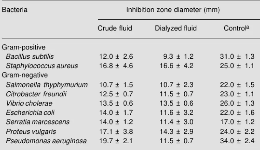
Both the crude (16.0 ± 0.7 mg protein/ ml) and the dialyzed purple fluid (10.1 ± 0.6 mg protein/ml) inhibited the growth of all species of Gram-positive and Gram-nega-tive bacteria tested, with Staphylococcus aureus, Pseudomonas aeruginosa and Pro-teus vulgaris being the most sensitive (Table 1). The antibacterial activity of the fluid against P. aeruginosa was reduced by 40% after dialysis, suggesting that low molecular mass components are involved in this activ-ity. The fluids stopped the growth of S. aureus
and P. aeruginosa but the bacteria grew again after their removal from culture, indi-cating a bacteriostatic and not a bactericidal action of the fluids, with a minimum inhibi-tory concentration of 0.625 mg protein/ml. The heat treatment of the fluid at 80oC for 2
min eliminated the inhibitory activity against
P. aeruginosa and S. aureus (the species selected as target cells). Similarly, after acid treatment at pH 2.0 the inhibitory action against the two species was completely lost. Nevertheless, after alkaline treatment (pH 12.0) the inhibitory action against P. aerugi-nosa was lost while that against S. aureus
was only reduced (the inhibition zone of 16.8 ± 4.6 mm was reduced to 9.0 ± 0.9 mm). Taken together, these data suggest that the active factor(s) probably is(are) protein. In fact, Yamazaki et al. (29) have reported that a glycoprotein is responsible for the antibac-terial activity of the purple fluid of Aplysia kurodai. Likewise, other antibacterial glyco-proteins have been reported to be present in different secretions of sea hares (14,30,31). The fluid was not active against the yeasts
Candida albicans or Saccharomyces cerevi-siae nor against the filamentous fungi As-pergillus niger, Penicillium herguei and
Trichophytum mentagrophytes.
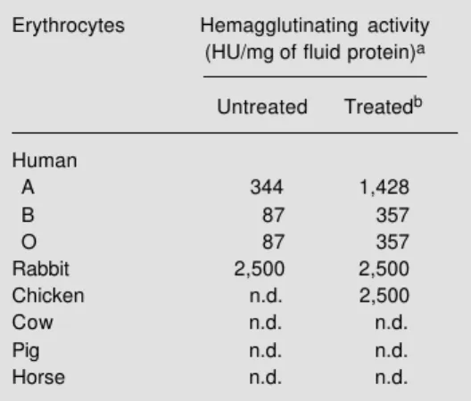
The results of the hemagglutination as-says are shown in Table 2. The dialyzed fluid preferentially agglutinated rabbit erythrocytes and, to a lesser extent, human erythrocytes
Table 1 - Antibacterial activity of the crude (0.48 mg protein) and dialyzed (0.30 mg protein) purple fluid of Aplysia dactylomela.
Data are reported as the mean ± SD for 3 experiments carried out in duplicate. aNitrofurantoin for Vibrio cholerae and tobramicin for all the others.
Bacteria Inhibition zone diameter (mm)
Crude fluid Dialyzed fluid Controla
Gram-positive
Bacillus subtilis 12.0 ± 2.6 9.3 ± 1.2 31.0 ± 1.3
Staphylococcus aureus 16.8 ± 4.6 16.6 ± 4.2 25.0 ± 1.1
Gram-negative
Salmonella thyphymurium 10.7 ± 1.5 10.7 ± 2.3 22.0 ± 1.5
Citrobacter freundii 12.5 ± 0.7 11.5 ± 0.7 23.0 ± 1.1
Vibrio cholerae 13.5 ± 0.6 13.5 ± 0.6 26.0 ± 1.3
Escherichia coli 14.0 ± 1.7 11.6 ± 3.2 22.0 ± 1.6
Serratia marcescens 14.0 ± 1.2 11.4 ± 3.0 17.0 ± 1.2
Proteus vulgaris 17.1 ± 3.8 14.3 ± 2.9 24.0 ± 2.2
(ABO). Treatment of the cells with trypsin revealed the agglutinating activity of the fluid against chicken erythrocytes and increased the sensitivity of human cells. Nevertheless, no activity was detected when the fluid was tested against cow, pig and horse erythro-cytes, even when using enzyme-treated cells. This selective agglutination may be due to the different nature of the glycoproteins pro-truding on the cell surface of the erythro-cytes tested. The activity of the fluid varied from 0.4 to 11.4 µg/ml depending on the cell used, thus being comparable in potency to the agglutinin purified from Aplysia kurodai
eggs, which was shown to react with B cells and rabbit blood cells at concentrations as low as 0.06 µg/ml (32). These authors also reported agglutinins in the serum of Aplysia dactylomela which strongly agglutinated human erythrocytes, but reacted weakly with rabbit blood cells, contrary to that observed in the present study. These findings suggest that the hemagglutinating activity may be due to different proteins or that other con-stituents of these fluids may interfere with this activity. Various simple sugars were reported to be potent hemagglutinin inhibi-tors (33). In the present study, the agglutina-tion of rabbit erythrocytes by the fluid was inhibited by the glycoprotein fetuin, but not by glucose, mannose, galactose, N-acetyl-glucosamine, N-acetyl-galactosamine or sialic acid (Table 3).
The purple fluid was shown to be toxic to the brine shrimp nauplii, with a calculated LD50 of 141.25 µg protein/ml. This effect
was dose dependent and despite its unknown mechanism, may well involve the fluid lec-tin as observed for leclec-tins of plant origin, such as those of Dioclea guianensis (23) and
Cratylia floribunda (34).
The fluid was also highly toxic to mice when injected intraperitoneally (ip), within 1 to 12 h, depending on the dose used. The LD50 found was 201.8 ± 8.6 mg protein/kg
body weight (20 ml of fluid/kg body weight). The typical effects observed invariably
in-cluded dyspnea and convulsions preceding the death of the animals. These acute effects were very similar to those produced by soyatoxin (SYTX), a seed protein purified from mature commercial soybean sold in Brazil, which is a mixture of undefined cul-tivars (35). The toxic activity present in the fluid was susceptible to inactivation by heat-ing at 92oC for 5 min.
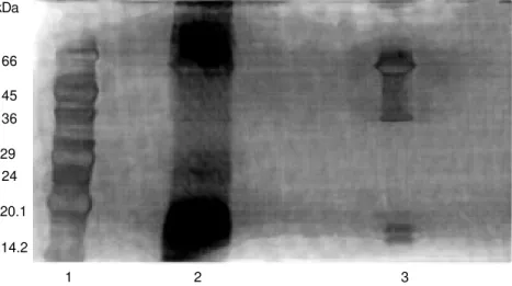
The electrophoretic profile of the crude and dialyzed fluids (Figure 1) showed a
simi-Table 2 - Agglutination of erythrocytes from vari-ous species by the purple fluid ofAplysia dactylo-mela.
aOne hemagglutination unit (HU) was defined as
the minimum protein concentration required to produce visible agglutination. bTrypsin treated. n.d., Not detected.
Erythrocytes Hemagglutinating activity
(HU/mgof fluid protein)a
Untreated Treatedb
Human
A 344 1,428
B 87 357
O 87 357
Rabbit 2,500 2,500
Chicken n.d. 2,500
Cow n.d. n.d.
Pig n.d. n.d.
Horse n.d. n.d.
Table 3 - Inhibition of hemagglutinating activity.
Assays were done using untreated rabbit cells. aMinimal concentration of sugar or glycoprotein
required to inhibit 1 HU. n.d., Not detected with the initial concentration of 1 mg/ml.
Sugar or glycoprotein Minimal concentration (mg/ml)a
Galactose n.d.
Glucose n.d.
Mannose n.d.
N-acetyl-galactosamine n.d.
N-acetyl-glucosamine n.d.
Sialic acid n.d.
lar distribution of the protein bands. In both fluid samples there was predominance of proteins with apparent molecular mass of 66.0 kDa and below 20.1 kDa. Nevertheless, in the dialyzed fluid proteins between 18.0 kDa and 36 kDa were not observed. This presumably could be due to the different amounts of protein applied in the electro-phoresis, since it was our intention to main-tain the same volume (30 µl) used in the antibacterial assay.
These preliminary data do not allow us to establish that all of these interesting activi-ties presented by the purple fluid of Aplysia dactylomela are due to the same component (s). Further studies are needed on this point. Nevertheless, the protein nature of the active component(s) is clear, as also is the presence of a lectin whose hemagglutinating activity is inhibitable by fetuin, a specific
glycopro-Figure 1 - SDS-polyacrylamide gel electrophoresis of the crude and dialyzed purple fluid of the sea hare Aplysia dactylomela. Lane 1, Standard protein mark-ers; lane 2, crude fluid; lane 3, fluid dialyzed against water.
kDa
66
45 36
29 24
20.1
14.2
1 2 3
tein. This is the first time that lectins are reported to be present in the purple fluid of sea hares and also that the purple fluid is toxic to living systems such as brine shrimp nauplii and mice. This study supports the role of the fluid as part of a chemical defense mechanism since it inhibited the growth of many Gram-positive and Gram-negative bac-teria and showed toxicity to other living systems.
Acknowledgments
References
1. Cooper EL (1980). Phylogeny of cytotoxic-ity. Endeavour, 4: 160-165.
2. Faulkner DJ, Stallard MO, Fayos J & Clardly J (1973). (3R, 4S, ‘S)-trans, trans-3,7-dimethyl-1-8, 8-tribromo-1, 5-octadi-ene, a novel monoterpene from the sea hare, Aplysia california. Journal of the American Chemical Society, 95: 3413-3414.
3. Kato Y & Scheuer PJ (1975). The aplysiatoxins. Pure and Applied Chemis-try, 41: 1-4.
4. Kinnel R, Duggan AJ, Eisner T, Melnwals J & Miura U (1977). Panacene: an aro-matic bromallene from a sea hare (Aplysia kurodai ). Tetrahedron Letters, 18: 3913-3916.
5. Ichida T & Higa T (1986). New cuparene-derived sesquiterpenes with unprece-dented oxygenation patterns from the sea hare Aplysia dactylomela. Journal of Or-ganic Chemistry, 51: 3364-3366. 6. Rinehart KL, Shaw PD, Shield LS, Gloer
JB, Harbour GC, Koker MES, Samain D, Schwart RE, Tymiak AA, Weller GT, Carter GT & Munro MHG (1981). Marine natural product as sources of antiviral, antimicro-bial and antineoplastic agents. Pure and Applied Chemistry, 53: 795-817. 7. Ireland C, Faulkner DJ, Finer J & Clardly J
(1976). A novel diterpene from Dolabella california. Journal of the American Chem-ical Society,98: 4664-4665.
8. Schmitz FJ, Michaud DP & Schmidt PG (1982). Marine natural products: pargue-rol, deoxyparguepargue-rol, and isoparguerol. New brominated diterpenes with modi-fied pimarane skeletons from the sea hare Aplysia dactylomela. Journal of the Ameri-can Chemical Society, 104: 6415-6423. 9. Pettit GR, Kamano Y, Herald CL, Tumman
AA, Boettner EE, Kizu H, Schmidt JM, Baczynsky L, Tomer KB & Bontems RJ (1987). The isolation and structure of a remarkable marine animal antineoplastic constituent: dolastatin 10. Journal of the American Chemical Society, 109: 6883-6885.
10. Pettit GR, Kamano Y, Herald CL, Dufresne C, Bates RB, Tumman AA, Schmidt JM, Cerny RL & Kizu H (1990). Antineoplastic agents. 190. Isolation and structure of the cyclodepsipeptide dolastatin 14. Journal of Organic Chemistry, 55: 2989-2990. 11. Kisugi J, Kamiya H & Yamazaki M (1987).
Purification and characterization of aplysianin E, an antitumor factor from sea hare eggs. Cancer Research, 47: 5649-5653.
12. Yamazaki M, Kimura K, Kisugi J, Muramoto K & Kamiya H (1989). Isolation and characterization of a novel cytolytic factor in purple fluid of the sea hare, Aplysia kurodai. Cancer Research, 49: 3834-3838.
13. Kamiya H, Muramoto K, Goto R & Yamazaki M (1988). Characterization of the antibacterial and antineoplastic glyco-protein in a sea hare Aplysia juliana. Nippon Suisan Gakkaishi, 54: 773-777. 14. Kamiya H, Muramoto K, Goto R, Sakai M,
Endo Y & Yamazaki M (1989). Purification and characterization of an antibacterial and antineoplastic protein secretion of a sea hare, Aplysia juliana. Toxicon, 27: 1269-1277.
15. Kisugi J, Yamazaki M, Ishii Y, Tansho S, Muramoto K & Kamiya H (1989). Purifica-tion of a novel cytolytic protein from albu-men gland of the sea hare, Dolabella au-ricularia. Chemical and Pharmaceutical Bulletin, 37: 2773-2776.
16. Kisugi J & Yamazaki M (1989). Purification of dolabellanin-C, an antineoplastic glyco-protein in the body fluid of a sea hare, Dolabella auricularia. Developmental and Comparative Immunology, 13: 3-8. 17. Rios EC (1985). Seashells of Brazil.
Fundação Universitária do Rio Grande. Museu Oceanográfico, Rio Grande. 18. Baethgen WE & Alley MM (1989). A
manual colorimetric procedure for meas-uring ammonium nitrogen in soil and plant Kjeldahl digests. Soil and Plant Analysis, 20: 961-969.
19. Bauer AW, Kirby WMM, Sherris JC & Turck M (1966). Antibiotic susceptibility testing by a standardized single disk method. American Journal of Clinical Pa-thology, 45: 493-495.
20. Collins CH, Lyne PM & Grange JM (1989). Antimicrobial sensitivity and assay tests. In: Collins CH, Lyne PM & Grange JM (Editors), Collins and Lyne’s Microbiologi-cal Methods. 8th edn. Butterworths, Lon-don.
21. Roberts WK & Selitrennikoff CP (1990). Zeamantin, an antifungal protein from maize with membrane-permeabilizing ac-tivity. Journal of General Microbiology, 136: 1771-1778.
22. Mirelman D, Galun E, Sharon N & Lotan R (1975). Inhibition of fungal growth by wheat germ agglutinin. Nature, 256: 414-416.
23. Vasconcelos IM, Cavada BS, Moreira RA & Oliveira JTA (1991). Purification and par-tial characterization of a lectin from the
seeds of Dioclea guianensis. Journal of Food Biochemistry,15: 137-154. 24. Carvalho AFFU, Melo VMM, Aguiar LMBA
& Matos FJA (1988). Avaliação da toxici-dade de extratos de plantas medicinais através de bioensaio com Artemia salina Leach. Ciência e Cultura, 40: 1109-1111. 25. Dawson-Saunders B & Trapp RG (1994).
Basic and Clinical Biostatistics. Appleton & Lange, E. Norwalk, CT.
26. Litchifield Jr JT & Wilcoxon F (1949). A simplified method for evaluation of dose-effect experiments. Journal of Pharmaco-logical and Experimental Therapy, 96: 99-104.
27. Laemmli UK (1970). Cleavage of struc-tural protein during the assembly of the head of bacteriophage T4. Nature, 227: 680-685.
28. Oakley B, Kirsch D & Morris N (1980). A simplified ultrasensitive silver staining for detecting proteins in polyacrylamide gels. Analytical Biochemistry, 105: 361-364. 29. Yamazaki M, Ohye H, Kisugi J & Kamiya H
(1990). Bacteriostatic and cytolytic activ-ity of purple fluid from the sea hare. De-velopmental and Comparative Immunol-ogy, 14: 379-383.
30. Yamazaki M (1993). Antitumor and antimi-crobial glycoproteins from sea hares. Comparative Biochemistry and Physiolo-gy, 105: 141-146.
31. Kamiya H, Muramoto K & Yamazaki M (1986). Aplysianin-A, an antibacterial and antineoplastic glycoprotein in the albu-men gland of a sea hare, Aplysia kurodai. Experientia, 42: 1065-1067.
32. Kamiya H & Shimizu Y (1981). A natural agglutinin inhibitable by D-galacturonic acid in the sea hare Aplysia eggs: charac-terization and purification. Bulletin of the Japanese Society of Fisheries, 47: 255-259.
33. Lis H & Sharon N (1986). Lectins as mol-ecules and as tools. Annual Review of Biochemistry, 55: 35-67.
34. Vasconcelos IM (1994). Soyatoxina - uma nova proteína tóxica purificada da soja (Glycine max L.). Doctoral thesis, Univer-sidade Federal do Rio de Janeiro, Rio de Janeiro, Brasil.