Angle Class II correction with MARA appliance
Kelly Chiqueto1, José Fernando Castanha Henriques2, Sérgio Estelita Cavalcante Barros3, Guilherme Janson4
How to cite this article: Chiqueto K, Henriques JFC, Barros SEC, Janson G. Angle Class II correction with MARA appliance. Dental Press J Orthod. 2013 Jan-Feb; 18(1):35-44.
» The author reports no commercial, proprietary or inancial interest in the prod-ucts or companies described in this article.
» Patients displayed in this article previously approved the use of their facial and intraoral photographs.
Contact address: Kelly Chiqueto
Av. Bento Gonçalves, 1515 - apto 1904C, Santo Antônio – Porto Alegre/RS CEP: 90.650-002 – E-mail: [email protected]
1 MSc and PhD in Orthodontics, FOB-USP. Coordinator of the Specialization
Course in Orthodontics, ABCD-BA.
2 Full Professor, Pediatric Dentistry, Orthodontics and Social Health Department,
FOB-USP.
3 Post-Doc in Orthodontics, FOB-USP. Adjunct Professor of Orthodontics,
Federal University of Rio Grande do Sul, UFRGS.
4 Full Professor and Head of the Pediatric Dentistry, Orthodontics and Social
Health Department, FOB-USP.
Submitted: March 31, 2009 - Revised and accepted: May 22, 2009
Objective:To assess the efects produced by the MARA appliance in the treatment of Angle’s Class II, division 1 malocclu-sion. Methods: The sample consisted of 44 young patients divided into two groups: The MARA Group, with initial mean age of 11.99 years, treated with the MARA appliance for an average period of 1.11 years, and the Control Group, with initial mean age of 11.63 years, monitored for a mean period of 1.18 years with no treatment. Lateral cephalograms were used to compare the groups using cephalometric variables in the initial and inal phases. For these comparisons, Student’s t test was employed.
Results: MARA appliance produced the following efects: Maxillary growth restriction, no change in mandibular develop-ment, improvement in maxillomandibular relationship, increased lower anterior facial height and counterclockwise rotation of the functional occlusal plane. In the upper arch, the incisors moved lingually and retruded, while the molars moved distally and tipped distally. In the lower arch, the incisors proclined and protruded, whereas the molars mesialized and tipped mesially. Finally, there was a signiicant reduction in overbite and overjet, with an obvious improvement in molar relationship. Con-clusions: It was concluded that the MARA appliance proved efective in correcting Angle’s Class II, division 1 malocclusion while inducing skeletal changes and particularly dental changes.
Keywords: Angle’s Class II malocclusion. Functional orthodontic appliances. Mandibular advancement.
Objetivo:avaliar os efeitos proporcionados pelo aparelho MARA no tratamento da má oclusão de Classe II, 1ª divisão.
Métodos: utilizou-se uma amostra de 44 jovens, divididos em dois grupos — Grupo MARA, com idade inicial média de 11,99 anos e tratado com o aparelho MARA por um período médio de 1,11 ano; e Grupo Controle, com idade inicial média de 11,63 ano e observado por um período médio de 1,18 ano, sem nenhum tratamento. Utilizou-se as telerradiograias em norma lateral para comparar os grupos quanto às variáveis cefalométricas das fases inicial e inal. Para essas comparações, aplicou-se o teste t de Student. Resultados: o aparelho MARA proporcionou efeitos na restrição do crescimento maxilar, sem nenhuma alteração do desenvolvimento mandibular, com melhora da relação maxilomandibular, aumento da altura facial anteroinferior e inclinação anti-horária do plano oclusal funcional. Na arcada superior, os incisivos foram lingualizados e retruídos, e os molares foram distalizados e inclinados para distal. Na arcada inferior, ocorreu vestibularização e protrusão nos incisivos, e mesialização e inclinação mesial dos molares. Por im, observou-se uma redução signiicativa nos trespasses horizontal e vertical, e uma melhora evidente na relação molar. Conclusão: pode-se concluir que o aparelho MARA foi eicaz na correção da má oclusão de Classe II, 1ª divisão, promovendo alterações esqueléticas e, principalmente, dentárias.
INTRODUCTION AND LITERATURE REVIEW
Although functional appliances have been around for quite some time, their use, mode of action and efects are still shrouded in controversy. Deciding on the most efective technique to use in the treatment of growing patients with Class II malocclusion has been the subject of much debate in orthodontic literature. Advocates of functional appliances highlight their role in stimulating mandibular growth as a result of positioning the
man-dible anteriorly.16 Histological studies in animals have
consistently shown a signiicant increase in cell activity
when the mandible is kept in an advanced position.34,35
In this context, it is speculated that a similar efect can be seen in humans using functional appliances, thereby
helping to correct Class II malocclusion.16
The fact that functional appliances are not suc-cessful is generally attributed to a lack of patient com-pliance in the use of the apcom-pliances and also to
sever-ity of the malocclusion.8 Therefore, to be effective
in treating Angle’s Class II, division 1 malocclusion, an appliance should generate the skeletal and dental effects necessary to correct the discrepancy between the basal bones while reducing overjet, thus eliminat-ing the need for patient compliance. Such appliance would also ideally allow the simultaneous (orthope-dic and orthodontic) placement of a fixed orthodon-tic appliance in one single step, thereby speeding up treatment. Thus, Class II correction would be
facili-tated since it would perform aligning and leveling, while at the same time correcting the anteroposterior relationship. This advantage is not observed in non-extraction Class II treatment using a fixed orthodon-tic appliance combined with headgear or elasorthodon-tics, since patient compliance would still be an issue, as is the case with removable functional appliances.
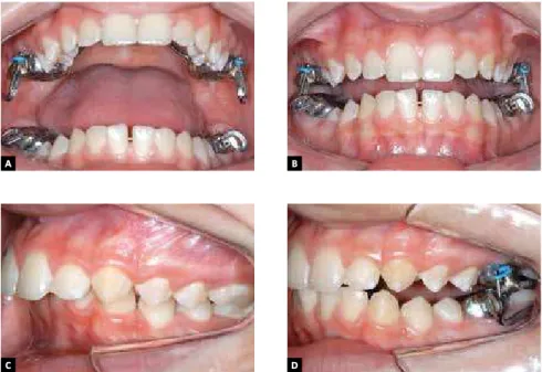
Mandibular Anterior Repositioning Appliance (MARA) is a fixed functional appliance, which therefore works irrespective of patient cooperation. It comprises four steel crowns cemented on the permanent first molars (Fig 1). These crowns have
loops that connect only when the patient occludes.2,10
Given that the MARA does not feature any systems involving telescopic tubes or springs connecting the jaws permanently, it allows greater freedom of
man-dibular movement.10 A lingual arch and transpalatal
bar are incorporated to the appliance to stabilize the upper and lower molars, respectively.
Once installed, the appliance prevents the mandible from closing in a more retruded or in a Class II position, quickly teaching the patient to position the mandible anteriorly both during function and at rest. Mandibu-lar advancement can be accomplished by inserting steel bands in the loop of the upper crown. There are four band sizes ranging from 1 to 4 mm in length. Thus, advancement can be gradual, while the patient is given the opportunity to adapt to the appliance.
The MARA allows concur-rent use with a rapid maxillary expansion appliance and a total or partial ixed orthodontic ap-pliance. To achieve orthopedic efects, a treatment time of 12
months is recommended.25
Some studies describe the skeletal and dental efects
pro-duced by the MARA.10,11,13,26,28
However, only two systematic studies have been published on the dental and skeletal changes observed in the correction of Class II, division 1. In 2003,
Pangrazio-Kulbersh et al26
eval-uated the cephalometric efects produced by the MARA in 30 patients (12 male and 18 female)
Figure 1 - A) MARA appliance in place with its components. B) The loops on the crowns create occlusal interference and hinder Class II occlusion. C, D) Photos taken before and after placement of the appliance.
A B
with initial ages ranging from 9.5 to 15.8 years, ater a mean treatment time of 10.7 months (8-17 months), and compared these with patients treated using the Herbst and Fränkel appliances and with patients with untreated Class II malocclusion. Results showed that the MARA appliance was efective in correcting Class II malocclusion by means of skeletal and dental chang-es. Proper molar relationship was obtained by means of 47% of skeletal changes and 53% of dental chang-es. Skeletal changes showed an increase in mandibu-lar length and in anterior and posterior facial heights, but were inefective in redirecting the maxilla. On the other hand, the dental efects included distalization of maxillary molars, mesialization of molars and incisors, and mild proclination of the lower incisors. In com-paring the MARA with the Fränkel and Herbst appli-ances, the former showed greater dentoalveolar efects than Fränkel, and less maxillary redirecting and less inclination of maxillary incisors than the Herbst.
Another study11 only assessed the effects of the
MARA appliance on the lower incisors in children (10.6 years), adolescents (14.9 years) and adults (33.7 years). It was used concurrently with a fixed orthodontic appliance for a period of 1.7 years in chil-dren, 1.3 years in adolescents, and 1.5 years in adults. In children, it was observed that the incisors protrud-ed by 0.4 mm and inclinprotrud-ed labially by 1.7°. Adoles-cents showed a 1.0 mm protrusion and a 3.6° procli-nation, whereas in adults, there was a 1.7 mm protru-sion and 4.5° proclination. They therefore concluded that the MARA appliance was effective in treating Class II patients in all groups, and the changes in the lower incisors were more substantial in adults than in adolescents and children. These changes were regard-ed as negligible comparregard-ed to other fixregard-ed functional appliances, whereas the use of the MARA combined with a fixed orthodontic appliance allowed a good control of lower incisor inclination.
More conclusive studies about the major dental and skeletal changes that result from the use of the MARA appliance are warranted as evaluations to support the evidence of such changes are extremely important. The main reason being that MARA is a ixed oral appliance designed to correct Class II malocclusion irrespective of patient compliance. It is thus an extremely
efec-tive and rapid solution for this kind of malocclusion.32
Therefore, this study aimed to evaluate through lateral
cephalograms the skeletal and dental efects produced by the MARA appliance during correction of Angle’s Class II, division 1 malocclusion.
MATERIAL AND METHODS
This research project was approved by the Ethics Committee of the School of Dentistry of Bauru (FOB-USP) and all patients signed an informed consent form before participating in the study.
Inclusion criteria were as follows: Bilateral Angle’s Class II, division 1 malocclusion, mandibular retrusion, no agenesis or loss of permanent teeth, no supernumerary teeth, no crowding or only mild crowding in the upper and lower dental arches, moderate or severe overjet, no previous orthodontic treatment. Thus, the sample con-sisted of 44 young patients divided into two groups.
The MARA group comprised 22 patients, 15 male and 7 female, with initial mean age of 11.99 years ± 1.20 years (minimum = 10.30, maximum = 15 years) treated with the MARA orthopedic appliance for an average of 1.00 year (minimum = 0.77, maximum = 1.25 years).
Patients started orthopedic treatment with the MARA appliance and were treated by the same student of the Doctoral Course in Dentistry, area of concen-tration: Orthodontics, FOB-USP. Care was taken to insert the appliance within one month ater the initial radiograph was taken. The MARA was installed with a transpalatal bar and a lingual arch in all patients. Only one patient presented initially with posterior crossbite involving only the irst molars and was then subjected to rapid maxillary expansion with a Hyrax appliance. The patients in this group were not subjected in ad-vance to tooth alignment and leveling, nor interproxi-mal stripping. All were treated until 2 mm, on average, beyond Class I molar relationship was obtained. Mal-occlusion correction was deemed successful when an occlusion in centric relation was achieved, i.e., when the mandible was positioned in a centric relation (CR) that matched the position of maximum intercuspa-tion (MI). Ater achieving this relationship, the appli-ance was kept in place for 6 months for retention pur-poses. The MARA appliance was thereater removed and the patient’s inal radiograph taken.
The initial mean age was 11.63 years ± 1.03 years (minimum = 10.16, maximum = 13.88 years); they were then monitored for a mean period of 1.18 years (minimum = 0.80, maximum = 2.01 years).
The patients were selected from a sample provided by the Center for the Study of Growth, FOB-USP, where a group of children was X-rayed and checked annually by the Department of Orthodontics with the purpose of developing a longitudinal sample of occlu-sions in children spanning from primary to permanent dentition. Subsequently, all patients were referred for orthodontic treatment, but some either chose to post-pone intervention to a later date, or showed no interest in the treatment, which allowed the authors to deine a control group.
The 44 children in the study sample met the follow-ing criteria:
» Bilateral Angle’s Class II, division 1 malocclusion. » Mandibular retrusion.
» No agenesis or no permanent teeth missing. » No supernumerary teeth.
» No crowding, or mild crowding in the upper and lower arches.
» Moderate or severe overjet.
» No previous orthodontic treatment.
Cephalometric method
The cephalometric tracing was performed on ac-etate tracing paper (Ultraphan) by the same researcher and then digitized (Numonics AccuGrid xnT, mod-el A30TL.F - Numonics Corporation, Montgom-eryville, Pa). Data were analyzed with Dentofacial Planner 7.2 sotware (Dentofacial Planner Sotware Inc., Toronto, Ontario, Canada); a 9.8% magniica-tion factor was corrected in the radiographs of the MARA Group MARA and 6% in the radiographs of the Control Group, since they were taken by diferent X-ray machines.
The lines and reference planes used in the study are shown in Figure 2A, comprising:
A) Line SN. B) Frankfort plane. C) Palatal plane.
D) Functional occlusal plane. E) Mandibular plane - GoGn. F) Mandibular plane - GoMe. G) Long axis of the upper incisor.
H) Long axis of lower incisor. I) Long axis of the molar. J) Long axis of the molar. K) NA line.
L) NB line. M) ANSperp line. N) Pogperp line.
The skeletal cephalometric measures are shown in Figure 2B, the dental measures are shown in Figure 2C and the dental relations corresponding to overjet (OJ), overbite (OB) and molar ratio (MR) are shown in Fig-ure 2D.
Superimposition of initial and inal tracings in the MARA group.
Statistical analysis and method error
Statistical analysis was performed with Statistica for Windows 6.0 sotware (StatSot Inc.). All results were considered statistically signiicant at p < 0.05.
To evaluate the method error, 20 randomly select-ed radiographs were once again tracselect-ed and measurselect-ed.
Paired t test was applied in order to estimate systematic
error. To evaluate random error, Dahlberg’s test was
used with the following formula: Se2 = ∑d2/2n, where
Se stands for Dahlberg’s error; d2 is the sum of the
squares of the diferences between the irst and second measurements, and 2n represents twice the number of cases in which the measurements were repeated.
Before starting the comparisons between groups, Kolmogorov-Smirnov test was applied and revealed that all variables had a normal distribution, thus allowing the application of parametric statistical tests.
Initial cephalometric compatibility and comparison of
cephalometric changes were assessed using Student’s t test.
RESULTS
Figure 2 - Cephalometric tracings. A) Refer-ence lines and planes. B) Skeletal cephalometric measures. C) Dental cephalometric measures. D) Dental relations: overjet (OJ), overbite (OB) and molar relationship (MR).
A
SN.POF SN.
GoGn
Co-Gn Co-A
SNB NSGn
B
C
M H
N
E
RM
Initial Final
TH TV 6.PP
6.GoMe
F K L G I
D
J
Figure 3 -Superimposition of initial and inal tracings in the MARA group.
A
C
B
D
SNA
ANB
Wits
AFAI
6-ENAperp 1-NB 6-Pogperp
1.NB
IMPA
1-NA
the MARA group, the maxilla showed a slight retru-sion and less growth. The mandible showed no sig-nificant differences in length and position, the maxil-lomandibular relationship improved significantly, the growth pattern was not affected, the lower anterior facial height increased and the occlusal plane experi-enced a counterclockwise rotation. The dentoalveo-lar changes found in the MARA Group were: Greater lingual inclination and retrusion of upper incisors, crown distalization and distal tipping of the long axis of the upper molars, considerable buccal inclination and protrusion of the lower incisors, and mesializa-tion and mesial tipping of mandibular molars. A re-duction in overjet and overbite was noted in the den-tal relations as well as a significant improvement in the molar relationship in the MARA Group.
DISCUSSION
The initial degree of compatibility between study groups directly inluences the reliability of the results of any cephalometric study. In this study, the groups were compatible in terms of initial age, observation time, gender distribution, initial severity and all cephalomet-ric variables, except overjet, which appeared greater in the MARA Group. A diference between groups can be ascribed to the need for a greater overjet that enabled mandibular advancement until the total correction of anteroposterior discrepancy between the arches was reached. Since the Control Group did not follow this protocol, some anterosuperior crowding was to be ex-pected, which may have contributed to a smaller overjet. Considering the skeletal changes produced by the MARA appliance, there was a restriction in maxillary
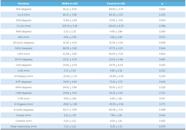
Table 1 - Results of ttest to assess initial cephalometric compatibility between groups.
Variables MARA (n=22) Control (n=22) p
SNA (degrees) 81.12 ± 3.03 80.64 ± 3.74 0.645
Co-A (mm) 85.12 ± 3.89 83.33 ± 5.23 0.203
SNB (degrees) 75.89 ± 2.83 75.95 ± 3.93 0.954
Co-Gn (mm) 105.54 ± 5.36 104.10 ± 6.33 0.366
ANB (degrees) 5.23 ± 1.20 4.69 ± 1.88 0.264
Wits (mm) 4.06 ± 2.09 3.06 ± 1.46 0.072
SN.GoGn (degrees) 31.30 ± 4.53 31.39 ± 5.34 0.949
NSGn (degrees) 68.39 ± 2.82 67.73 ± 4.25 0.544
LAFH (mm) 61.08 ± 3.82 60.83 ± 3.50 0.822
SN.FH (degrees) 20.51 ± 4.33 21.54 ± 5.48 0.495
1.NA (degrees) 25.82 ± 5.03 24.79 ± 6.03 0.542
1-NA (mm) 5.71 ± 2.19 4.90 ± 2.16 0.222
6-ENAperp (mm) -29.18 ± 2.14 -29.99 ± 2.40 0.244
6.PP (degrees) 74.45 ± 4.94 73.41 ± 3.72 0.434
IMPA (degrees) 94.91 ± 3.84 93.95 ± 6.27 0.539
1.NB (degrees) 24.63 ± 3.44 24.14 ± 5.45 0.722
1-NB (mm) 4.90 ± 1.68 4.49 ± 1.82 0.437
6-Pogperp (mm) -29.67 ± 1.48 -30.55 ± 2.56 0.173
6.GoMe (degrees) 81.57 ± 3.99 80.58 ± 3.55 0.388
Overjet (mm) 9.12 ± 1.78 7.99 ± 1.81 0.042
Overbite (mm) 5.02 ± 2.12 4.09 ± 2.41 0.180
growth due to a decrease in the SNA angle (0.6°) and much lessened growth in Co-A (0.8 mm) compared to the control group (2.4 mm). It should be remembered that functional appliances exert upward and backward forces on the maxilla. This “headgear efect” is caused by tension in the facial muscles in an attempt to reposition the mandible back to its uppermost and posterior-most
position.5,7,14,22 Given that the appliance contacts the
up-per arch, forces arising from the muscles and sot tissues are delivered by the appliance to the teeth and maxilla.
On the other hand, Pangrazio-Kulbersh et al26
eval-uated the efects of the MARA and although the SNA and A-Nperp values indicated maxillary retrusion, this change did not prove statistically signiicant compared to controls, despite a decrease of 0.4° and 0.2 mm in
an-nualized SNA and A-Nperp values. Almeida et al4 also
found no signiicant change in the sagittal position of the jaw ater treatment with the Herbst appliance, despite a greater decrease observed in the SNA angle (-0.9°) in the Herbst group compared with controls (-0.5°).
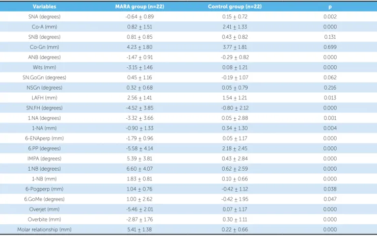
Table 2 - Results of comparing changes in cephalometric variables of the MARA and Control groups.
Variables MARA group (n=22) Control group (n=22) p
SNA (degrees) -0.64 ± 0.89 0.15 ± 0.72 0.002
Co-A (mm) 0.82 ± 1.51 2.41 ± 1.33 0.000
SNB (degrees) 0.81 ± 0.85 0.43 ± 0.82 0.131
Co-Gn (mm) 4.23 ± 1.80 3.77 ± 1.81 0.699
ANB (degrees) -1.47 ± 0.91 -0.29 ± 0.82 0.000
Wits (mm) -3.15 ± 1.46 0.08 ± 1.21 0.000
SN.GoGn (degrees) 0.45 ± 1.16 -0.19 ± 1.07 0.062
NSGn (degrees) 0.32 ± 0.68 0.05 ± 0.79 0.216
LAFH (mm) 2.56 ± 1.41 1.54 ± 1.21 0.013
SN.FH (degrees) -4.52 ± 3.85 -0.80 ± 2.12 0.000
1.NA (degrees) -3.32 ± 3.66 0.05 ± 2.88 0.001
1-NA (mm) -0.90 ± 1.33 0.34 ± 1.30 0.004
6-ENAperp (mm) -1.79 ± 0.96 0.05 ± 1.17 0.000
6.PP (degrees) -5.58 ± 4.14 2.18 ± 2.45 0.000
IMPA (degrees) 5.39 ± 3.81 0.43 ± 2.84 0.000
1.NB (degrees) 6.60 ± 4.07 0.62 ± 2.59 0.000
1-NB (mm) 1.83 ± 0.81 0.10 ± 0.66 0.000
6-Pogperp (mm) 1.04 ± 0.76 -0.42 ± 1.12 0.038
6.GoMe (degrees) 1.00 ± 2.62 -0.42 ± 1.95 0.047
Overjet (mm) -5.46 ± 2.01 0.07 ± 1.17 0.000
Overbite (mm) -2.87 ± 1.76 0.30 ± 1.11 0.000
Molar relationship (mm) 5.41 ± 1.38 0.22 ± 0.66 0.000
No statistically signiicant diference was found be-tween groups regarding changes in the mandible. Al-though a greater protrusion of the mandible was expect-ed with the orthopexpect-edic treatment due to its permanent anterior position, changes in the Control Group were found to be similar.
The efect of functional orthopedic treatment during mandibular growth currently poses ierce controversy and disagreement among authors. Pangrazio-Kulbersh
et al26 found a statistically signiicant increase in length
and some protrusion ater treatment with the MARA and with Herbst in terms of control.
Some studies also report a signiicant protrusion af-ter treatment with other types of ixed functional
appli-ances such as Jasper Jumper12 and Herbst,4,14,18 whereas
other studies found no signiicant changes in growth or
sagittal position of the mandible.6,15,20
It is noteworthy that most scientiic
publica-tions7,17,24,31 report an increase in the length of the
i.e., an increase in sagittal growth. Ruf and Pancherz30 concluded that the anterior-most position of the man-dible ater treatment appears to result from remodeling of the condylar joint and mandibular fossa. Popowich,
Nebbe and Major27 conducted a review of the skeletal
efects of the Herbst appliance and concluded that most studies using Magnetic Resonance Imaging (MRI) or Computed Tomography (CT) are not conclusive.
Functional appliances induce a rapid, if temporary, anterior mandibular displacement during the irst phase of treatment. This anterior repositioning of the mandi-ble extends the retrodiscal tissues which, in turn, deliver forces to the condyle and articular fossa, thereby
stimu-lating the process of bone remodeling in this region.34
Once the stimulus is removed, the process gradually
loses intensity until it reaches baseline levels.33
DeVincenzo9 reported in their study that a major
re-lapse of mandibular length increase occurs as a result of functional orthopedic treatment during the early phase of orthodontic treatment.
It is speculated that treatment with the MARA ap-pliance may have generated this greater stimulus towards growth in the irst six months or until a centric occlusion relationship was attained (CR=MI). Thereater, while keeping the appliance for retention, growth may have de-clined, and eventually the total sum was equivalent to the total mandibular growth found in the Control Group. No values were found above those genetically programmed.
There was a signiicant improvement in the relation-ship between basal bones. These changes can result from a combination of several efects on the dento-skeletal structures associated with normal craniofacial growth. In this study, the improvement observed in the relation-ship between basal bones may have occurred as a result of maxillary retrusion combined with normal growth and anterior displacement of the mandible.
A statistically signiicant increase in lower anterior
facial height was observed. Pancherz21 demonstrated
that the Herbst appliance caused a temporary increase in lower anterior facial height. McNamara Jr, Howe and
Dischinger17 reported an increase in anterior and
poste-rior facial height, which did not negatively inluence the
mandibular plane angle. Nahás19 also found that the
cra-niofacial growth pattern was not afected by treatment with the Herbst, as observed in this study.
There was a counterclockwise rotation in the functional occlusal plane (FOP) due to extrusion of the premolars, which were used as reference in con-structing the FOP. The MARA appliance allowed this extrusion due to a posterior open bite (Fig 1D), thus helping to correct the curve of Spee, and help-ing to determine a new FOP position.
The differences between groups are more evident and significant for the dentoalveolar variables, as
noted by Almeida et al,3 Neves20 and Lima.15
Regarding changes in inclination and anteropos-terior positioning of the upper incisors, these teeth exhibited lingual inclination and retrusion. The up-per molars showed crown distalization and distal tipping. Distalization of first molars is advocated by
some authors.7,17,19,24 Valant and Sinclair31 found that
the effects of the Herbst appliance on the maxilla (re-stricted displacement) and on the upper teeth (molar distalization) were similar to those of a headgear.
The lower incisors tipped labially signiicantly in pa-tients treated orthopedically. The MARA appliance is used as a lingual arch to stabilize mandibular molars. Since the resultant force is applied anteriorly, the efects of molar mesialization are relected mostly in the incisors.
Procli-nation therefore occurs in these teeth. Gönner et al11
ob-served a 3.6° labial inclination in adolescents, and 4.5° in adults treated with the MARA appliance.
Neves20 and Lima15 found a 2.6° incisor proclination
at the end of treatment with the Jasper Jumper. However, as noted by the authors, increased proclination must have occurred in the lower incisors during the period when the Jasper Jumper was in place. Later, ater the Jasper Jumper was removed and during the inishing phase, there may have been some lingual inclination (retroclination) of these teeth, resulting from both natural tendency to relapse and lingual torque placed in the antero-inferior region of the rectangular archwire. In assessing the efects of the Herbst
appliance on the mixed dentition, Almeida et al4 found a
sig-niicant proclination of the incisors, relected in a 5° increase
in IMPA. Pancherz and Hansen23 compared ive types of
lower anchorage provided by the Herbst appliance and con-cluded that none was efective in controlling lower incisor
proclination. However, Ruf, Hansen and Pancherz29
corroborating other authors who argue that such increased
inclination has not been shown to be harmful.1
Lower molars in the MARA Group experienced mesialization and mesial tipping statistically higher than in the Control Group. In correcting Angle’s Class II malocclusion it is desirable to move the molars mesially. This molar mesialization effect has
been reported in several studies.17,21,31
Regarding dental relationships, overbite and over-jet were significantly reduced by the MARA appli-ance. Furthermore, molar relationship showed sig-nificant improvement. The pronounced proclination noted in the lower incisors probably resulted from an evident correction of the molar relationship, and also contributed to a greater reduction in overbite.
CONCLUSIONS
MARA appliance was efective in correcting An-gle’s Class II, division 1 malocclusion, producing more dentoalveolar than skeletal efects, with skeletal chang-es occurring predominantly in the maxilla — where maxillary growth was restrained —, and no signiicant efects on the mandible. In addition, the MARA ap-pliance increased the vertical dimension of the face. Regarding dental changes, the upper incisors were inclined lingually and retruded. The upper molars showed distalization and distal tipping. The lower inci-sors inclined labially and protruded. The lower molars showed mesialization and mesial tipping. The MARA caused some signiicant improvement in dental rela-tions (overbite and overjet, and molar relarela-tionship).
1. Allais D, Melsen B. Does labial movement of lower incisors inluence the level of the gingival margin? A case-control study of adult orthodontic patients. Eur J Orthod. 2003;25(4):343-52.
2. Allen-Noble PS. Clinical management of the MARA: A manual for orthodontists and staf. Sturtevant: Ormco Corporation; 2002. 3. Almeida MR, Henriques JF, Almeida RR, Almeida-Pedrin RR, Ursi W.
Treatment efects produced by the Bionator appliance. Comparison with an untreated Class II sample. Eur J Orthod. 2004;26(1):65-72.
4. Ameida MRA, Henriques JFC, Almeida RR, Ursi W, Almeida-Pedrin RR, McNamara JA Jr. Efeitos dentoesqueléticos produzidos pelo aparelho de Herbst na dentadura mista. Rev Dental Press Ortod Ortop Facial. 2006;11(5):21-34.
5. Angelieri F. Comparação dos efeitos cefalométricos promovidos pelos aparelhos extrabucal cervical e pendulum [tese]. Bauru (SP): Universidade de São Paulo; 2005.
6. Covell DA Jr, Trammell DW, Boero RP, West R. A cephalometric study of Class II division 1 malocclusions treated with the Jasper Jumper appliance. Angle Orthod. 1999;69(4):311-20.
REFERENCES
7. Croft RS, Buschang PH, English JD, Meyer R. A cephalometric and tomographic evaluation of Herbst treatment in the mixed dentition. Am J Orthod Dentofacial Orthop. 1999;116(4):435-43.
8. Cureton SL, Regennitter F, Orbell MG An accurate, inexpensive headgear timer. J Clin Orthod. 1991;25(12):749-54.
9. DeVincenzo JP. Changes in mandibular length before, during, and after successful orthopedic correction of Class II malocclusions, using a functional appliance. Am J Orthod Dentofacial Orthop. 1991;99(3):241-57. 10. Eckhart JE, White LW. Class II therapy with the Mandibular Anterior
Repositioning Appliance. World J Orthod. 2003;4(2):135-44.
11. Gönner U, Ozkan V, Jahn E, Toll DE. Efect of the MARA appliance on the position of the lower anteriors in children, adolescents and adults with Class II malocclusion. J Orofac Orthop. 2007;68(5):397-412. 12. Karacay S, Akin E, Olmez H, Gurton AU, Sagdic D. Forsus Nitinol
13. Kinzinger G, Ostheimer J, Förster F, Kwandt PB, Reul H, Diedrich P. Development of a new ixed functional appliance for treatment of skeletal Class II malocclusion irst report. J Orofac Orthop. 2002;63(5):384-99.
14. Küçükkeleş N, Ilhan I, Orgun IA. Treatment eiciency in skeletal Class II patients treated with the jasper jumper. Angle Orthod. 2007;77(3):449-56. 15. Lima KJS. Comparação das alterações dentoesqueléticas promovidas
pelos aparelhos Jasper Jumper e Ativador combinado à ancoragem extrabucal seguido de aparelho ixo, no tratamento da má oclusão de Classe II, 1ª divisão [tese]. Bauru (SP): Universidade de São Paulo; 2007. 16. McNamara JA Jr, Bookstein FL, Shaughnessy TG. Skeletal and dental
changes following functional regulator therapy on Class II patients. Am J Orthod. 1985;88(2):91-110.
17. McNamara JA Jr, Howe RP, Dischinger TG. A comparison of the Herbst and Frankel appliances in the treatment of Class II malocclusion. Am J Orthod Dentofacial Orthop. 1990;98(2):134-44.
18. Moro A, Janson G, de Freitas MR, Henriques JF, Petrelli NE, Lauris JP. Class II Correction with the Cantilever Bite Jumper. A variant of the Herbst. Angle Orthod. 2009;79(2):221-9.
19. Nahás ACR. Estudo cefalométrico das alterações dento-esqueléticas da má oclusão de Classe I, divisão 1 tratada com o aparelho de Herbst e o aparelho extrabucal de tração occipital [tese]. Bauru (SP): Faculdade de Odontologia de Bauru, Universidade de São Paulo; 2004.
20. Neves LS. Estudo comparativo dos efeitos do tratamento da má oclusão de Classe II, 1ª divisão com os aparelhos Jasper Jumper e Bionator, associados ao aparelho ixo [tese]. Bauru (SP): Faculdade de Odontologia de Bauru, Universidade de São Paulo; 2007.
21. Pancherz H. The Herbst appliance--its biologic efects and clinical use. Am J Orthod. 1985;87(1):1-20.
22. Pancherz H, Anehus-Pancherz M. The headgear efect of the Herbst appliance: a cephalometric long-term study. Am J Orthod Dentofacial Orthop. 1993;103(6):510-20.
23. Pancherz H, Hansen K. Occlusal changes during and after Herbst treatment: a cephalometric investigation. Eur J Orthod. 1986;8(4):215-28. 24. Pancherz H, Zieber K, Hoyer B. Cephalometric characteristics of Class II
division 1 and Class II division 2 malocclusions: a comparative study in children. Angle Orthod. 1997;67(2):111-20.
25. Pangrazio-Kulbersh V. Entrevista. Rev Dental Press Ortod Ortop Facial. 2008;13(2):29-33.
26. Pangrazio-Kulbersh V, Berger JL, Chermak DS, Kaczynski R, Simon ES, Haerian A. Treatment efects of the mandibular anterior repositioning appliance on patients with Class II malocclusion. Am J Orthod Dentofacial Orthop. 2003;123(3):286-95.
27. Popowich K, Nebbe B, Major PW. Efect of Herbst treatment on temporomandibular joint morphology: a systematic literature review. Am J Orthod Dentofacial Orthop. 2003;123(4):388-94.
28. Rondeau B. MARA appliance. Funct Orthod. 2002;19(2):4-12, 14-6. 29. Ruf S, Hansen K, Pancherz H. Does orthodontic proclination of lower
incisors in children and adolescents cause gingival recession? Am J Orthod Dentofacial Orthop. 1998;114(1):100-6.
30. Ruf S, Pancherz H. Does bite-jumping damage the TMJ? A prospective longitudinal clinical and MRI study of Herbst patients. Angle Orthod. 2000;70(3):183-99.
31. Valant JR, Sinclair PM. Treatment efects of the Herbst appliance. Am J Orthod Dentofacial Orthop. 1989;95(2):138-47.
32. Von Bremen J, Pancherz H. Eiciency of early and late Class II Division 1 treatment. Am J Orthod Dentofacial Orthop. 2002;121(1):31-7. 33. Voudouris JC, Kuftinec MM. Improved clinical use of Twin-block and
Herbst as a result of radiating viscoelastic tissue forces on the condyle and fossa in treatment and long-term retention: growth relativity. Am J Orthod Dentofacial Orthop. 2000;117(3):247-66.
34. Voudouris JC, Woodside DG, Altuna G, Angelopoulos G, Bourque PJ, Lacouture CY, Kuftinec MM. Condyle-fossa modiications and muscle interactions during Herbst treatment, Part 2. Results and conclusions. Am J Orthod Dentofacial Orthop. 2003;124(1):13-29.