Epithelial rests of Malassez: from latent cells to active
participation in orthodontic movement
Bianca Silva e Silva1, Nathalia Carolina Fernandes Fagundes1, Bárbara Catarina Lima Nogueira1,
José Valladares Neto2, David Normando3, Rafael Rodrigues Lima1
1 Laboratory of Functional and Structural Biology, Institute of Biological
Science, Universidade Federal do Pará (Belém/PA, Brasil).
2 School of Dentistry, Universidade Federal de Goiás (Goiânia/GO, Brasil). 3 School of Dentistry, Universidade Federal de Pará (Belém/PA, Brasil).
Contact address: Rafael Rodrigues Lima
Laboratório de Biologia Estrutural e Funcional, Instituto de Ciências Biológicas, Universidade Federal do Pará, Rua Augusto Correa 1, Campus Básico, Belém/PA – CEP: 66.075-110 – E-mail: [email protected]
Introduction: The epithelial rests of Malassez (ERM) represent a group of cells in the periodontal ligament classi-cally consisting of latent or quiescent structures associated with pathological processes. However, recent evidence shows that these structures cannot be considered only as cellular debris. The ERM is a major tissue structure, with functions in maintaining the homeostasis of periodontal tissue, including the maintenance of orthodontic movement. Objec-tive: The present literature review aims at presenting the potential functions of ERM, with emphasis on orthodon-tic movement and the functional structure of the periodontium. Conclusion: ERM cells have a functional activity in modulation of orthodontic movement, trough their potential for differentiation, maintenance functions and the capacity of repairing periodontium.
Keywords: Epithelial cells. Periodontium. Tooth movement.
DOI: https://doi.org/10.1590/2177-6709.22.3.119-125.sar
How to cite this article: Silva e Silva B, Fagundes NCF, Nogueira BCL, Valladares Neto J, Normando D, Lima RR. Epithelial rests of Malassez: from latent cells to active participation in orthodontic movement. Dental Press J Orthod. 2017 May-June;22(3):119-25.
DOI: https://doi.org/10.1590/2177-6709.22.3.119-125.sar
Submitted: March 07, 2017 Revised and accepted: April 10, 2017
» The authors report no commercial, proprietary or financial interest in the products or companies described in this article.
» Patients displayed in this article previously approved the use of their facial and in-traoral photographs.
Introdução: os remanescentes epiteliais de Malassez (REM) se configuram como um grupo de células epiteliais pre-sentes no ligamento periodontal, classicamente consideradas estruturas latentes ou quiescentes, associadas a processos patológicos. Entretanto, ao longo dos anos, esse paradigma vem sendo rompido e hoje não mais são consideradas apenas como restos celulares, mas sim uma importante estrutura tecidual, com funções na manutenção da homeostase do perio-donto, inclusive durante a movimentação ortodôntica. Objetivo: na presente revisão da literatura, buscou-se apresentar as funções dessas estruturas, com ênfase nelas durante a movimentação ortodôntica, rompendo o conceito errôneo de que são meros restos celulares, e defendendo sua compreensão como uma estrutura funcional do periodonto. Conclu-são: os REM possuem uma atividade funcional na modulação da movimentação ortodôntica, por meio de seu potencial para diferenciação, de suas funções de manutenção e de sua capacidade de reparação periodontal.
INTRODUCTION
For a long time, the scientiic literature has not even hypothesized a role for the epithelial rests of Malas-sez (ERM). Initially, its latent or quiescent role was associated only to the formation of cysts and periapi-cal granulomas. However, over the last 50 years, stud-ies have gradually revealed the participation of ERM in the synthesis of mediators linked to the maintenance of
periodontal homeostasis.1,2
The functional aspects of ERM are afected by me-chanical forces in periodontal tissues during orthodontic tooth movement. Thus, this review aims at demystify-ing the idea that ERM constitute residual cells of odon-togenesis with no deined function, but rather represent cells with responsibilities in maintaining periodontal space and orthodontic movement.
FIRST STUDIES:
MORPHOLOGICAL CHARACTERIZATION
The ERM are resulting from cell fragmentation of the Hertwig epithelial root sheath (HERS), an api-cal extent of internal and external epithelial enamel, which stimulates the diferentiation of ectomesenchy-mal cells into odontoblasts, secreting root dentin. Ec-tomesenchymal cells also induce the diferentiation of the insertion periodontium. Ater these events, cells undergo fragmentation and remain as dispersed islands of epithelial cells in the periodontal ligament and
even-tually in the pulp.3,4
Initially described by Serres,5 these remaining cells
of the periodontal ligament are portrayed as enamel
or-gan debris. Decades later, Legros and Magiot6 pointed
to the epithelial origin of these remaining cells and sug-gested that they could be associated with the forma-tion of cysts, as dentigerous cysts and odontomas.
In-terestingly, Serres5 had approached the occurrence of
HERS degeneration in the same work that described the enamel organ debris, but did not relate these events
with each other.5,6
Malassez7 was the irst to detail the histological
char-acteristics of epithelial rests and their distribution in the periodontal ligament. In longitudinal and transverse slic-es of human specimens, Malassez noted the prslic-esence of epithelial cells that persisted around the roots (Figs 1, 2
and 3). Ater the irst descriptions made by Malassez,7
other authors described the distribution of ERM and its relation to the dental element in a more detailed manner.
These structures thus came to be known as the "epithe-lial rests of Malassez", receiving the eponymous name of
its principal investigator.7
Subsequently, ERM were considered as lymphatic
channels coated by epithelium and illed with lymph8.
In subsequent research, Black8 started to understand
them as glandular tissue,8,9 indings that have been
prov-en wrong by recprov-ent histological studies.
Von Brunn,through the study of the development
of the roots of teeth in rats, clariied the relationship
be-tween HERS and ERM.1 It was shown that the HERS
consists of the union between the outer and inner epi-thelia of the enamel organ in the future region of the
cementum-enamel junction.1 It was also suggested that
ERM produce endocrine glands and hormones that would prevent the union of cementum to the alveolar
bone.9 Orban10 stated that the epithelium rests contain
a pseudotubular structure, indicating the possibility of endocrine function.
Regarding the location, the ERM oten occurs in
the periodontal ligament,11,12 with a predilection for the
apex region, especially in the furca and cervical area
of the tooth, in ascending order of frequency.12 Later
studies also reported the presence of ERM in the pulp
and cement.13,14 Wentz et al.15 analyzed the type and
prevalence of ERM in the periodontal ligaments of rat molars. Three morphological variations were observed: small, proliferative and diferentiated. Regarding preva-lence, ERM were present in 50% of rats. ERM inci-dence decreased with increasing age. Regarding loca-tion, the results were similar to those reported by Reeve
and Wentz:16 the supralveolar area (47%), middle third
(30%), bifurcation region (15%) and apical region (8%).
Reeve and Wentz16 determined the frequency of
epithelial rests in patients aged 1–77 years, classifying diferent ERM types and locations on the periodontal ligament. The results showed that the ERM were pres-ent in all 31 specimens analyzed (regardless of age), but the incidence decreased with increasing age. As for the most frequent types of REM, small and diferentiated ones decrease with age, while proliferative ones increase with age. Among young patients, most ERM were lo-cated in the cervical area and accumulated at the apex with increasing age. The authors suggested that ERM were persistent vestigial structures in the periodontal ligament with a potential role in the development of
Loe and Waerhaug17 were the i rst to suggest a
com-patible function for ERM, having described the spatial distribution of these structures around the tooth root. According to the authors, the ERM were not isolated structures, forming a structure similar to a i shing net around tooth roots (Fig 1), giving ERM a role in main-taining the periodontal space and preventing
alveolo-dental ankylosis.17
Through scanning electron microscopy and
immu-nohistochemistry, Ten Cate18 reported lower metabolic
activity in cells of remaining epithelial components, suggesting a possible role of ERM should be disregard-ed in adult patients. However, later Trowbridge and
Shibata19 identii ed mitotic activity in epithelium rests
in experimental studies with animal models. Such evi-dence created possibilities for further investigations.
Using staining and electron microscopy to distin-guish ERM in the surrounding region, Valderhaug and
Zander20 noted the presence of numerous blood vessels
between the REM and dental cementum, with a larger number of ERM close to the cementoenamel junction, without, however, touching the cementum. This result suggests the participation of ERM in the regeneration and repair of cementum.
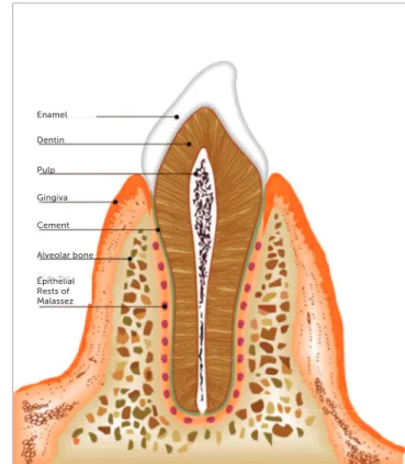
Figure 3 - Scheme showing the conformation of ERM in longitudinal slice. Figure 1 - Morphology of ERM in human tooth: ERM distributed around the tooth
root, forming something like a fishing net. Figure 2 - Conformation of ERM in a longitudinal slice in human tooth.
Epithelial Rests of Malassez Periodontal Ligament
Cement
Dentin
Pulp
Alveolar Bone Epithelial
Rests of Malassez
Periodontal Ligament
Bone Gingiva
Alveolar
Epithelial Rests of Malassez Alveolar bone Cement Gingiva Pulp Dentin Enamel
The bone morphogenetic proteins, especially BMP-2, as well as bone matrix proteins, act in regulating osteo-genesis, with an initial rise in the dental follicle. These molecules have proven potential for bone remodelling
throughout life.34,35
The prostaglandins, speciically prostaglandin E2, have a role in activating osteoclasts and can stimulate bone remodelling. Orthodontic treatment, in turn, is es-sentially a periodontal event that involves an acute inlam-matory response during its initial phase. Furthermore, prostaglandins are important mediators of inlamma-tion that play a key role in orthodontic movement, being synthesized by cyclooxygenases (COX). Prostaglandins stimulate bone resorption by increasing the number and activity of osteoclasts. Thus, prostaglandins have a major
inluence on the speed of orthodontic movement.36,37
In ERM, the release of epidermal growth factor (EGF)
was veriied as well.1,38 The EGF, compared to
proxim-ity to the periodontal apparatus, acts to lead resorption of the alveolar bone surface while keeping the human peri-odontal space in appropriate measure. The tooth move-ment induced by EGF causes an increase in periodon-tal tissue proliferation, which in turn is mediated by the continuous release induced by ERM. EGF plays an im-portant role in the prevention of ankylosis and
osteoclas-togenesis and cemenosteoclas-togenesis processes.1,36,38 Thus,
dur-ing orthodontic treatment, there is no ankylosis, for the orthodontic movement does not promote the destruction of ERM. EGF also stimulates ERM to proliferate and
maintain their organizational structure.39
Regarding the role of these receptors in
osteoclas-togenesis, Yi et al40 demonstrated the inhibition of the
signalling pathways afected in this process, which is also mediated by the receptor activator of nuclear factor kappa-B ligand (RANKL) and causes apoptosis through
caspase activation.40
From their potential in the context of maintaining periodontal insertion through the release of inlamma-tory mediator structures, proteins and growth factors, the ERM were identiied as key igures in maintaining homeostasis, assisting in the maintenance of space in the periodontal ligament, cementum repair and
regenera-tion processes and collagen renewal.1,14
Maintenance of the periodontal ligament space The ERM represent the only odontogenic epithe-lial structure able to persist in the periodontal ligament
However, Ten Cate21 stated that ERM had a role in the
formation of dental cysts, because the remaining lial cells from ERM behave the same way as other epithe-lial cells when the local tissue is changed. With support from deep periodontal ligament tissue, inlammation can
proliferate within such tissue, giving rise to dental cysts.21
Brice et al22 published a study that contributed to
change the paradigm of ERM as cell rests, reporting the presence of cells ultrastructurally similar to ERM in areas of root resorption with contiguous repair ater orthodontic movement at the root surface of human premolars (in patients undergoing maxillary expansion). The ultrastructural characteristics of these cells provid-ed evidence that they could be involvprovid-ed in mprovid-ediating cementogenesis in the root resorption process, opening
precedence for further research in this direction.22
FUNCTIONAL CHARACTERISTICS OF EPITHELIAL RESTS OF MALASSEZ
The ERM assists with the homeostasis of the peri-odontium, with a role in the renewal of collagen in the periodontal ligament, as well as the secretion of enamel proteins and bone matrix protein by these epithelial
cells and speciic enzymes.23,24 As part of this process,
the ERM release inlammatory mediators such as pros-taglandins and enamel proteins such as amelogenin and amelin. The latter also promote release of bone matrix proteins, BMP-2, osteopontin, osteoprotegerin and sia-loprotein, proteins that aid in the repair and periodontal regeneration phase, aside from contributing to the
re-pair of cementum itself.25,26,27
Enamel proteins have not limited participation in amelogenesis and are identiied as active in inducing mantle dentin formation, the deposition of cellular and acellular cementum, cementum integrity maintenance,
as well as a possible role as biological mediators.13,28,29
The presence of mechanical stresses along the periodon-tal ligament stimulates the release of bone matrix pro-teins, particularly osteopontin, which is associated with
the prevention of ankylosis and root resorption.22,27,30,31
In this process, ater the rupture of periodontal integ-rity, ERM release proteins of the enamel matrix and start to play a leading role in the early events of peri-odontal regeneration. The modulation of enamel pro-tein expression by epithelial remnants in the ERM thus suggests inherent regenerative ability, through directed
in adulthood, as that is the only structure able to per-sist in situations in which the periodontal ligament tis-sue is afected. These resistant structures are extremely important for maintaining homeostasis. ERM are able to promote the maintenance and regeneration of the
periodontal ligament space.41,42
This process is triggered by EGF and prostaglandins released from bone resorption of the alveolar surface and preserves the human periodontal space, which
var-ies in thickness from 0.20 to 0.40 mm.1 Such mediators
have been shown to be important in the bone resorption process, thus contributing to maintenance of the peri-odontal ligament.
The regeneration of periodontal tissue occurs through assistance in the renewal of cells that sufered some sort of damage. Damage to periodontal tissue is directly associated with orthodontic movement, where hyalinizing outbreaks have been recorded on the pres-sure side in the periodontal ligament, mainly during the
irst hours ater the application of force.43,44
ERM are structures that maintain the balance of the environment, contributing to the regeneration of the cementoblastic process. Unlike bone cells, cemento-blasts do not have receptors for remodelling mediators, requiring the beginning of a cascade of events that causes the pH to normalize. This promotes the inclusion of new collagen ibres at the damaged cementoblastic sur-face, induced by ERM, allowing the root surface to
re-generate and return to its normal state.27,36
The maintenance of this balance by REM allows for
maintenance of the periodontal ligament space45,46 and
prevents the occurrence of dentoalveolar ankylosis, con-tributing to reorganization of the periodontal ligament
during orthodontic tooth movement and resorption.47
Regeneration and repair of cement
From the release of substances and diferentiation of its epithelial cells, the remaining ERM play an im-portant role in the formation and repair of cementum, particularly in the diferentiation of cementoblasts with mesenchymal origin, preferably through the direct
transformation of epithelial cells to cementoblasts.1,48,49
This role is contrary to the events resulting from the induced orthodontic tooth movement. After the application of force, the change in the levels of periodontal capillary pressure can trigger blood sup-ply dysfunction until its collapse, according to the
force applied. This process can result in cementoblast
degradation and cementoid tissue layer formation.50
According to Hasegawa et al,25 the ERM are related
to the repair of cementum due to the release of enamel matrix proteins expressed during tooth development, which is directly associated with epithelial-mesenchy-mal interactions. In this procedure, epithelial cells as-sociated with ERM modify their distribution and ex-press BMP-2, osteopontin and ameloblastin.
Cementoblasts are involved in the process of
re-modelling and repair of the root surface.51 The
ap-plication of mechanical forces to the tooth occurs based on changes in vascular flow and the synthe-sis of prostaglandins, cytokines and growth factors. The actions of these mediators result in bone remod-elling, which involves resorption or bone deposition
on opposite side of the periodontal ligament.52,53
Prevention of alveolodental ankylosis
Alveolodental ankylosis is characterized by the ab-sence of tooth elastin due to the loss of periodontal
lig-ament cells associated with root resorption.27
This sit-uation occurs ater the removal of ERM, followed by the resorption of dental tissue, which is resorbed and replaced by bone. ERM keep the bone away from the root surface through the release of mediators such as EGF, which induces osteoclasia. In cases of trauma, where the ERM are damaged, leading to necrosis, mediators that promote osteoclasia may not be present in the required concentration, which, as a result, may
lead to alveolodental ankylosis.54,55
Orthodontic movement, however, does not pro-mote ERM necrosis, but, rather, just the reverse. When properly executed, the orthodontic movement is able to stimulate the secretory power of these cells, which prevents the recurrence of ankylosis. The growth in size of ERM during tooth movement indicates the level of the response to mechanical stimulation and its
possible role in tooth remodelling.36,37,47,56
Collagen renewal
the extracellular matrix, allowing for the transforma-tion of active collagenase enzymatic cleavage, resulting
in the synthesis of collagen ibres.43,52,53
The synthesis of collagen as well as changes in the matrix of the periodontal ligament contribute to the bone remodelling process, which induces orthodontic
forces57,58. During the early inlammatory events that
compose the induced movement process, the constric-tion of the microvasculature of the periodontal liga-ment results in focal areas of necrosis, with
histologi-cal features of hyalinization.34,59 During this process,
the synthesis of new collagen, in a process induced by ERM, promotes homeostasis refurbishment and the
maintenance of periodontal tissue.52
CONCLUSIONS
The ERM cells indicated to present many functions at tooth movement ater orthodontic forces. The capacity to repair periodontium and to prevent cementum dam-ages were connected with this process. Moreover, an in-crease in ankylosis events related to the processes and the hyalinization of periodontal tissue ater the application of mechanical forces during the aging process can be associ-ated to the potential of ERM. Otherwise, more studies are necessary to clarify these functions and consequences in daily orthodontics practice.
Authors' contribution
Conception/design of the study: NCFF, BCLN. Data acquisition, analysis or interpretation: BSS, NCFF, BCLN, JVN, RRL. Writing of the article: BSS, NCFF, BCLN, DN, RRL. Critical revision of the article: BSS, NCFF, BCLN, JVN, DN, RRL. Final approval of the article: BSS, NCFF, BCLN, JVN, DN, RRL. Obtained funding: BSS, NCFF, BCLN, DN, RRL. Overall re-sponsibility: BSS, NCFF, BCLN, JVN, DN, RRL.
1. Consolaro A, Consolaro MFM. ERM functions, EGF and orthodontic movement or Why doesn't orthodontic movement cause alveolodental ankylosis? Dental Press J Orthod. 2010 Mar-Apr;(2):24-32.
2. Keinan D, Cohen RE. The signiicance of epithelial rests of Malassez in the periodontal ligament. J Endod. 2013 May;39(5):582-7.
3. Becktor KB, Nolting D, Becktor JP, Kjaer I. Immunohistochemical localization of epithelial rests of Malassez in human periodontal membrane. Eur J Orthod. 2007 Aug;29(4):350-3. Epub 2007 July 2.
4. Xiong J, Mrozik K, Gronthos S, Bartold PM. Epithelial cell rests of Malassez contain unique stem cell populations capable of undergoing epithelial-mesenchymal transition. Stem Cells Dev. 2012 July 20;21(11):2012-25.
5. Serres A. Essai sur l’anatomie et la physiologie des dents ou nouvelle théorie de la dentition. Paris: Méquignon-Marvis; 1817.
6. Legros C, Magiot E. The origin and formation of dental follicle. Chicago: Jensen M. Clurg; 1850. 122 p.
7. Malassez L. Sur l’existence d'amas épithéliaux autour de la racine des dents chez l'homme adulte et a l'état normal. Arch Physiol. 1885;5:129-48.
8. Black GV. The periosteum and peridental membranes. Dent Rev (Wien). 1886-1987;1:1, 57, 113, 169, 233, 289, 353, 411.
9. Black G V. The ibers and glands of the periodontal membrane. Dent Cosmos. 1889;41:101-22.
10. Orban B. Oral histology and embryology. St. Louis: C V Mosby; 1953. 11. Meyer W. Lehrbuch der normalen Histologie und Entwicklungsgeschichte der
Zähne des Menschen. Munchen: Lehmanns Verlag; 1932.
12. Fisher G. Uber die bedeutung des epithels im periodontalen raum menschlicher und tierischer zahne. Vrljsschr F Zahn. 1932;48:413-25.
13. Hamamoto Y, Nakajima T, Ozawa H, Uchida T. Production of amelogenin by enamel epithelium of Hertwig’s root sheath. Oral Surg Oral Med Oral Pathol Oral Radiol Endod. 1996 June;81(6):703-9.
14. Cho MI, Garant PR. Expression and role of epidermal growth factor receptors during diferentiation of cementoblasts, osteoblasts, and periodontal ligament ibroblasts in the rat. Anat Rec. 1996 June;245(2):342-60.
15. Wentz FM, Weinmann JP, Schour I. The prevalence, distribution, and morphologic changes of the epithelial remnants in the molar region of the rat. J Dent Res. 1950;29(5):637-46.
16. Reeve CM, Wentz FM. The prevalence, morphology, and distribution of epithelial rests in the human periodontal ligament. Oral Surg Oral Med Oral Pathol. 1962 July;15:785-93.
17. Loe H, Waerhaug J. Experimental replantation of teeth in dogs and monkeys. Arch Oral Biol. 1961 Apr;3:176-84.
18. Ten Cate AR. The histochemical demonstration of speciic oxidative enzymes and glycogen in the epithelial cell rests of Malassez. Arch Oral Biol. 1965 Mar-Apr;10:207-13.
19. Trowbridge HO, Shibata F. Mitotic activity in epithelial rests of Malassez. Periodontics. 1967 May-June;5(3):109-12.
20. Valderhaug J, Zander HA. Relationship of “epithelial rests of Malassez” to other periodontal structures. Periodontics. 1967 Sept-Oct;5(5):254-8. 21. Ten Cate AR. The epithelial cell rests of Malassez and the genesis of the
dental cyst. Oral Surg Oral Med Oral Pathol. 1972 Dec;34(6):956-64. 22. Brice GL, Sampson WJ, Sims MR. An ultrastructural evaluation of the
relationship between epithelial rests of Malassez and orthodontic root resorption and repair in man. Aust Orthod J. 1991;12(2):90-4. 23. Bykov VL. [Epithelial cell rests of Malassez: tissue, cell, and molecular
biology]. [Article in Russian]. Morfologiia. 2003;124(4):95-103. 24. Luan X, Ito Y, Diekwisch TGH. Evolution and development of Hertwig’s
epithelial root sheath. Dev Dyn. 2006 May;235(5):1167-80. 25. Hasegawa N, Kawaguchi H, Ogawa T, Uchida T, Kurihara H.
Immunohistochemical characteristics of epithelial cell rests of Malassez during cementum repair. J Periodontal Res. 2003 Feb;38(1):51-6. 26. Wang Y, Lv L, Yu X, Zhang T, Li S. The characteristics of epithelial cell rests
of Malassez during tooth eruption of development mice. J Mol Histol. 2014 Feb;45(1):1-10.
27. Xiong J, Gronthos S, Bartold PM. Role of the epithelial cell rests of Malassez in the development, maintenance and regeneration of periodontal ligament tissues. Periodontol 2000. 2013 Oct;63(1):217-33.
28. Hasegawa N, Kawaguchi H, Ogawa T, Uchida T, Kurihara H.
Immunohistochemical characteristics of epithelial cell rests of Malassez during cementum repair. J Periodontal Res. 2003 Feb;38(1):51-6. 29. Fong CD, Hammarström L. Expression of amelin and amelogenin in
epithelial root sheath remnants of fully formed rat molars. Oral Surg Oral Med Oral Pathol Oral Radiol Endod. 2000 Aug;90(2):218-23.
30. Wallace JA, Vergona K. Epithelial rests’ function in replantation: is splinting necessary in replantation? Oral Surg Oral Med Oral Pathol. 1990 Nov;70(5):644-9.
31. Rincon JC, Young WG, Bartold PM. The epithelial cell rests of Malassez—a role in periodontal regeneration? J Periodontal Res. 2006 Aug;41(4):245-52.
32. Hamamoto Y, Hamamoto N, Nakajima T, Ozawa H. Morphological changes of epithelial rests of Malassez in rat molars induced by local administration of N-methylnitrosourea. Arch Oral Biol. 1998 Nov;43(11):899-906. 33. Nishio C, Wazen R, Kuroda S, Mofatt P, Nanci A. Disruption of periodontal
integrity induces expression of apin by epithelial cell rests of Malassez. J Periodontal Res. 2010 Dec;45(6):709-13.
34. Wise GE, King GJ. Mechanisms of tooth eruption and orthodontic tooth movement. J Dent Res. 2008 May;87(5):414-34.
35. Hasegawa N, Kawaguchi H, Ogawa T, Uchida T, Kurihara H.
Immunohistochemical characteristics of epithelial cell rests of Malassez during cementum repair. J Periodontal Res. 2003 Feb;38(1):51-6. 36. Consolaro A. The concept of root resorptions or root resorptions are not
multifactorial, complex, controversial or polemical! Dental Press J Orthod. 2011 July-Aug;16(4):19-24.
37. Kale S, Kocadereli I, Atilla P, Aşan E. Comparison of the efects of 1,25 dihydroxycholecalciferol and prostaglandin E2 on orthodontic tooth movement. Am J Orthod Dentofacial Orthop. 2004;125(5):607-14. 38. Wang HM. Detection of lysosomal enzymes derived from pig periodontal
ligament ibroblasts and their ability to digest collagen ibrils and proteoglycan. Arch Oral Biol. 1982;27(9):715-20.
39. Theslef I. Epithelial cell rests of Malassez bind epidermal growth factor intensely. J Periodontal Res. 1987;22(5):419-21.
40. Yi T, Lee HL, Cha JH, Ko SI, Kim HJ, Shin HI, et al. Epidermal growth factor receptor regulates osteoclast diferentiation and survival through cross-talking with RANK signaling. J Cell Physiol. 2008 Nov;217(2):409-22. 41. Wesselink PR, Beertsen W. The prevalence and distribution of rests of Malassez
in the mouse molar and their possible role in repair and maintenance of the periodontal ligament. Arch Oral Biol. 1993;38(5):399-403.
42. Mutoh N, Nakatomi M, Ida-Yonemochi H, Nakagawa E, Tani-Ishii N, Ohshima H. Responses of BrdU label-retaining dental pulp cells to allogenic tooth transplantation into mouse maxilla. Histochem Cell Biol. 2011;136(6):649-61.
43. von Böhl M, Maltha JC, Von Den Hof JW, Kuijpers-Jagtman AM. Focal hyalinization during experimental tooth movement in beagle dogs. Am J Orthod Dentofacial Orthop. 2004;125(5):615-23.
44. von Böhl M, Kuijpers-Jagtman AM. Hyalinization during orthodontic tooth movement: a systematic review on tissue reactions. Eur J Orthod. 2009 Feb;31(1):30-6.
45. Shimonishi M, Hatakeyama J, Sasano Y, Takahashi N, Komatsu M, Kikuchi M. Mutual induction of noncollagenous bone proteins at the interface between epithelial cells and ibroblasts from human periodontal ligament. J Periodontal Res. 2008 Feb;43(1):64-75.
46. Shimono M, Ishikawa T, Ishikawa H, Matsuzaki H, Hashimoto S, Muramatsu T, et al. Regulatory mechanisms of periodontal regeneration. Microsc Res Tech. 2003 Apr 1;60(5):491-502.
47. Talic NF, Evans CA, Daniel JC, Zaki AEM. Proliferation of epithelial rests of Malassez during experimental tooth movement. Am J Orthod Dentofacial Orthop. 2003 May;123(5):527-33.
48. Kagayama M, Sasano Y, Zhu J, Hirata M, Mizoguchi I, Kamakura S. Epithelial rests colocalize with cementoblasts forming acellular cementum but not with cementoblasts forming cellular cementum. Acta Anat (Basel). 1998 Jan;163(1):1-9.
49. Nam H, Kim J, Park J, Park JC, Kim JW, Seo BM, et al. Expression proile of the stem cell markers in human Hertwig’s epithelial root sheath/Epithelial rests of Malassez cells. Mol Cells. 2011;31(4):355-60.
50. Roscoe MG, Meira JBC, Cattaneo PM. Association of orthodontic force system and root resorption: A systematic review. Am J Orthod Dentofacial Orthop. 2015 May;147(5):610-26.
51. Diercke K, König A, Kohl A, Lux CJ, Erber R. Human primary cementoblasts respond to combined IL-1β stimulation and compression with an impaired BSP and CEMP-1 expression. Eur J Cell Biol. 2012 May;91(5):402-12. 52. Retamoso LB, Cunha TDMA, Knop LAH, Shintcovsk RL, Tanaka OM.
Organization and quantiication of the collagen ibers in bone formation during orthodontic tooth movement. Micron. 2009;40(8):827-30. 53. Talic NF, Evans CA, Daniel JC, Zaki AEM. Proliferation of epithelial rests of
Malassez during experimental tooth movement. Am J Orthod Dentofacial Orthop. 2003 May;123(5):527-33.
54. Takahashi K, Shimonishi M, Wang R, Watanabe H, Kikuchi M. Epithelial-mesenchymal interactions induce enamel matrix proteins and proteases in the epithelial cells of the rests of Malassez in vitro. Eur J Oral Sci. 2012 Dec;120(6):475-83.
55. Lindskog S, Blomlöf L, Hammarström L. Evidence for a role of odontogenic epithelium in maintaining the periodontal space. J Clin Periodontol. 1988 July;15(6):371-3.
56. Yamanaka T, Sakamoto A, Tanaka Y, Zhang Y, Hayashido Y, Toratani S, et al. Isolation and serum-free culture of epithelial cells derived from epithelial rests of Malassez in human periodontal ligament. In Vitro Cell Dev Biol Anim. 2000 Sept;36(8):548-53.
57. Krasny M, Zadurska M, Cessak G, Fiedor P. Analysis of efect of non-steroidal anti-inlammatory drugs on teeth and oral tissues during orthodontic treatment. Report based on literature review. Acta Pol Pharm. 2013 May-June;70(3):573-7.
58. von Böhl M, Kuijpers-Jagtman AM. Hyalinization during orthodontic tooth movement: a systematic review on tissue reactions. Eur J Orthod. 2009 Feb;31(1):30-6.