Abstract
Submitted: December 24, 2016
Accepted: April 19, 2017
Systemic effect of mineral
aggregate-based cements: histopathological
analysis in rats
Objective: Several studies reported the local tissue reaction caused by mineral aggregate-based cements. However, few studies have investigated the systemic effects promoted by these cements on liver and kidney when directly applied to connective tissue. The purpose of this in vivo study was to investigate the systemic effect of mineral aggregate-based cements on the livers and kidneys of rats. Material and Methods: Samples of Mineral Trioxide Aggregate (MTA) and a calcium aluminate-based cement (EndoBinder)
After 7 and 30 d, samples of subcutaneous, liver and kidney tissues were submitted to histopathological analysis. A score (0–3) was used to grade the
reaction (2) observed for 7 d in the subcutaneous tissue decreased with
for MTA, persisting until the end of the analysis. Liver functions increased
the liver and kidney. For MTA, the reactions were more accentuated.
Ke yw or ds:
Kidney. Lucas da Fonseca Roberti GARCIA1
Claudia HUCK2
Fernando Augusto Cintra
MAGALHÃES3
Pedro Paulo Chaves de SOUZA3
Carlos Alberto de SOUZA COSTA3
http://dx.doi.org/10.1590/1678-7757-2016-0634
1Universidade Federal de Santa Catarina, Centro de Ciências da Saúde, Departamento de
Odontologia, Área de Endodontia, Florianópolis, SC, Brasil.
2Univ. Estadual Paulista, Faculdade de Odontologia de Araraquara, Departamento de Odontologia
Restauradora, Araraquara, SP, Brasil.
3Univ. Estadual Paulista, Faculdade de Odontologia de Araraquara, Departamento de Fisiologia e
Patologia, Araraquara, SP, Brasil.
Corresponding address: Carlos Alberto de Souza Costa Departamento de Fisiologia e Patologia -Faculdade de Odontologia de Araraquara UNESP -
Introduction
Mineral Trioxide Aggregate (MTA) is one of the most
used biomaterials in endodontics currently3. It is a
Portland cement (75.0%) (wt) added to dehydrated
CaSO4 (5.0%) and Bi2O3 (20.0%), the latter being
responsible for its radiopacity2. Because of its hydraulic
nature, after the addition of water, the setting process
of the cement begins by initially forming a hydrated
silica gel3.
There is few information regarding the MTA cement manufacturing process. However, the companies
the cement under conditions that guarantee its safe use. Portland cement, the main raw material of
MTA, has several compounds in its formulation, the
following being the mostly used ones: SiO2 (21.2%);
CaO (68.1%); Al2O3 (4.7%); MgO (0.48%) and Fe2O3 (1.89%)3
process before being used, studies have proved the
presence of heavy metals in the composition of MTA,
among them As, Cr and Pb11,12. Nevertheless, it is
12.
In spite of its proven biocompatibility and
bioactivity20,21, undesirable effects of Bi
2O3 have
been pointed out on the rate of Ca++ ions released
from MTA, thus compromising its performance as a
reparative material3,25. Furthermore, studies have
reported that heavy metal salts, mainly Bi and Cd, are responsible for the expression of the heme
oxygenase-1 enzyme (HO-1) in different cell lineages,
compromising their regulatory functions and protective
mechanisms4,22.
In a recent study, Khalil and Eid18 (2013) reported
liver and kidneys of rats, questioning the systemic
compatibility of this type of cement. Therefore, new biomaterials have been developed to minimize adverse
reactions caused by reparative cements when in
contact with living tissues13.
A novel calcium aluminate-based cement
(EndoBinder - Patent Number PI0704502-6) with
similar clinical applications of MTA was developed at
the Federal University of São Carlos (UFSCar - Brazil) to address the deficiencies of the silicate-based
cement3,11,12,25
as ZnO, have been proposed as a feasible alternative
to the compounds currently used for this purpose26.
Therefore, the aim of this in vivo study was to investigate the local (subcutaneous) and systemic (liver and kidney) effects of MTA and a new reparative
calcium aluminate-based cement (EndoBinder)
hypothesis tested was that there would be no
cements in the different evaluated tissues.
Material and methods
Animals
The entire study was developed according to the guidelines of the Research Ethics Committee on the
Use of Animals (Process CEUA No. 3/2013), and the
National Institutes of Health guide for the care and
use of laboratory animals (NIH Publications No. 8023, revised 1978). For this study, 40 male rats (Rat t us novergicus, Wistar), weighing 300 g, were selected. The number of specimens per group was determined based on other biological studies that used a similar quantity of animals1,13,14. In addition, sample size was
calculated to set a number of needed specimens to
among control and experimental groups. Animals were kept in plastic cages (40x32x17 cm) especial for this
purpose, accommodated in an acclimatized bioterium
(temperature: 21–23°C/relative humidity: 60±5%/12 h light-dark cycle), and received balanced rations (Nuvilab, Colombo, PR, Brazil) and water ad libit um
during the experiment.
Experimental design and mineral
aggregate-based cements treatment
The tested cements were distributed as it follows: EndoBinder (Binderware, São Carlos, SP, Brazil) +
20% (wt) Bi2O3 (EBBO Group), EndoBinder + 20%
ZnO (EBZnO Group), and White MTA (Ângelus,
Londrina, PR, Brazil) (WMTA Group). Then, they were manipulated according to the manufacturers’
recommendations. For EndoBinder, the proportion of
1 g of powder to 0.21 mL of distilled water was used,
whereas for White MTA, the recommended proportion was of one dose of powder (0.15 mg) to 1 drop (0.5
mL) of distilled water.
After manipulating the cements, pre-sterilized
g of cements using a sterile Lentulospiral (Dentsply/
Maillefer, Ballaigues, Switzerland)13
tubes with the tested cements, one of their extremities
was heat-sealed to avoid cement extravasation13.
The animals were anesthetized by intraperitoneal
administration with a 10% solution of ketamine chloride (Ketamina Agener, União Química Farmacêutica
Nacional S/A, Embu-Guaçu, SP, Brazil) (75 mg/kg) and
xylazine(Dopazer, Laboratórios Calier S.A, Barcelona, Spain) (10 mg/kg). After trichotomy of the animal’s
dorsum, to perform the surgical procedure, the area
was disinfected with a 5% Povidone-iodone (PVP-I)
solution. A 5 mm-long incision was made at the center of the animal’s dorsum with a No.15 scalpel blade.
The subcutaneous tissue was laterally divulsed with
a blunt-tipped scissor, from the center of the incision.
A surgical pocket with an average depth of 20 mm was obtained, in which the polyethylene tube with
the cement under test was implanted longitudinally,
in parallel to the incision, letting the open extremity
in direct contact with the subcutaneous tissue. Each cement was implanted in groups of 10 animals.
Each group of 10 animals was distributed into two
sub-groups (n=5) according to the time interval of
analysis (7 and 30 d). In other 10 animals, surgical pockets were prepared, in which empty polyethylene
tubes were implanted, thus establishing the negative
control group. After concluding the polyethylene tubes
implants, the wound was sutured with a 3/0 silk thread (Ethicon, São José dos Campos, SP, Brazil).
After experimental time intervals of 7 and 30
d, animals were anesthetized for blood sample
collection (n=5). Blood (5 mL) was collected under vacuum by saphenous vein puncture18 of the
retro-orbital plexus with the aid of heparinized capillary
tubes (BD Vacultainer, BD - Benton, Dickinson and Company, Franklin Lakes, NJ, USA). Afterwards,
to perform biopsies of subcutaneous tissues, livers
and kidneys. To standardize the sampling, only the left kidney of each animal was collected for analysis.
After biopsies, the tissues and organs collected were
immediately immersed in a 10% formalin solution
(Merck, Darmstadt, Germany), where they remained for 24 hours at room temperature. After this period,
all surgical parts were submitted to routine laboratory
processing. Polyethylene tubes were removed before
tissue sectioning, then the tissues were sectioned
longitudinally, in 5 μm-thick semi-serial cuts,
considering 10 cuts were discarded and 5 were stained
with hematoxylin-eosin (Merck).
Histopathological examination
Histopathological examination was performed
blindly and double-checked by two previously
calibrated examiners with a concordance index of
94% between them, using an optical light microscope (Axio Star Plus, Carl Zeiss, Oberköchen, Germany).
The local (subcutaneous tissue) and systemic
(liver and kidney) biocompatibility of the cements were evaluated considering the presence of the
following histopathological events: inflammatory
(fibroblasts and blood vessels) and macrophagic
activity (mononuclear phagocytes and multinucleated
giant cells)5,13. In addition to these tissue events, the
presence of micro and macrovesicular steatosis, and the occurrence of apoptosis in hepatic cells were also
the liver5, whereas, for the kidney, the presence of
hypercellularity in the cortex was used18. According
17 (2008), and
based on the tissue responses stimulated by different
cements and the control group, a score was used to
quantify the extension of these events, with tissue
(1) discrete, (2) moderate, and (3) severe. In the case
of subcutaneous implants, the thickness (μm) of the
Axio Vision 4.6 software (Axio Star Plus).
Blood sample analyses
The blood samples previously collected in
heparinized tubes were transferred to Eppendorf tubes (Eppendorf do Brasil Ltda., São Paulo, SP, Brazil), and
then centrifuged to separate the plasma and the solid
constituents of the blood. After centrifugation, 1 mL
of plasma was collected and submitted to biochemical analysis to quantify alanine aminotransferase
(ALT) and aspartate aminotransferase (AST) serum
enzymes, which are indicators of liver functions, and
urea and creatinine levels (of renal function).
Statistical analysis
After testing the normality of the sample
(Shapiro-Wilk test - P>.05), values obtained in different
two-way analysis of variance (ANOVA) and the Bonferroni’s
post hoc test (P<.05). Statistical analysis was performed with the OriginPro 8 SRO program (OriginLab Corp., Northampton, MA, USA).
Results
Subcutaneous tissue
F r e q u e n c i e s o b s e r v e d f o r e a c h o f t h e
histopathological events evaluated in different
experiment time intervals can be seen in Table 1.
EBBO group
In the initial time interval of 7 d, we observed an
adjacently to the tube opening where the material was
placed in contact with the tissue. This capsule had
by the presence of polymorphous and mononuclear cells. We noted discrete collagenization associated with
observed giant multinuclear cells and macrophages
Histopathological events
Experimental periods
7 Days 30 Days
EBBO EBZnO WMTA *C EBBO EBZnO WMTA *C
(polymorphonuclear and mononuclear)
2 2 2 1 1 1 1 1
Fibroblasts 2 2 2 1 2 2 2 1
Blood vessels 2 2 2 1 1 1 2 1
Macrophages 2 2 2 1 1 1 1 1
Giant multinuclear cells
2 2 2 1 0 0 1 0
2 2 2 1 1 1 1 1
n=5
Table 1- Frequencies of histopathological events observed in the subcutaneous tissue in each group at different experimental periods
Figure 1- Photomicrographs of histological sections (5 μm) of subcutaneous tissue stained with hematoxylin & eosin. (A) EBBO 7 d: We
phagocyting biomaterial residues dispersed at the
tube opening. Small areas of tissue necrosis could be
activity. The inflammatory capsule at the tube
a characteristic of the tissue repair process (Figures
1A and E). In the time interval of 30 d, we considered
EBZnO group
As described in the previous group, in the initial
time interval (7 d), it was possible to observe
opening, the main area of analysis. We also noted
presence of macrophages phagocyting dispersed biomaterial residues and mononucleated giant cells in
the region. In addition, we noted an area of necrosis
similar to the EBBO Group in this initial time interval
of analysis. After 30 d, there was a reduction in
blood vessels in the tissue. No areas of mineralization
analysis (Figures 1B and F). As observed in EBBO
Group, after a period of 30 d, we established a general
Group.
WMTA group
After 7 d, at the tube opening, it was possible
to identify the formation of a disorganized capsule
with moderate fibroangioblastic proliferation and numerous blood vessels associated with moderate
giant multinucleated cells phagocyting dispersed
material residues, as well as areas of tissue necrosis and absence of locally mineralized foci
at the end of analysis, but there was the formation of
in a normal repaired tissue (Figures 1C and G). The
interval of 30 d was also 1 (discrete).
Control group
In the time interval of 7 d, there was a formation of a thin inflammatory capsule with a discrete
quantity of polymorphous and mononuclear. Discrete
occurred with neoformation of blood vessels and without signs of congestion. A discrete quantity
of mononuclear phagocytes was noted without
signs of tissue necrosis. After 30 d, we detected a
local macrophagic activity, but we did not see giant
small caliber blood vessels, which characterizes
complete tissue repair (Figures 1D and H). For the
(discrete) at 30 d.
capsule of different groups with distinct experimental
times can be seen in Figures 2A and B.
decreasing extent for all groups (P<.05). The capsule
formed adjacently to the different tested cements in
to the Control Group (P<.05). We observed this trend
at the end of analysis Thickness values were as it
follows: EBBO (86.09 μm); EBZnO (82.80 μm); and
WMTA (85.43 μm), with no statistical difference among them (P>.05). However, these groups were statistically
different from the Control Group (46.75 μm)(P<.05).
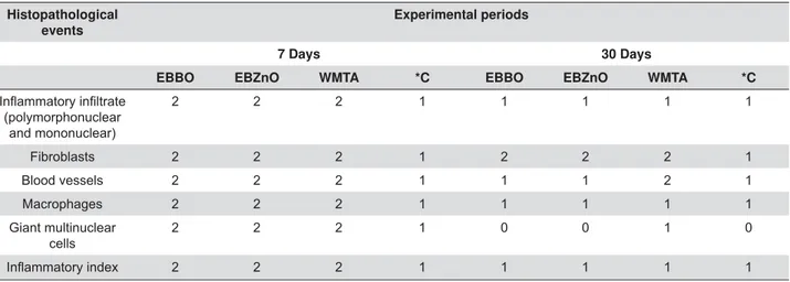
Liver and kidney
The frequencies observed for each of the histopathological events evaluated in the different
experimental time intervals can be seen in Table 2.
EBBO group
close to the blood vessels, and mononuclear cells
inside the tributary vessels of the hepatic portal
system. Most of these vessels seemed distorted,
dilated and had a ruptured endothelial layer. In addition, we noted the congestion of these vessels and
the presence of microvesicular steatosis, characteristic
of a degenerative process. After 30 d, there was still
Figure 2- μ
Histopathological events
Liver Kidney
Experimental periods
7 Days 30 Days 7 Days 30 Days
EBBO EBZnO WMTA *C EBBO EBZnO WMTA *C EBBO EBZnO WMTA *C EBBO EBZnO WMTA *C
(polymorphonuclear and mononuclear)
2 2 2 0 2 2 2 0 2 2 2 0 1 1 2 0
Fibroblasts 2 2 2 1 1 1 2 0 2 2 2 1 2 2 2 0
Blood vessels 3 3 3 1 2 2 2 0 2 2 2 1 1 1 2 1
Macrophages 2 2 2 0 1 1 2 0 1 1 1 0 1 1 1 0
Giant multinuclear cells
1 1 2 0 1 1 1 0 1 1 1 0 1 1 1 0
Steatosis 2 2 2 0 1 1 2 0 - - -
-Apoptosis 1 1 2 0 1 1 1 0 - - -
-Hipercellularity - - - - - - - - 2 2 2 0 1 1 2 0
2 2 2 0 1 1 2 0 2 2 2 0 1 1 2 0
n=5
notable reduction in mononuclear phagocytes. It was
also possible to observe hepatocytes in a process of
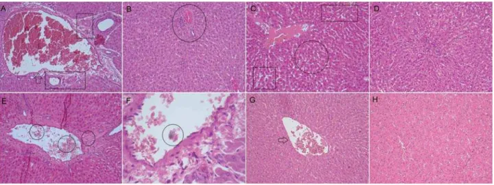
apoptosis; in a smaller quantity, however (Figures 3A and B). At 7 d, in the kidney, the main observed events
were vascular congestion and moderate increase of
local cellularity, triggering an increase in renal cortex
density, typical of a tissue degenerative process. However, at the period of 30 d, we noted a reduction
in the extent of these events in the affected areas
(Figures 4A and B). At the end of the analysis, we
index for the liver and kidney.
EBZnO group
The histopathological events observed in both
time intervals of analysis were similar to those from the previous groups, both for livers and kidneys. We
polymorphous and mononuclear cells of moderate
extent in the initial period. Most blood vessels seemed congested, and had a ruptured endothelial layer, which
the formation of various accessory vessels. In addition,
we noted the presence of moderate microvesicular steatosis and several areas of intracellular edema. At
the period of 30 d, we also noted a small quantity of
hepatocytes in mitosis and apoptosis and a decrease
the kidney, we observed vascular congestion and hypercellularity with an increase in the renal cortex
density, followed by reduction of these features at the
1 (discrete).
WMTA group
At the period of 7 d, despite involving larger
areas than the ones observed in the groups treated with different versions of EndoBinder, we noted the
extent in the liver. Blood vessels seemed congested
cells within and adjacently to them. In addition, we
observed microvesicular steatosis and several areas
of regional necrosis. In only one of the samples, we
detected a foreign body in one of the tributary vessels of the hepatic portal system, associated with the
dispersed residual material tested in this group. After
and associated with moderate fibroangioblastic proliferation, with several hepatocytes that seemed
to be undergoing mitosis and apoptosis. We also
Figure 3- Photomicrographs of histological sections (5 μm) of liver stained with hematoxylin and eosin. (A) EBBO 7 d: Details of tributary vessel of distorted, dilated and congested hepatic portal system with ruptured endothelial layer (arrow). The stroma presents moderate
the increased size of the hepatocytes, characterizing a cell degeneration process (circle). We could also detect several intracellular edema
treated with EndoBinder. Note the presence of polymorphous and mononuclear cells within and surrounding the blood vessels (circle).
detected punctual areas of necrosis and hydropic
degeneration in the tissue (Figures 3E and F). After 7 d, we observed vascular congestion and hypercellularity
in the kidney, with an increase in renal cortex density
and the presence of malformed glomerulus. In
some samples, it was possible to detect residues of degenerated glomerulus. After 30 d, we observed
the intensity of diminished renal response with tissue
reorganization, a characteristic of a repair process.
However, this decrease in the extent of the observed histological events was lower than that observed in
the groups treated with EndoBinder (Figures 4E and
F). After 30 d, we considered 2 (moderate) as the
Control group
In the control group, animals underwent the same
operative procedures, but we did not expose them
to mineral aggregate-based cements. We observed tributary vessels of the hepatic portal system with
normal morphology in the liver. In the kidney, the
renal cortex outline exhibited no signs of thickening
or rupture. Histological sections of the liver showed no hepatic microvesicular steatosis in any of the
time intervals of analysis and no sign of necrosis. We
which was not present at the period of 30 d (Figures
3G and H, and Figures 4G and H). The general
Blood sample analyses
The mean values obtained for ALT, AST, urea, and
creatinine levels can be seen in Figure 2C-F.
levels between time intervals of 7 and 30 d (P<.05).
AST levels for WMTA were statistically different
compared to the EBBO Group in both time intervals.
between the studied groups (P>.05). Whereas for
difference compared to the Control Group (30 d)
(P<.05). For creatinine, there were no differences between the studied groups and time intervals of
analysis (P>.05).
Figure 4- Photomicrographs of histological sections (5 μm) of kidney stained with hematoxylin and eosin. (A) EBBO 7 d: We observed renal corpuscles containing preserved glomeruli, as well as intact proximal and distal tubule. However, note dilated and congested
of hypercellularity and congested blood vessels (arrow) associated with the formation of numerous capillaries among the few mononuclear
7 d: The histological section shows several dilated blood vessels and endothelial disruption (box), characterizing areas of hemorrhage and wide edema formation. Note hydropic degeneration of cells that comprise the distal and proximal tubules, which appear distorted
Discussion
This study aimed to investigate the local and
systemic effect of different mineral-aggregate-based cements (MTA and EndoBinder - containing different
were able to state that we partially accepted the
tested hypothesis as all cements caused a similar
subcutaneous tissue. However, the systemic results
were adverse when we analyzed the action of these
materials on the liver and kidney.
Diverse previous studies have evaluated the
tissue reactions caused by calcium silicate-based
cements, such as MTA, and new calcium
aluminate-based cements, such as EndoBinder1,13,24. However,
few studies have evaluated the systemic effect of
these cements when applied directly with connective
tissue9,10,18 15 (2006)
determines that systemic toxicity of all biomaterials
that contacted blood, such as those evaluated in this
study, must be assessed. According to Culliton, et al.6
(1981), the subcutaneous implantation protocol is a reliable method for determining the systemic toxic
effects of biomaterials.
Despite the reliability of the systemic toxicity
in this study should not be considered for humans18.
However, these preliminary results are important and
must be considered regarding the clinical use of the
tested materials.
Bismuth oxide (Bi2O3), a compound used as a
of its deleterious effects on important biological
properties of the cement, such as its reparative capacity7. Bi
2O3 is known for interfering directly in the
hydration mechanism of MTA3, reducing the release of
Ca++ ions25. This fact appears to harm the good biologic
performance of MTA as direct capping agent, because it inhibits the synthesis and deposition of reparative
hard tissue on exposed pulps, and the consequent
formation of mineral barrier3,25.
For this reason, in this study we evaluated different
versions of EndoBinder containing two distinct types
dental implants because of its adequate biological
compatibility and because it does not promote
mechanical properties of these materials26. In addition
to the version containing 20% of ZnO in its composition,
we tested a version of EndoBinder containing 20% of Bi2O3. Therefore, we were able to answer questions
regarding the effect of this compound on the biological
compatibility of these cements.
Over a decade ago, Yaltirik, et al.30(2004) reported
d after its subcutaneous implantation, in which they
could observe macrophages and giant multinucleated around the particles of cement dispersed at the middle
of the tissue. On the other hand, in the present
study, we characterized the tissue reaction caused
by the different materials tested, including MTA, by subcutaneous implantation protocol, by the presence
30 d of analysis. These data are by Garcia, et al.13
(2014), who showed that both MTA and EndoBinder
implantation, which is associated with the formation
of a well-organized capsule, a characteristic of the
tissue repair process. At the end of 90 d, the authors
the main area of analysis. According to the ISO 7405
17 (2008), a certain material presents
acceptable compatibility when it causes moderate
decreasing tissue reaction after 90 d. It is known that
all materials applied directly with living tissues cause
some type of local irritant action. However, more important than irritation caused by these materials is
its persistence29. Thus, the results of recent research
appear to indicate the local biocompatibility of MTA
and EndoBinder cement. Moreover, in this study, this positive biological property of EndoBinder did not
Although previous studies have shown that Bi2O3 diminished the release of Ca++ ions from MTA3,25, good
tissue repair observed at the end of 30 d showed that,
at least locally, the Ca++ ions released by it did not
change28; this was one of the main reasons for the
positive results on tissue biocompatibility obtained
in this study. However, we observed significant
changes in the liver and kidney of animals when we
compared the experimental groups to the Control group, demonstrating that despite the good local repair
results, the tested cements presented some systemic
toxic potential.
microvesicular steatosis in the liver, a degenerative
phenomenon that could lead to macrovesciular
steatosis if we did not remove the pathogenic agent18,27. In the groups treated with the cements,
such phenomenon can represent a certain degree of
liver toxicity18,27.
in the liver and kidney when compared to the control
group, the intensity of the events observed was greater
of analysis; and the areas of tissue necrosis and
hidropic degeneration can explain the slower process
of recovery in the liver with the implementation of this cement. The analysis of the blood samples
proved these results because the functions of the liver
a possible hepatotoxicity18,27. Histologically, we could
observe the same in the kidney; we could still observe
residues of degenerated glomerulus at the end of the
time interval of 30 d.
Diverse heavy metals have been detected in the composition of MTA, among them arsenic11,12. However,
the quantity released by the cement is many times
lower than the value considered safe (2 mg/kg-1) by
16 (2007), which it does
not compromise its clinical application11,12. It has also
been demonstrated that EndoBinder releases diverse
heavy metals, such as lead, chrome and arsenic12. As
observed for MTA, the levels found for EndoBinder were lower than those considered safe by the ISO
16 (2007). In the case of arsenic,
the dose considered lethal ranges from 2 to 3 mg/Kg
of body weight. Thus, 140 to 210 mg of arsenic would be required to poison an individual weighing 70 Kg23.
Considering that the quantity of MTA used during a
clinical procedure is less than 1.0 g, the quantity of arsenic present in the cement is much lower than
the lethal dose23. However, from the clinical point of
view, despite the low levels of arsenic released by the
cements12
changes in tissues and vital organs that are at a certain
distance from the site of application has not yet been
elucidated.
Clinical procedures that involve the treatment of exposed living tissues, such as cases of pulpotomy,
direct pulp protection, treatment of root perforations
and furca, as well as parendodontic surgeries are
part of daily routine, and in most cases, require the
use of cements such as EndoBinder and MTA14. When
particles of arsenic come into direct contact with blood,
red globules absorb them and take them to the liver and kidneys by the blood stream19. We point out in
this study that the cements applied in surgical pockets
created in the subcutaneous connective tissue of rats,
which led to the contact of the tested materials with the animals’ blood. Other in vivo studies demonstrated that once pentavalent arsenic is absorbed by the liver,
it becomes methylated into its trivalent form, which is less toxic and more easily eliminated8,11. However,
if the liver is not capable of metabolizing the arsenic
in a reasonable time, it causes irreparable damage to
the organ, even when the doses are not considered lethal8,11. The results observed in the histopathological
analysis of the liver and kidney led us to believe that
these heavy metals in the composition of EndoBinder
and MTA, particularly arsenic can play a relevant clinical role in the systemic toxicity caused by these
cements8, and that their action is time-dependent.
In a recent research, Demirkaya, et al.9 (2016)
have reported that the levels of aluminium in blood plasma and liver were higher in rats with MTA-like
cements implanted into their dental extraction
sockets, indicating that aluminium could be released
from calcium silicate cements into the blood stream. Moreover, in another study, Demirkaya, et al.10
(2016) demonstrated that aluminium levels increased
in the brains of rats submitted to these cements
implantation. According to the authors, heavy metals released from mineral aggregate-based cements can
promote a potential neurological damage to brains
because of oxidative stress induction by transitional
upregulation of antioxidant enzymes. Similarly, it is possible to considered that the presence of aluminium
in liver and kidney would lead to changes in ALT, AST,
urea, and creatinine levels, in addition to the damage histologically observed in these organs10.
Conclusion
In summary, the different mineral
aggregate-based cements investigated in this study promoted
tissue, which decreased with time. Thus, it could be
stated that all cements were locally biocompatible.
Systemically, all cements caused adverse histological
accentuated reactions for MTA that persisted at the end
of 30 d. In addition, it is worth to emphasize that these
systemic reactions can not be considered irreversible, and further researches using longer periods of analysis
must be conducted.
Acknowledgements
This study was supported by the Foundation for University Estadual Paulista Development -
and Technological Development - CNPq. (grants:
443153/2014-0 and 303599/2014-6).
interest during the elaboration of this study.
References
1- Aguilar FG, Roberti Garcia LF, Panzeri Pires-de-Souza FC. Biocompatibility of new calcium aluminate cement (EndoBinder). J Endod. 2012;38:367-71.
2- Belío-Reyes IA, Bucio L, Cruz-Chavez E. Phase composition of ProRoot mineral trioxide aggregate by X-ray powder diffraction. J. Endod. 2009;35:875-8.
3- Camilleri J. Characterization of hydration products of mineral trioxide aggregate. Int Endod J. 2008;41:408-17.
4- Cavicchi M, Gibbs L, Whittle BJ. Inhibition of inducible nitric oxide synthase in the human intestinal epithelial cell line, DLD-1, by the inducers of heme oxygenase 1, bismuth salts, heme, and nitric oxide donors. Gut. 2000;47:771-8.
steatosis associated with hepatitis C virus infection. Rom J Morphol Embryol. 2005;46:283-9.
6- Culliton CR, Meenaghan MA, Sorensen SE, Greene GW, Eick JD. A critical evaluation of the acute systemic toxicity test for dental alloys using histopathologic criteria. J Biomed Mater Res. 1981;15:565-75. 7- Dammaschke T, Gerth HUV, Zuchner H, Schafer E. Chemical and physical surface and bulk material characterization of white ProRoot MTA and two Portland cements. Dent Mater. 2005;21:731-8. 8- Datta S, Saha DR, Ghosh D, Majumdar T, Bhattacharya S, Mazumder S. Sub-lethal concentration of arsenic interferes with the proliferation of hepatocytes and induces in vivo apoptosis in Clarias bat rachus L. Comp Biochem Physiol C Toxicol Pharmacol. 2007;145:339-49.
S, Akay C. I n vivo evaluation of the effects of hydraulic calcium silicate dental cements on plasma and liver aluminium levels in rats. Eur J Oral Sci. 2016;124:75-81.
Tunca YM. Brain aluminium accumulation and oxidative stress in the presence of calcium silicate dental cements. Hum Exp Toxicol. 2016. pii: 0960327116679713. Epub ahead of print.
11- Duarte MA, Oliveira Demarchi AC, Yamashita JC, Kuga MC, Campos Fraga S. Arsenic release provided by MTA and Portland cement. Oral Surg Oral Med Oral Pathol Oral Radiol Endod. 2005;99:648-50. 12- Garcia LF, Chinelatti MA, Rossetto HL, Pires-de-Souza FC. Solubility and disintegration of new calcium aluminate cement (EndoBinder) containing different radiopacifying agents. J Endod. 2014;40:261-5. 13- Garcia LF, Huck C, Menezes de Oliveira L, Souza PP, Souza Costa CA. Biocompatibility of new calcium aluminate cement: tissue reaction
2014;40:2024-9.
14- Garcia LF, Huck C, Scardueli CR, Souza Costa CA. Repair of bone
Endod. 2015;41:864-70.
15- International Standards Organization. ISO 10993-11:2006: Biological evaluation of medical devices, Part 11. Tests for acute systemic toxicity. Geneva: ISO; 2006.
16- International Standards Organization. ISO 9917-1:2007: Water-based cements - Part 1: Powder/liquid acid-base cements. Geneva: ISO; 2007.
17- International Standards Organization. 7405:2008: Dentistry - Evaluation of biocompatibility of medical devices used in dentistry. Geneva: ISO; 2008.
18- Khalil WA, Eid NF. Biocompatibility of BioAggregate and mineral trioxide aggregate on the liver and kidney. Int Endod J. 2013;46:730-7. 19- Laskin DL. Sinusoidal lining cells and hepatotoxicity. Toxicol Pathol. 1996;24:112-8.
20- Leites AB, Baldissera EZ, Silva AF, Tarquinio S, Botero T, Piva E, et
pulp capping in pig primary teeth with mineral trioxide aggregate or calcium hydroxide. Oper Dent. 2011;36:448-56.
21- Leye Benoist F, Gaye Ndiaye F, Kane AW, Benoist HM, Farge P. Evaluation of mineral trioxide aggregate (MTA) versus calcium hydroxide cement (Dycal®) in the formation of a dentine bridge: a randomised controlled trial. Int Dent J. 2012;62:33-9.
22- Min KS, Park HJ, Lee SK, Park SH, Hong CU, Kim HW, et al. Effect of mineral trioxide aggregate on dentin bridge formation and expression of dentin sialoprotein and heme oxygenase-1 in human dental pulp. J Endod. 2008;34:666-70.
23- Minotti PG, Ordinola-Zapata R, Midena RZ, Marciano MA, Cavenago BC, Bramante CM, et al. Rat subcutaneous tissue response to calcium silicate containing different arsenic concentrations. J Appl Oral Sci. 2015;23:42-8.
24- Naghavi N, Ghoddusi J, Sadeghnia HR, Asadpour E, Asgary S. Genotoxicity and cytotoxicity of mineral trioxide aggregate and calcium
J. 2014;33:64-9.
25- Ozdemir HO, Ozçelik B, Karabucak B, Cehreli ZC. Calcium ion diffusion from mineral trioxide aggregate through simulated root resorption defects. Dent Traumatol. 2008;24:70-3.
26- Piconi C, Maccauro G. Zirconia as a ceramic biomaterial. Biomaterials. 1999;20:1-25.
27- Ramachandran R, Kakar S. Histological patterns in drug-induced liver disease. J Clin Pathol. 2009;62:481-92.
28- Sarkar NK, Caicedo R, Ritwik P, Moiseyeva R, Kawashima I. Physicochemical basis of the biologic properties of mineral trioxide aggregate. J Endod. 2005;31:97-100.
connective tissue. J Endod. 2006;32:776-80.