Abstract
Submitted: January 15, 2017 0RGL¿FDWLRQ0DUFK Accepted: April 19, 2017
I n vit r o effect of amorphous
calcium phosphate paste applied for
extended periods of time on enamel
remineralization
Dental applications based on the unique characteristics of amorphous calcium phosphate stabilized by casein phosphopeptides (CPP-ACP) have been proposed, as well as the improvement of its properties. Objectives: The objective of this study was to determine the ability of topically applied CPP-ACP from a commercial product to remineralize subsurface lesions when applied for extended periods of time (3 h and 8 h). Material and Methods:
previously selected by surface hardness. After treatments with gel without F and CPP-ACP applied for 1 minute (Placebo); 2% NaF neutral gel applied for 1 minute (Fluoride 1 min); CPP-ACP applied for 3 min (ACP 3 min); and CPP-ACP applied for 3 h (ACP 3 h) and for 8 h (ACP 8 h), the enamel blocks were submitted to the remineralization pH-cycling. Surface hardness and synchrotron micro-tomography were used to determine the percentage of surface hardness recovery (%SHR) and to calculate mineral concentration (gHAp.cm-3), respectively. The data were submitted to ANOVA followed by
the Student-Newman-Keuls test (p<0.05). Results: Fluoride gel presented higher %SHR followed by ACP 3 min (p<0.001). No difference (p=0.148) was found for Placebo, ACP 3 h and ACP 8 h groups for %SHR. Fluoride gel showed greater mineral concentration (p<0.001) when compared with the
from ACP 3 h and ACP 8 h. The ACP 3 h and 8 h presented a subsurface lesion with development of laminations in all blocks. Conclusion: In this in vit r o study the use of CPP-ACP for extended periods of time did not produce an additive effect in the remineralization process.
Ke y w or d s: X-ray microtomography. Tooth remineralization. Calcium phosphates. Fluoride. Dental enamel.
Ana Elisa de Mello VIEIRA1
Marcelle DANELON1
Danielle Mendes da CAMARA1
Eliana Rodrigues ROSSELLI1
Stuart R STOCK2
Mark L CANNON3
Xianghui XIAO4
Francesco DE CARLO4
Alberto Carlos Botazzo DELBEM1
http://dx.doi.org/10.1590/1678-7757-2016-0513
1Univ. Estadual Paulista, Faculdade de Odontologia de Araçatuba, Departamento de Odontologia
Infantil e Social, Araçatuba, SP, Brasil.
2Northwestern University, Feinberg School of Medicine, Department of Molecular Pharmacology and
Biological Chemistry, Chicago, IL USA.
3Northwestern University, Feinberg School of Medicine, Ann and Robert Lurie Children’s Hospital,
Chicago, IL, USA.
4Argonne National Laboratory, Advanced Photon Source, Argonne, IL, USA.
Corresponding address: Alberto Carlos Botazzo Delbem Universidade Estadual Paulista (UNESP). Departamento de Odontologia Infantil e Social.
Rua José Bonifácio, 1193 - Araçatuba - SP 16015-050 - Brazil. Phone: +55 18 3636 3235 - Fax +55 18 3636 3332.
Introduction
Dental applications based on the unique
characteristics of casein phosphopeptide-amorphous calcium phosphate (CPP-ACP) have been proposed,
as well as the improvement of its properties and
associations with similar products that have
anti-demineralizing and remineralizing potential. The developed systems use a special milk-derived protein
(casein phosphopeptides; CPP) to stabilize the calcium
phosphate ions from amorphous calcium phosphate
(ACP)4. The CPP-ACP may then act as a calcium phosphate reservoir, buffering the free calcium and
phosphate ion activities, thereby helping to maintain a
state of supersaturation with respect to tooth mineral depressing enamel demineralization and enhancing
remineralization12.
CPP-ACP has been incorporated into various
products in order to exert a topical effect. These products include commercially available sugar-free
chewing gum, mints and topical gels12. The indications
for use are during bleaching, following professional
for caries active patients, and those suffering from
erosion or xerostomia. In view of its broad methods of
application for dental care, the CPP-ACP can be used
safely from infants through the elderly12.
The application of the pastes containing CPP-ACP
as a home-care product is commercially available.
CPP-ACP has been used as re-mineralizing agent in
caries active patients, dentin sensitivity, post bleaching sensitivity, and for tooth erosion12,20. For these
patients, the extended topical application of CPP-ACP
may bring a greater reduction in the severity of these
dental problems. Since the CPP-ACP formulation is non-toxic12 it could be swallowed instead of spat out,
allowing the overnight use in a tray by the patient,
mainly after dental bleaching. Nevertheless, there is
no published research using CPP-ACP with different approaches such as a longer treatment time15.
Therefore, further research is required to provide a
clinical applications.
supported clinical recommendation for its use, the aim
of this in vit r o study was to determine the ability of a commercial product containing CPP-ACP to remineralize
subsurface lesions when applied for extended periods
of time (3 h and 8 h). The null hypothesis was that
the product with CPP-ACP applied for 3 minutes would
present the same ability to produce remineralization
when applied for 3 h or 8 h.
Material and Methods
Experimental design
Enamel blocks (4x4 mm) were obtained from
bovine incisor teeth that were stored in 2%
formaldehyde solution with a pH of 7.0 for 30
days7. The enamel surface of the blocks was then serially polished, followed by their selection through
surface hardness analysis (SH0; 330 up to 376 KHN;
p=0.119). The blocks were then demineralized and
submitted to post demineralization surface hardness analysis (SH1). The blocks were randomly allocated
to 5 groups of 10 blocks each: Placebo – gel without
F and CPP-ACP applied for 1 minute; Fluoride 1 min
– application of neutral F gel (2% NaF, Vigodent, Rio de Janeiro, RJ, Brasil) for 1 minute7; ACP 3 min –
application of CPP-ACP (MI Paste, GC America Inc,
Alsip, IL, USA) for 3 minutes; ACP 3 h – application
of ACP for 3 h; and ACP 8 h – application of CPP-ACP for 8 h. The blocks were rinsed with deionized
water jet for 30 seconds and kept in non-stimulated
human saliva (2 ml/block) for 30 minutes prior to the
pH cycling. Then, the blocks were submitted to pH cycling at 37°C for 6 days19. Surface hardness was
determined (SH2) and the blocks were submitted to
synchrotron micro-computed tomography (SMCT)
analysis, to calculate the integrated loss of subsurface mineral and analyze the lesion depth (μm). The
variation factors were the materials and the variables
were SH2, 'gHAp.cm-3, thickness of enamel surface layer and lesion depth (μm).
Products formulations
The gels had the following ingredients: glycerol,
propylene glycol, hydroxyethyl cellulose, sodium
methyl-p-hydroxybenzoate, sodium saccharin,
Vigodent, Rio de Janeiro, RJ, Brasil). The MI Paste
was formulated with: casein phosphopeptides and amorphous calcium phosphate, glycerol,
D-sorbitol, sodium carboxymethyl cellulose, silicon
dioxide, titanium dioxide, xylitol, phosphoric acid,
p-hydroxybenzoate, magnesium oxide, guar gum,
propyl p-hydroxybenzoate, butyl p-hydroxybenzoate,
water and pH 7.8 (MI Paste, GC America Inc, Alsip, IL, USA).
Subsurface enamel demineralization
The surface of each specimen, except enamel,
was coated with acid-resistant varnish and subsurface
enamel demineralization was produced, after SH0, by immersing each enamel block in 32 mL of a
solution with 1.3 mmol/l Ca, 0.78 mmol/l P in 0.05
mol/l acetate buffer, pH 5.0; 0.03 ppm F; for 16 h at 37°C16,18. The surface hardness of the enamel blocks
was measured again (SH1). Mean (SD) of SH1 was 62.7
(24.7) KHN; the lowest mean value was 56.5 (25.1)
and the highest was 68.0 (26.7) (p=0.946).
pH-cycling of remineralization
The effect of products promoting the remineralization
of carious lesions was evaluated using the model of
Vieira, et al.19 (2005). The surface of each specimen,
except enamel, was coated with acid-resistant varnish and the blocks were individually submitted
to pH cycling, at 37°C for 6 days. The remineralizing
solution (1.5 mmol/l Ca, 0.9 mmol/l P, 0.15 mmol/l
KCl, 0.02 mol/l cacodylate buffer, 0.02 ppm F, pH 7.0; 4 ml/block) was changed twice a day (8 a.m. and 4
p.m.). The cariogenic challenges were achieved using
a demineralizing solution (2.0 mmol/l Ca, 2.0 mmol/l
P, 0.075 mol/l acetate buffer, 0.03 ppm F, pH 4.7; 12 ml/block) for two hours (12 p.m. to 2 p.m.), which was
freshly changed once a day. The blocks were rinsed
with deionized water at each change of solution.
Analysis of surface hardness
The hardness (KHN) of the enamel surface
(SH) was determined using a Shimadzu HMV-2000
microhardness tester (Shimadzu Corp., Kyoto, Kyoto,
Japan) and a Knoop diamond under a 25 g load for 10
were made at the center of the enamel surface (SH0).
Indentations for SH1 and for SH2 were spaced 100
percentage of surface hardness recovery (%SHR) was
then calculated [%SHR=((SH2–SH1)/(SH0–SH1))x100].
Analysis of synchrotron micro-computed
tomography
Six blocks of each group were sectioned
longitudinally and the samples (2x4 mm) were
submitted to synchrotron micro-computed tomography (SMCT), at bending magnet station 2-BM of Advanced
Photon Source (APS), Argonne National Laboratory,
Argonne, IL, USA. X-ray photons with energy of 20 keV
were provided by a double multi-layer monochromator (DMM)2. The detector system consisted of a 12-bit,
2K x 2K CCD camera coupled with an optical lens
(2.5X) to a CdWO4 single-crystal phosphor. Views
were recorded every 0.25° from 0° to 180° and were normalized for detector and beam non-uniformities.
The specimens were reconstructed on a 2K x 2K grid
of isotropic voxels, side length ~2.8 μm. The analysis
was based on mineral concentrations calculated from
μ
-3) per
unit volume of tissue (gHAp.cm-3)9,10.
The analysis was performed up to 221.2 μm below the surface and the mean value of mineral
concentration (gHAp.cm-3) was determined for each
unit of depth (2.8 μm) in each slice. Three areas in
the central region of the slices were analyzed using Software Image J to determine the lesion depth (μm).
The integrated area under the curve (cross-sectional
using mineral concentration values (gHAp.cm-3), by the trapezoidal rule (GraphPad Prism, version 3.02,
GraphPad Software Inc., La Jolla, CA, USA) for each
depth (μm) from the lesion to sound enamel. This value was subtracted from the integrated area of sound
enamel to obtain the integrated area of subsurface
regions in enamel, which was named integrated loss
of subsurface mineral (IML, gHAp.cm-3 .μm)8.
To analyze the patterns of remineralization in
relation to placebo, differential mineral concentration
values of each group at each depth from the Placebo group (i.e., Fluoride 1 min, ACP 3 min, ACP 3 h and
ACP 8 h groups values minus the Placebo group).
Statistical analysis
Analyses were performed using the SigmaPlot software (version 12.0, Systat Software Inc., San
was established at 5%. Data from SH2, %SHR, IML and lesion depth showed normal and homogeneous
distributions and were submitted to ANOVA (One-way)
followed by Student-Newman-Keuls test. The 'IML
values were submitted to two-way ANOVA followed by the Student-Newman-Keuls test. Pearson’s correlation
2 and IML, and SH2 and lesion depth.
Results
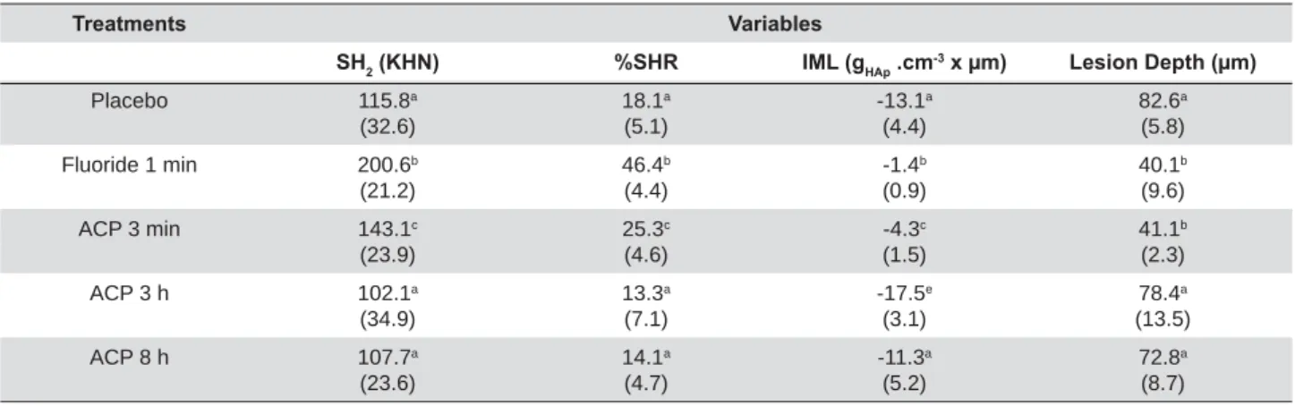
The Fluoride 1 min group presented the highest mean value for SH2 and %SHR (Table 1) compared
with the other groups (p<0.001). The Placebo, ACP 3
h and ACP 8 h showed similar results (SH2: p=0.599;
%SHR: p=0.148) with lower values compared with the other groups (p<0.001).
The mean (SD) mineral concentration value for
sound bovine enamel of the blocks analyzed was 2.47
(0.03) gHAp.cm-3 (2.40-2.53). The results from the SMCT analysis showed that the Fluoride 1 min group
presented the lowest value (p<0.001) for integrated
loss of subsurface mineral (IML) compared with the
other groups (Table 1). There was a statistically
3 min and the ACP 3 h and ACP 8 h groups (Table
between SH2 and IML (r value=0.839; R2=0.704).
The lesion depth (Table 1 and Figure 1) was higher
in the placebo and ACP 3 h and ACP 8 h groups
(p=0.126) when compared with the groups treated
1 min groups presented similar (p=0.849) depth
correlation between the lesion depth and SH2 (r value=-0.846, R2=0.716).
The mineral concentration profile in function
of the depth demonstrated an outer enamel hypermineralization for Fluoride 1 min group (Figure
was followed by an area of low mineralization in all
groups (Figure 1 and Figure 2a). The ACP 3 h and ACP 8 h groups presented a subsurface lesion with
development of laminations in all blocks of the two
groups (Figure 1b and 1c, Figure 2b).
The values integrated from differential mineral
patterns (Table 2 and Figure 1d). The remineralization
process for Fluoride 1 min group was ~74% lower at
zone B (19.6-33.6 μm) when compared with zone A (2.8-16.8 μm) (p<0.001). This relation was ~40% for
ACP group (p<0.019). There was different mineral loss
at zone B for ACP 3 h and ACP 8 h groups compared
with the treatments of the other groups (p<0.001). Although ring artifacts were observed in all
reconstructed images, they did not interfere with the
measurements of mineral concentrations in enamel
up to 221.2 μm of depth, as they were closer to the dentino-enamel junction (Figure 2).
Treatments Variables
SH2 (KHN) %SHR IML (gHAp .cm-3 x μm) Lesion Depth (μm)
Placebo 115.8a
(32.6) 18.1a (5.1) -13.1a (4.4) 82.6a (5.8)
Fluoride 1 min 200.6b
(21.2) 46.4b (4.4) -1.4b (0.9) 40.1b (9.6)
ACP 3 min 143.1c
(23.9) 25.3c (4.6) -4.3c (1.5) 41.1b (2.3)
ACP 3 h 102.1a
(34.9) 13.3a (7.1) -17.5e (3.1) 78.4a (13.5)
ACP 8 h 107.7a
(23.6) 14.1a (4.7) -11.3a (5.2) 72.8a (8.7) /RZHUFDVHOHWWHUVLQGLFDWHVWDWLVWLFDOO\VLJQL¿FDQWGLIIHUHQFHV6WXGHQW1HZPDQ.HXOVSEHWZHHQWKHJURXSVLQHDFKDQDO\VLV SH2=surface hardness after pH-cycling.
6+5 SHUFHQWDJHRIVXUIDFHKDUGQHVVUHFRYHU\ IML=integrated loss of subsurface mineral.
Discussion
The present in v it r o laboratory study evaluated
the effectiveness of CPP-ACP, as it would be applied for extended time in trays. As a null hypothesis, the
CPP-ACP applied for 3 minutes would present the
same remineralizing effect as compared with CPP-ACP
applications for 3 hours or 8 hours. The ACP 3 h group
was tested because it is stated by the manufacturer (instructions for use by GC Corporation, Tokyo, Japan)
that the longer the CPP-ACP stays in the mouth,
the more effective it will be releasing calcium and
phosphate continuously for the 3 hours. As there is the necessity of more research using CPP-ACP with Figure 1-'HSWKSUR¿OHVRIPLQHUDOFRQFHQWUDWLRQJHAp.cm-3LQOHVLRQVIRUHDFKWUHDWPHQWD&RPSDULVRQRIPLQHUDOSUR¿OHVIURP3ODFHER
)OXRULGHPLQDQG$&3PLQJURXSVE0LQHUDOSUR¿OHVIURP3ODFHER$&3KDQG$&3KF0LQHUDOSUR¿OHVIURP3ODFHER$&3 PLQ$&3KDQG$&3KG'LIIHUHQWLDOPLQHUDOFRQFHQWUDWLRQSUR¿OHVDVDIXQFWLRQRIGHSWKDFFRUGLQJWRWKHWUHDWPHQWV]RQH$ ±ȝPDQG]RQH%±ȝPPD[LPXPPLQHUDOFRQFHQWUDWLRQRQRXWHUHQDPHOOD\HU: maximum mineral concentration
through the lesion for each group.ØVXEVXUIDFHOHVLRQĻODPLQDWLRQ
different approaches, such as longer treatment time15,
the time of 8 hours was tested for comparisons.
Clinically, the patient would be instructed to apply the paste using a tray for 3 or 8 hours (overnight), mainly
after dental bleaching.
Application of CPP-ACP for 3 minutes (ACP 3 min)
was capable of improving enamel remineralization (Table 1,Table 2 and Figure 1), reducing mineral loss
(IML and 'IML) and lesion depth (μm). As described
in the literature, the CPP-ACP has the ability to adhere
to enamel and supersaturate the environment with free calcium and phosphate ion activities3,11,17. Both
the outer part and the inner part of the lesion showed
a more homogeneous remineralization ratio (Table 2). Probably the great diffusion of calcium phosphate
neutral ion pair (CaHPO40), produced by calcium
and phosphate supersaturation1,11, leads to a higher
mineral recovery in the inner part of the lesion (zone B). The diffusion of charged ions (Ca2+ and PO
43-) through a charged enamel surface layer is lower than
neutral ion2. Despite producing lower mineral recovery
than the Fluoride 1 min group, the greater diffusion of neutral calcium phosphate reduced the lesion depth
at the same level (Table 1 and Figure 1).
in the outer part of the lesion (zone A, Table 2 and Figure 1a) mainly due its strong deposition into the
outer zones of the enamel lesion13,14. In addition,
the presence of fluoride promoted higher ionic
activity to HF0 and greater degree of saturation with
on remineralization11
products in the outer part of enamel resulted in higher
surface hardness (Table 1) and mineral content (zone A, Table 2) than the products precipitated by CPP-ACP.
Surface hardness varied according to the integrated
loss of subsurface mineral (IML) and lesion depth,
allowing an evaluation of mineral loss through a faster
and more direct method. However, it did not identify
(Figure 1).
The increase in the time of contact with the
surface enamel (3 h and 8 h) simulating an overnight application did not show superior mineral gain
compared with the placebo group as well as for lesion
depth. It had been expected that by maintaining
prolonged contact of CPP-ACP with enamel would result in wider diffusion of neutral calcium phosphate
into the enamel reducing the lesion to a greater
extent. The greater time of contact with enamel produced laminations in the extension of the original
lesion with mineral deposition within the body lesion
(Figure 1b and 1c). The development of lamination is
due to alternating de-/remineralization episodes and
can be adsorbed in varying concentrations at various
depths, resulting in differences in acid susceptibility
of the mineral13,14. However, there is no previous report on the development of lamination produced
by supersaturation of calcium and phosphate from a
source of stabilized casein phosphopeptide-amorphous
calcium phosphate (CPP-ACP). But the zones of high and low mineral content through the lesion were
similar to those described by Lagerweij and ten
Cate13 (2006): the surface zone with high mineral
concentration, the original lesion with low mineral concentration, followed by a zone with a high mineral
density, then a secondary lesion which is formed during
the short demineralization of the pH-cycling model,
and underneath these lesion zones is the sound enamel (Figure 1a, 1b and 1c).
The prolonged contact with the enamel leads to a
Treatments HAp.cm-3 × μm)
zone A (2.8-16.8 μm) zone B (19.6-33.6 μm)
Fluoride 1 min 6.24a,A
(0.69)
1.60a,B
(0.40)
ACP 3 min 3.56b,A
(0.79)
2.11a,B
(0.61)
ACP 3 h 0.08d,A
(1.43)
-3.50b,B
(1.66)
ACP 8 h 1.71c,A
(2.29)
-1.59c,B
(2.47)
/RZHUFDVHOHWWHUVLQGLFDWHVWDWLVWLFDOO\VLJQL¿FDQWGLIIHUHQFHVEHWZHHQWKHJURXSVLQHDFK]RQH&DSLWDOOHWWHUVLQGLFDWHWKHGLIIHUHQFHV EHWZHHQ]RQH$DQG]RQH%IRUHDFKWUHDWPHQW6WXGHQW1HZPDQ.HXOVS
Table 2-0HDQYDOXHV6'RILQWHJUDWHGRIGLIIHUHQWLDOSUR¿OHVǻ,0/FDOFXODWHGIRUWZR]RQHV)LJXUHGLQWKHHQDPHOOHVLRQVDFFRUGLQJ
great precipitation of calcium phosphate on enamel
surface from CPP-ACP with formation of laminations
through the lesion during the pH cycling process (Figure 1b, 1c and Figure 2). With the development
of laminations, it seems likely that the mineral
precipitated from CPP-ACP is not capable of reducing
the acid diffusion through the lamination, increasing the mineral loss in the periphery of the lesion. It is
possible that the higher precipitation of CPP-ACP led
to obstruction of the enamel pores, hindering CaHPO40 diffusion into the enamel lesion. Nevertheless, the
precipitate from CPP-ACP does not prevent the acid
diffusion and it still occurs into the enamel producing
a new area of demineralization beyond the inner boundary of the outermost demineralized area (Figure
1b, 1c and Figure 2). As the ionic exchanges with the
medium are altered due to pore obstruction, part of the
demineralization product accumulates in the boundary between the outermost and innermost demineralized
areas, forming a zone with greater mineral density.
This may explain in part the lower remineralization
capacity of CPP-ACP applied for 3 minutes in relation
due to the short time in acid medium. Thus, the lower
resistance of mineral precipitated from the CPP-ACP
applied for extended time to acid dissolution and diffusion presented the formation of laminations. The
mineral (re)precipitation produced in the presence
acid diffusion and presented lower acid dissolution in the remineralization model with the short time of
demineralization. It is possible to observe in the region
between 50.4 to 61.6 μm a remnant of lamination in
Conclusion
Based on the results of this in vit r o study, the use
of CPP-ACP for extended periods of time as a way of
increasing the effectiveness of the product produced no additive effect on the remineralization process.
Additional studies should be performed using MI Paste
Acknowledgments
Use of the Advanced Photon Source was supported
DE-AC02-06CH11357.
interest.
References
1- Amaral JG, Sassaki KT, Martinhon CC, Delbem AC. Effect of
low-enamel demineralization in sit u. Am J Dent. 2013;26:75-80. 2- Chu YS, Liu C, Mancini DC, de Carlo F, Macrander AT, Shu D.
Performance of a double multilayer monochromator at 2-BM at the Advanced Photon Source. Rev Sci Instrum. 2002;73:1485-7.
3- Cochrane NJ, Saranathan S, Cai F, Cross KJ, Reynolds EC. Enamel subsurface lesion remineralisation with casein phosphopeptide
2008;42:88-97.
4- Cross KJ, Huq NL, Stanton DP, Sum M, Reynolds EC. NMR studies
nanocomplexes. Biomaterials. 2004;25:5061-9.
5- Danelon M, Takeshita EM, Peixoto LC, Sassaki KT, Delbem AC. Effect
demineralization. Clin Oral Investig. 2014;18:1119-27.
6- Danelon M, Takeshita EM, Sassaki KT, Delbem AC. I n sit u evaluation
enamel remineralization. Am J Dent. 2013;26:15-20.
7- Delbem AC, Cury JA. Effect of application time of APF and NaF gels
in vit r o enamel caries. Am J Dent. 2002;15:169-72.
8- Delbem AC, Danelon M, Sassaki KT, Vieira AE, Takeshita EM, Brighenti
FL, Rodrigues E. Effect of rinsing with water immediately after neutral
an in sit u study. Arch Oral Biol. 2010;55:913-8.
9- Delbem AC, Sassaki KT, Vieira AE, Rodrigues E, Bergamaschi M,
Stock SR, et al. Comparison of methods for evaluating mineral loss: hardness versus synchrotron microcomputed tomography. Caries Res.
2009;43:359-65.
10- Delbem AC, Vieira AE, Sassaki KT, Cannon ML, Stock SR, Xiao
X, et al. Quantitative analysis of mineral content in enamel using synchrotron microtomography and microhardness analysis. Proc SPIE.
2006;6318:631824-1.
11- Ferreira L, Pedrini D, Okamoto AC, Jardim Júnior EG, Henriques
TA, Cannon M, et al. Biochemical and microbiological characteristics of
in sit u
calcium phosphate. Am J Dent. 2013;26:207-13.
12- Gupta R, Prakash V. CPP-ACP complex as new adjunctive agent for remineralisation: a review. Oral Health Prev Dent. 2011;9:151-65.
13- Lagerweij MD, ten Cate JM. Acid susceptibility at various depths of pH-cycled enamel and dentine specimens. Caries Res. 2006;40:33-7.
baseline severity and mineral distribution on lesion progression. Caries
15- Pulido MT, Wefel JS, Hernandez MM, Denehy GE,
Guzman-Armstrong S, Chalmers JM, Qian F. Inhibitory effect of MI paste &
Oper Dent. 2008;33:550-5.
16- Queiroz CS, Hara AT, Leme FP, Cury JA. pH-cycling models to
remineralization. Braz Dent J. 2008;19:21-7.
17- Reynolds EC. Calcium phosphate-based remineralization systems:
18- Spiguel MH, Tovo MF, Kramer PF, Franco KS, Alves KM, Delbem AC.
the de/remineralization process: an in vit r o and in sit u study. Caries
Res. 2009;43:302-7.
19- Vieira AE, Delbem AC, Sassaki KT, Rodrigues E, Cury JA, Cunha
RF. Fluoride dose response in pH-cycling models using bovine enamel. Caries Res. 2005;39:514-20.